Adsorption site determination of a molecular monolayer via inelastic tunneling
Abstract
We have combined scanning tunneling microscopy with inelastic electron tunneling spectroscopy (IETS) and density functional theory (DFT) to study a tetracyanoethylene monolayer on Ag(100). Images show that the molecules arrange in locally ordered patterns with three non-equivalent, but undeterminable, adsorption sites. While scanning tunneling spectroscopy only shows subtle variations of the local electronic structure at the three different positions, we find that vibrational modes are very sensitive to the local atomic environment. IETS detects sizeable mode frequency shifts of the molecules located at the three topographically detected sites, which permits us to determine the molecular adsorption sites through identification with DFT calculations.
keywords:
scanning tunneling microscopy, density functional theory, hybrid organometallic interfaces, adsorption site determination, inelastic electron tunneling spectroscopy, vibrational modesUniversität Münster and CeNTech]Physikalisches Institut and Center for Nanotechnology (CeNTech), Westfälische Wilhelms-Universität Münster, 48149 Münster, Germany University of California at Berkeley]Department of Physics, University of California at Berkeley, and Materials Science Division, Lawrence Berkeley National Laboratory, Berkeley, California 94720-7300, USA University of California at Berkeley]Department of Physics, University of California at Berkeley, and Materials Science Division, Lawrence Berkeley National Laboratory, Berkeley, California 94720-7300, USA University of California at Berkeley]Department of Physics, University of California at Berkeley, and Materials Science Division, Lawrence Berkeley National Laboratory, Berkeley, California 94720-7300, USA University of California at Berkeley]Department of Physics, University of California at Berkeley, and Materials Science Division, Lawrence Berkeley National Laboratory, Berkeley, California 94720-7300, USA University of California at Berkeley]Department of Physics, University of California at Berkeley, and Materials Science Division, Lawrence Berkeley National Laboratory, Berkeley, California 94720-7300, USA Centre d’Investigació en Nanociència i Nanotecnologia]Centre d’Investigació en Nanociència i Nanotecnologia, CIN2 (CSIC-ICN), Campus de la UAB, 08193 Bellaterra, Spain
Many phenomena are determined by the way molecules adsorb on substrates, e.g., heterogeneous catalysis 1, 2, transport in molecular electronics 3, and competing many-body interactions 4. This has led to a large effort to apply surface science tools for adsorption analysis. Particularly powerful techniques for investigating large arrays of ordered molecules use diffraction and interference effects. For instance, low-energy electron diffraction 5 conveys information on molecular arrangements, X-ray standing waves reveal the adsorbate’s vertical geometrical structure 6, and surface X-ray diffraction can determine the structure of molecular adlayers by counteracting the penetration depth of X-rays using grazing-incidence angles 7. As the number of molecules inside the repeating pattern increases, these techniques grow more complex and the diffraction patterns become increasingly difficult to unravel. In such cases, local-probe techniques can be particularly useful. Indeed, the scanning tunneling microscope (STM) has proven to be a powerful tool to for structure determination of atomic surfaces and molecular layers 8, 9, 10. Recent progress in scanning probe techniques even permits the detection of chemical bonds within and between molecules 11, 12.
With the active search of molecular nanostructures, very complex arrays of dense molecular layers have become ubiquitous in surface science 13, 14, 15, 16, 17, 18. Structure analysis by diffraction techniques becomes hopeless when commensurability issues between molecule and substrate set in, impeding long-range order on the surface. While such structures can only be studied by local-probe techniques, exact determination of adsorption sites via STM alone remains very difficult due to the challenge to acquire molecular and surface atom positions simultaneously with high precision 19, 20, 21, 22, 23.
Analysis of the scanning tunneling spectroscopy (STS) structure by inelastic effects during electron tunneling allows a deeper understanding of molecular behavior at a surface. Such inelastic electron tunneling spectroscopy (IETS) has become a very successful tool to detect vibrational, photon, and even spin excitations 24, 25, 26. In its vibrational version, the extraordinary spatial resolution of IETS 27 allows chemical analysis capabilities at the atomic scale 28, 29, 30, 31, 32, 33. Moreover, IETS is generally below the meV energy resolution. Hence, we can explore small environment-induced frequency shifts with intra-molecular resolution. This naturally leads us to explore whether IETS can improve adsorption site determination through observation of systematic shifts in vibrational-mode frequencies for molecules locally bound at different adsorption sites on a surface.
Here, we demonstrate that IETS can indeed yield accurate information on molecular binding geometries within a defect-free densely-packed molecular monolayer that lacks long-range order and thus would be challenging to study using diffraction-based techniques. The studied system is tetracyanoethylene (TCNE, \ceC6N4) adsorbed on Ag(100). TCNE is known to be an electron acceptor showing strong interactions with metals due to the four cyano-group low-energy empty orbitals conjugated with the central C=C double bond 34. TCNE plays a crucial role in molecule-based room-temperature magnets and spin-injection devices 35, 36. Given the local character of the cyano groups and the non-commensurability of TCNE with the Ag(100) unit cell, a strong stressed adsorption is to be expected that can give rise to complex patterns 37, 38. Indeed, we found that TCNE forms a short-range ordered monolayer exhibiting three non-equivalent adsorption sites. While STM imaging and STS only show subtle differences in the local electronic density of states (LDOS) that cannot help to determine specific adsorption sites, we found that vibrational modes, as observed in IETS, give quantitative differences in the mode energy of a particular vibration due to local changes of molecule-substrate interaction. This inelastic information serves as a reliable input for comparisons with DFT calculations, permitting structural determination of the TCNE monolayer.
The experiments were performed in ultrahigh vacuum (UHV) using a home-built STM operated at . The Ag(100) single-crystal substrate was cleaned by standard sputter-annealing procedures, followed by TCNE deposition at room temperature through a leak valve 37. After deposition, the sample was transferred in situ to the cryogenic STM. Topography images were taken in constant-current mode, and STS and IETS were performed by measuring the differential conductance and the 2nd derivative as a function of the sample bias by standard lock-in techniques (modulation (rms), frequency ) under open-feedback conditions.

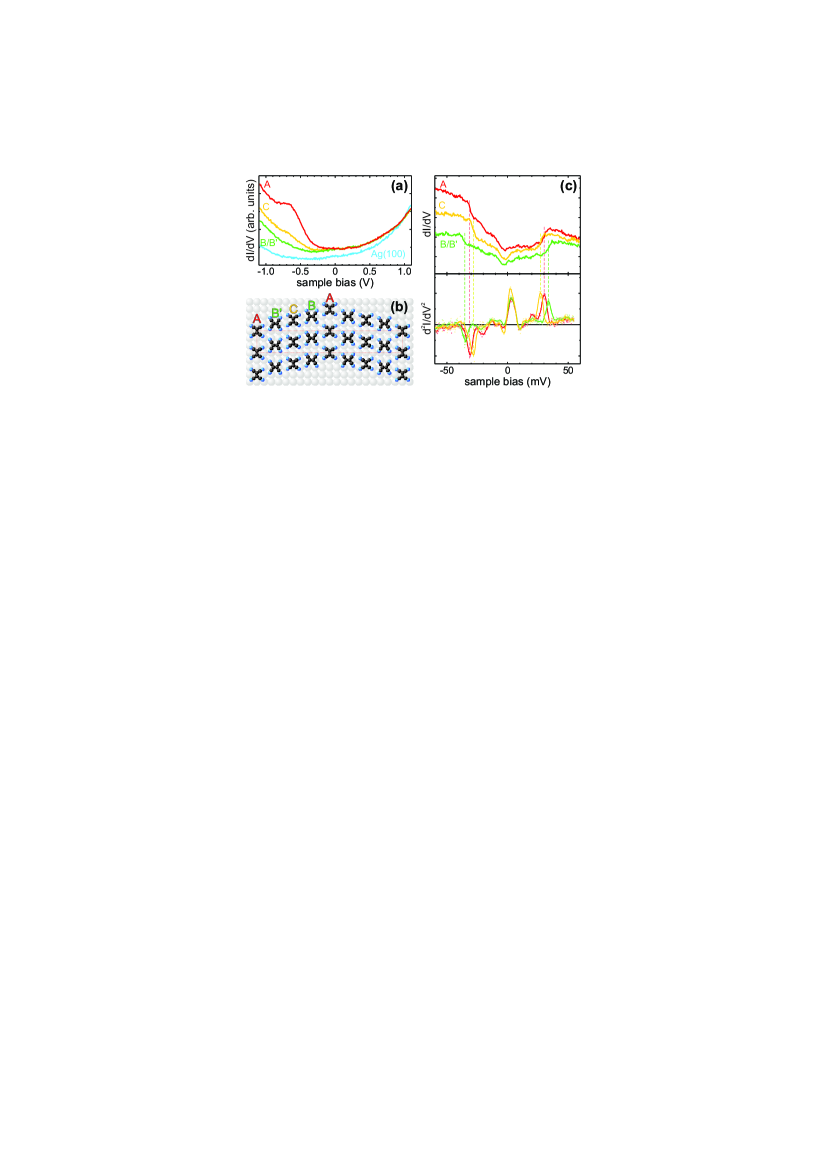
Fig. 1 shows STM images of the closed-packed TCNE monolayer on Ag(100) \bibnoteLocal monolayer formation can already be observed for coverages above 10% 37.. A clear pattern with local regular order can be observed. Molecules arrange in rows along the [110] direction with an intermolecular distance of Å, i.e., about three times the Ag(100) surface lattice constant. Neighboring rows do not form a rectangular arrangement since the molecules are shifted along the [110] direction in order to create a dense packing. Fig. 1a shows a highly resolved STM image of the monolayer pattern. The molecular shape seen here mainly reflects the lowest unoccupied molecular orbital (LUMO) of neutral TCNE and permits determining molecular orientations 37, 38. Within each row, all TCNEs orient identically with the C=C double bond being either parallel or perpendicular to the row. From row to row, the molecular orientation always changes by . When the double bond lies along the row direction, the LUMO shape looks slightly distorted with two of the four cyano legs appearing less pronounced.
The apparent height of the monolayer exhibits a significant bias dependence (Fig. 1c,d). For positive sample bias, each molecule is seen as an almost round protrusion with only slight height differences. However, when , molecular shapes are distorted and TCNEs in some rows appear much higher than in others. Overall, we identify three different heights and mark the corresponding rows of TCNE with “A”, “B”, and “C”, respectively. From the highly resolved STM images, we can already make a first connection between the different electronic properties and the structure: B molecules always have the C=C double bonds oriented perpendicular to the row direction, while A and C molecules always have it aligned parallel.
While this local order is observed throughout the entire monolayer, we do not observe long-range order. Rather, the pattern is easily perturbed by local defects or missing molecules. This results in a variation of combinations of A, B and C rows. In regions with more defects, the pattern consists of alternating A and B rows only (cf. Fig. 1a), leading to a chevron pattern with a rectangular unit cell that contains two TCNEs. In other areas, we observe a larger regular chevron structure with an A-B-C-B-A… sequence, resulting in a unit cell with four molecules. The largest chevron pattern observed consists of eight molecular rows, as seen in Fig. 1c. This pattern reveals a fourth row (B’) that, however, seems to behave similar to B rows. This patterns spans a unit cell of containing eight molecules.
In order to better understand the molecular LDOS variations, we have performed spatially resolved STS measurements on all TCNE molecules within the monolayer pattern. As expected from the topographical analysis, we find that all molecules within a row exhibit identical tunneling spectra. We can therefore summarize all spectroscopic features by showing a representative spectrum for each row (Fig. 2a). Compared to the spectrum on a bare Ag(100) terrace, all TCNE spectra exhibit only minor differences at positive bias (i.e., unoccupied states), while we observe a significant increase in the signal at negative sample bias (i.e., occupied states). B and B’ rows show identical spectra and exhibit a monotonous increase as we go to larger negative bias. Molecules in C rows show a similar monotonous LDOS increase, but with a larger slope. The largest slope is observed for TCNE in A rows, and the spectrum shows an additional broad peak at -0.66 V. This spectroscopic feature is reminiscent of that observed for isolated TCNE on Ag(100), where molecules were found to be adsorbed on top of Ag atoms 37.
The observed structural and electronic features described up to this point allow us to propose a rough structural model (Fig. 2b) where the rows of TCNE molecules form a locally commensurate structure on the Ag(100) surface with unit cells (). The spectral resemblance with isolated TCNE leads us to suggest that molecules in row A are likely to be adsorbed on top of Ag atoms 37. Further assuming an arrangement of molecules A-B-C-B’-A along a straight line, we find that for C molecules the bridge site is a probable adsorption position with high symmetry. However, B and B’ molecules would then lie in a position of very low symmetry. Within our experimental accuracy, we cannot rule out that these molecules relax laterally to a bridge or hollow site, both being only about 0.7 Å away. Consequently, our experimental structure model requires comparison with calculations which are rendered difficult due to the lack of quantifiable spectroscopic features.
The situation changes altogether when we take a look at the vibrational structure of the TCNE molecules by performing STS with high energy resolution (Fig. 2c). Again, we find that all molecules within a specific row exhibit identical spectra. In all cases spectra show pronounced step-like features at about and meV with a conductance change of 2-3%. This is a well-known signature of IETS (Fig. 2c, top) 24. The energy of this feature is in good agreement with reported values of the in-plane rocking mode as well as the out-of-plane wagging mode of TCNE 40, and it has been observed via STM-IETS for TCNE in various local environments 32, 41. Upon closer inspection, we find that the exact IETS energy depends on the location of TCNE within the monolayer pattern. This can be seen clearly in the spectra (Fig. 2c, bottom). Molecules exhibit a mode energy of meV in row A, meV in row B, and meV in row C. The error bars are much smaller than the frequency shifts and the actual measured frequencies for single measurements never overlapped among different types of molecules. These data are based on a statistical analysis of 32 molecules. Hence, the observed three different mode energies are clearly resolvable and distinguishable. Stiffening or softening of vibrational modes can be caused by local variations of intermolecular or molecule-substrate interactions. Therefore, the observed energies serve as quantitative fingerprints that can help to identify the three non-equivalent adsorption sites of TCNE within the monolayer. Despite both sitting on bridge sites, the B and C conformations are different because the molecular C–C axis in C molecules aligns along the row, while in B rows it is perpendicular to the row. Hence, an individual B molecule is rotated with respect to an individual C molecule. As a consequence, the reconstructed Ag layer below B and C molecules is different, affecting both local electronic structure and mode frequency.
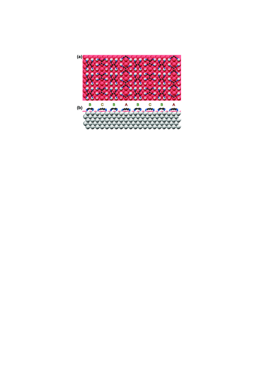
This adsorption-site determination can be achieved by comparing our experimental findings with DFT calculations. We show that comparing only the electronic structure is inconclusive, whereas the added information from IETS is decisive to permit the correlation between vibrational modes and the underlying geometrical structure. We first perform a structural analysis by calculating a () unit cell containing four molecules. The substrate was modeled using five Ag substrate layers. The first two layers and the molecules were relaxed until forces on the atoms were less than 0.01 eV/Å. We used the vasp code and the PBE approximation for the exchange and correlation functional 42, 43, 44. Fig. 3 shows the geometry of the simulated TCNE monolayer on Ag(100). A transparent plane was added to emphasize the degree of distortion of Ag atoms in the surface layer due to molecule-substrate interactions. Type A molecules sit on top of a silver atom with elastic substrate-mediated intermolecular interactions among A molecules since the substrate distortion aligns parallel to the molecular rows of the same species. Type B molecules are on bridge sites and are completely surrounded by high-lying Ag atoms. Type C molecules also adsorb on bridge sites but with an orientation similar to A-type molecules, which also leads to an aligned substrate distortion parallel to the molecular row. Thus, the local adsorption environment is indeed different for all three types of TCNE molecules. All molecules are adsorbed via the N–Ag local interactions, and the arrangement provides denser packing. The simulated Tersoff-Hamann image (Fig. 1b) is in very good agreement with the experimental STM topography at small sample bias 45. Calculations of the density of states projected onto the molecular orbitals (not shown), demonstrate that the LUMO of all molecules is broadened and shifted slightly below the Fermi energy. While this explains the LUMO character in STM images at small sample bias, the differences in the electronic structure between A, B and C sites are too small to account for the experimentally observed image contrast at larger bias (Fig. 1c,d) as well as the STS differences seen in Fig. 2a. Thus, a comparison between the experimentally derived and the calculated structural model via electronic structure is not conclusive, likely due to the reliance of the simulations on the non-physical Kohn-Sham orbitals.
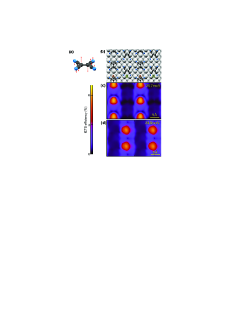
On the other hand, the vibrational structure simulations do not suffer from these deficiencies. For the analysis of vibrational modes, we restrict our discussion to the smallest observed () unit cell containing only molecules A and B (Fig. 4a). The IETS simulation is performed using a many-body perturbation extension of the Bardeen tunneling theory 46, 47. Unfortunately, DFT calculations of vibrational energies of adsorbed molecules on a surface are generally shifted and at best within 10% of the experimental values. Hence, while absolute frequency values are not reliable, the frequency difference between modes is very accurate 48. Our IETS simulations predict that only one mode dominates the signal within the experimental energy range. This is the out-of-plane wagging mode illustrated in Fig. 4c. Two different frequencies are found for molecules A and B. The out-of-plane wagging mode of type A molecules is located at meV, while that of type B molecules is found at meV. The maximum fraction of computed inelastic electrons is 7%, whereas all other modes yield inelastic fractions below 1%. The direction and magnitude of the energy shift meV are in good agreement with the experimentally determined shift of meV. The frequency shift of the other molecular modes does not always follow this trend. Especially, the second possible candidate, the in-plane rocking mode, is found at 30.8 meV for molecule A and at 28.3 meV for molecule B in the simulations, i.e., the shift is opposite to the experimental observation. Using DFT-D2 to account for dispersion forces does not alter this conclusion \bibnoteNeglecting dispersion corrections can lead to wrong vertical molecule-substrate distances and intramolecular distortions 52, 53, 54. Using the DFT-D2 approach as coded in VASP 55, the N atoms move 0.03 Å and the C=C double bond moves 0.25 Å closer to the surface. However, the vibrational modes exhibit only minor softening. In particular, the wagging mode is shifted by 2 meV, well within the error bar of the calculations.. The simulations hence show that only the out-of-plane wagging mode is excited in the experiment.
We have performed an equivalent calculation for a fictitious CBC structure in order to prove that indeed our geometrical assignment is correct. The CBC structure is energetically less favorable than the ABA structure and indeed, it is not found experimentally \bibnoteOur calculations show that a molecule adsorbed with its axis on a top site (such as the A molecules) are 0.6 eV more bound to the substrate than molecules on a bridge site (such as B and C molecules). Hence stable structures contain A molecules in their periodic pattern.. Nevertheless, due to the locality of vibrational properties, we expect that the CBC structure captures the frequency shifts of C with respect to B molecules, although we emphasize that a quantitative comparison with the ABA calculations is not valid. Despite this, B molecules show a wagging mode frequency of 29.2 meV in very good agreement with the value of the ABA structure. Furthermore, we find that the wagging mode of C molecules is 1.9 meV lower in energy than that of B molecules, i.e., the trend is in agreement with the experimental finding. Thus, the mode frequency is not only determined by the adsorption site but also by the local molecular environment.
Due to the localization of the vibrations to each molecule, the IETS signal is also well localized. This permits detecting the different molecular adsorption sites. Localization is a general feature of intramolecular modes. In fact, it leads to very weakly dispersing optical-like phonons in molecular adlayers that can be individually excited within a molecule 51. Fig. 4c shows a map of conductance change with bias when the mode at is excited. The IETS simulations clearly reflect the spatial distribution of type A molecules with a maximum IETS signal at the molecular center, in agreement with the experimental findings. When the mode at is excited, it is localized to type B molecules, as shown in Fig. 4d. Thus, we find very good agreement between the experimental IETS results and the calculated vibrational structure and IETS simulations, which permits to confirm the DFT-calculated structure conclusively. Particularly, we conclude that type A molecules adsorb on top and type B and C molecules on bridge sites of the Ag(100) surface.
This proof-of-principle study shows that IETS combined with DFT can be used to discriminate between non-equivalent molecular adsorption environments in a dense complex molecular monolayer. The high chemical sensitivity of IETS enables the detection of small variations in molecular environments that easily lead to meV spectroscopic changes, i.e., well within the typical IETS energy resolution, while the high spatial resolution of IETS displays intramolecular localization when intrinsic molecular modes are excited. These unique properties combined with DFT reveal hidden geometrical structure not attainable by the usual structural methods.
This work was supported by the Director, Office of Science, Office of Basic Energy Sciences of the US Department of Energy under contract no. DE-AC02-05CH11231 (STM instrumentation development and measurements), and by the Deutsche Forschungsgemeinschaft (DFG) project WE 4104/2-1 (numerical simulations and analysis). D.W. acknowledges support by the Alexander von Humboldt Foundation (data acquisition) and the North-Rhine Westphalian Academy of Sciences and Arts (data analysis). N.L. is supported by the ICT-FET Integrated Project AtMol (http://www.atmol.eu) (IETS simulation code development).
References
- Hofmann et al. 1994 Hofmann, P.; Schindler, K.-M.; Bao, S.; Bradshaw, A. M.; Woodruff, D. P. Nature 1994, 368, 131
- Frank and Bäumer 2000 Frank, M.; Bäumer, M. Phys. Chem. Chem. Phys. 2000, 2, 3723
- Reuter et al. 2011 Reuter, M. G.; Seideman, T.; Ratner, M. A. J. Chem. Phys. 2011, 134, 154708
- Franke et al. 2011 Franke, K. J.; Schulze, G.; Pascual, J. I. Science 2011, 332, 940
- Heinz 1995 Heinz, K. Rep. Prog. Phys. 1995, 58, 637
- Hauschild et al. 2005 Hauschild, A.; Karki, K.; Cowie, B. C. C.; Rohlfing, M.; Tautz, F. S.; Sokolowski, M. Phys. Rev. Lett. 2005, 94, 036106
- Robinson and Tweet 1992 Robinson, I. K.; Tweet, D. J. Rep. Prog. Phys. 1992, 55, 599
- Binnig et al. 1983 Binnig, G.; Rohrer, H.; Gerber, C.; Weibel, E. Phys. Rev. Lett. 1983, 50, 120–123
- De Feyter and De Schryver 2003 De Feyter, S.; De Schryver, F. Chem. Soc. Rev. 2003, 32, 139–150
- Heinz and Hammer 2004 Heinz, K.; Hammer, L. J. Phys. Chem. B 2004, 108, 14579–14584
- Gross et al. 2009 Gross, L.; Mohn, F.; Moll, N.; Liljeroth, P.; Meyer, G. Science 2009, 325, 1110
- Temirov et al. 2008 Temirov, R.; Soubatch, S.; Neucheva, O.; Lassise, A. C.; Tautz, F. S. New J. Phys. 2008, 10, 053012
- Barth et al. 2005 Barth, J. V.; Constantini, G.; Kern, K. Nature 2005, 437, 671–679
- Wang et al. 2007 Wang, Y.; Yamachika, R.; Wachowiak, A.; Grobis, M.; Khoo, K. H.; Lee, D.-H.; Louie, S. G.; Crommie, M. F. Phys. Rev. Lett. 2007, 99, 086402
- Otero et al. 2008 Otero, R.; Lukas, M.; Kelly, R. E. A.; Xu, W.; Lægsgaard, E.; Stensgaard, I.; Kantorovich, L. N.; Besenbacher, F. Science 2008, 319, 312
- Wang et al. 2010 Wang, Y.-L.; Ren, J.; Song, C.-L.; Jiang, Y.-P.; Wang, L.-L.; He, K.; Chen, X.; Jia, J.-F.; Meng, S.; Kaxiras, E.; Xue, Q.-K.; Ma, X.-C. Phys. Rev. B 2010, 82, 245420
- Schöll et al. 2010 Schöll, A.; Kilian, L.; Zou, Y.; Ziroff, J.; Hame, S.; Reinert, F.; Umbach, E.; Fink, R. H. Science 2010, 329, 303
- McGuiness et al. 2010 McGuiness, C. L.; Diehl, G. A.; Blasini, D.; Smilgies, D.-M.; Zhu, M.; Samarth, N.; Weidner, T.; Ballav, N.; Zharnikov, M.; Allara, D. L. ACS Nano 2010, 4, 3447–3465
- Weiss and Eigler 1993 Weiss, P. S.; Eigler, D. M. Phys. Rev. Lett. 1993, 71, 3139–3142
- Meyer et al. 1996 Meyer, G.; Zöphel, S.; Rieder, K.-H. Phys. Rev. Lett. 1996, 77, 2113
- Böhringer et al. 1998 Böhringer, M.; Schneider, W.-D.; Glöckler, K.; Umbach, E.; Berndt, R. Surf. Sci. 1998, 419, L95
- Lagoute et al. 2004 Lagoute, J.; Kanisawa, K.; Fölsch, S. Phys. Rev. B 2004, 70, 245415
- Tautz 2007 Tautz, F. S. Prog. Surf. Sci. 2007, 82, 479
- Stipe et al. 1998 Stipe, B. C.; Rezaei, M. A.; Ho, W. Science 1998, 280, 1732
- Ćavar et al. 2005 Ćavar, E.; Blüm, M.-C.; Pivetta, M.; Patthey, F.; Chergui, M.; Schneider, W.-D. Phys. Rev. Lett. 2005, 95, 196102
- Heinrich et al. 2004 Heinrich, A. J.; Gupta, J. A.; Lutz, C. P.; Eigler, D. M. Science 2004, 306, 466
- Ho 2002 Ho, W. J. Chem. Phys. 2002, 117, 11033–11061
- Lee and Ho 1999 Lee, H. J.; Ho, W. Science 1999, 286, 1719–1722
- Pascual et al. 2001 Pascual, J. I.; Jackiw, J. J.; Song, Z.; Weiss, P. S.; Conrad, H.; Rust, H.-P. Phys. Rev. Lett. 2001, 86, 1050–1053
- Lauhon and Ho 2000 Lauhon, L. J.; Ho, W. Phys. Rev. Lett. 2000, 84, 1527–1530
- Kim et al. 2002 Kim, Y.; Komeda, T.; Kawai, M. Phys. Rev. Lett. 2002, 89, 126104
- Wegner et al. 2009 Wegner, D.; Yamachika, R.; Zhang, X.; Wang, Y.; Baruah, T.; Pederson, M. R.; Bartlett, B. M.; Long, J. R.; Crommie, M. F. Phys. Rev. Lett. 2009, 103, 087205
- Okabayashi et al. 2010 Okabayashi, N.; Paulsson, M.; Ueba, H.; Konda, Y.; Komeda, T. Phys. Rev. Lett. 2010, 104, 077801
- Miller 2006 Miller, J. S. Angew. Chem. Int. Ed. 2006, 45, 2508
- Manriquez et al. 1991 Manriquez, J. M.; Yee, G. T.; McLean, R. S.; Epstein, A. J.; Miller, J. S. Science 1991, 252, 1415–1417
- Yoo et al. 2010 Yoo, J.-W.; Chen, C.-Y.; Jang, H. W.; Bark, C. W.; Prigodin, V. N.; Eom, C. B.; Epstein, A. J. Nat. Mater. 2010, 9, 638
- Wegner et al. 2008 Wegner, D.; Yamachika, R.; Wang, Y.; Brar, V. W.; Bartlett, B. M.; Long, J. R.; Crommie, M. F. Nano Lett. 2008, 8, 131
- Bedwani et al. 2008 Bedwani, S.; Wegner, D.; Crommie, M. F.; Rochefort, A. Phys. Rev. Lett. 2008, 101, 216105
- 39 Local monolayer formation can already be observed for coverages above 10% 37.
- Michaelian et al. 1982 Michaelian, K. H.; Rieckhoff, K. E.; Voigt, E. M. J. Mol. Spectrosc. 1982, 95, 1
- Choi et al. 2010 Choi, T.; Bedwani, S.; Rochefort, A.; Chen, C.-Y.; Epstein, A. J.; Gupta, J. A. Nano Lett. 2010, 10, 4175
- Kresse and Furthmüller 1996 Kresse, G.; Furthmüller, J. Phys. Rev. B 1996, 54, 11169–11186
- Kresse and Joubert 1999 Kresse, G.; Joubert, D. Phys. Rev. B 1999, 59, 1758–1775
- Perdew et al. 1996 Perdew, J. P.; Burke, K.; Ernzerhof, M. Phys. Rev. Lett. 1996, 77, 3865–3868
- Tersoff and Hamann 1983 Tersoff, J.; Hamann, D. R. Phys. Rev. Lett. 1983, 50, 1998–2001
- Lorente and Persson 2000 Lorente, N.; Persson, M. Phys. Rev. Lett. 2000, 85, 2997–3000
- Lorente 2004 Lorente, N. Appl. Phys. A 2004, 78, 799–806
- Bocquet et al. 2006 Bocquet, M.-L.; Lesnard, H.; Lorente, N. Phys. Rev. Lett. 2006, 96, 096101
- 49 Neglecting dispersion corrections can lead to wrong vertical molecule-substrate distances and intramolecular distortions 52, 53, 54. Using the DFT-D2 approach as coded in VASP 55, the N atoms move 0.03 Å and the C=C double bond moves 0.25 Å closer to the surface. However, the vibrational modes exhibit only minor softening. In particular, the wagging mode is shifted by 2 meV, well within the error bar of the calculations.
- 50 Our calculations show that a molecule adsorbed with its axis on a top site (such as the A molecules) are 0.6 eV more bound to the substrate than molecules on a bridge site (such as B and C molecules). Hence stable structures contain A molecules in their periodic pattern.
- Bocquet and Lorente 2009 Bocquet, M.-L.; Lorente, N. J. Chem. Phys. 2009, 130, 124702
- Romaner et al. 2009 Romaner, L.; Nabok, D.; Puschnig, P.; Zojer, E.; Ambrosch-Draxl, C. New J. Phys. 2009, 11, 053010
- Atodiresei et al. 2009 Atodiresei, N.; Caciuc, V.; Lazić, P.; Blügel, S. Phys. Rev. Lett. 2009, 102, 136809
- Ruiz et al. 2012 Ruiz, V. G.; Liu, W.; Zojer, E.; Scheffler, M.; Tkatchenko, A. Phys. Rev. Lett. 2012, 108, 146103
- Grimme 2006 Grimme, S. J. Comput. Chem. 2006, 27, 1787
![[Uncaptioned image]](/html/1306.0463/assets/x5.png)