Morphotropic interfaces in Pb(Mg1/3Nb2/3)O3-PbTiO3 single crystals
Abstract
The paper describes heterostructures spontaneously formed in (1-)Pb(Mg1/3Nb2/3)O3-PbTiO3 (PMN-xPT) single crystals cooled under bias electric field applied along [001]pc and then zero-field-heated in the vicinity of the so-called depoling temperature . In particular, formation of lamellar structures composed of tetragonal-like and rhombohedral-like layers extending over macroscopic (mm) lengths is demonstrated by optical observations and polarized Raman investigations.
pacs:
77.80.-e,77.80.Dj,68.37.PsLarge electromechanical coupling constant, piezoelectric coefficient and strain level of poled Pb(Mg1/3Nb2/3)1-xTixO3 (PMN-PT) and similar single crystals have attracted a considerable interest because of their excellent performance in various solid state electromechanical sensors and actuators.Par97 ; Dam03 ; Zha12 It is well understood that the piezoelectricity results from the ferroelectric ordering and that the best piezoelectric figures of merit are found in materials with compositions at the so-called morphotropic phase boundary () - a boundary separating stability domains of rhombohedral-like titanium-poor phase () from the tetragonal-like titanium-rich () phase in the temperature-concentration phase diagram of the material.Zha12 This phase boundary is only very weakly temperature dependent, but in a narrow concentration region around (which precisely comprises the materials of technological interest), one may typically pass from the rhombohedral-like phase to the tetragonal-like phase at a certainFeng04 temperature , i.e. one may cross the MPB also upon heating.
In these materials, the rhombohedral-like phase has in fact a very complicated micro and nano-scale domain texture. In fact, it is usually described as a lower symmetry phase, most often as monoclinic (also denoted as ), or even as a fine mixture of several phases of different symmetry.Zha12 ; Jin03 ; Kam06 ; Chi06 ; Han03 ; comment1
Typically, to achieve high piezoelectric figures of merit, one uses rhombohedral-like material poled in a strong electric field applied along 100-direction (i.e., by a frustrativeOnd09 ; Par97 poling which favors several domain states at a time). When such poled single crystals are heated above the temperature, their ferroelecric domain structure imposed by the frustrative poling is destroyed, and their macroscopic piezoelectric properties are degraded. Therefore, numerous efforts have been undertaken to increase of the temperature (often denoted as depoling temperature).Zha12
Here we have investigated the passage across the temperature in a PMN-0.32PT single crystal, and, in the course of these investigations, we have observed formation of macroscopic planar interfaces between the rhombohedral-like and tetragonal-like phase, or, in other words, interfaces that could be denoted as ”real-space morphotropic phase boundaries”. The optical observation has been complemented by local-probe polarized Raman spectroscopy investigations what allowed us to distinguish between the two phases.
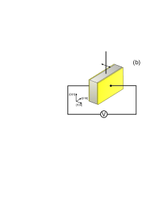
The sample was a 0.5 mm thick platelet of PMN-0.32PT single crystalpurchased with main facets perpendicular to the [100]pc direction. These facets were covered by evaporated gold electrodes. Optical observations and Raman scattering experiments were performed in a reflection geometry from an optically polished (001)pc facet (perpendicular to the principal (100)pc facet, see Fig. 1b).
For Raman scattering investigations, we have used a Renishaw RM 1000 micro-Raman spectrometer with 514.5 nm Ar laser excitation line. Samples were placed into Linkam TS 1200 high temperature cell where they were heated above the phase transition temperature. The design of the cell allows applying electrical field to the sample, so both electrical field and the temperature were controlled during the experiment. The cell was mounted on the rotation microscope table, and the sample position with respect to polarization of incident light could be manually set to the desired angle. With this experimental setup, we could use the same microscope objective for Raman scattering as well as for optical imaging in situ.
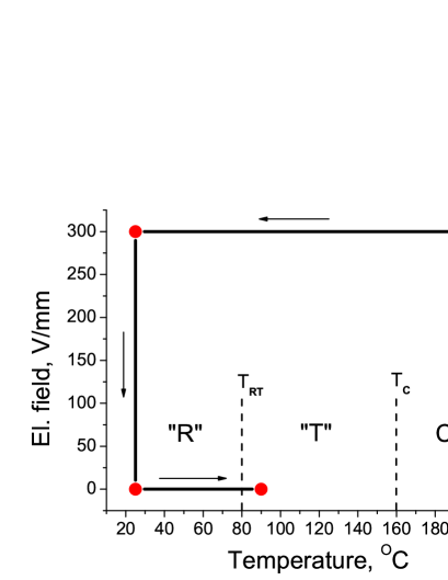
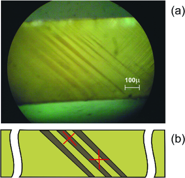
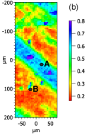
Prior to the Raman scattering measurement, the sample was field-cooled (5K /min) from the annealing temperature of about 470 K under a bias electric field of about 300 V/mm. This thermal treatment was done with a sample already mounted in the optical cell, but up to this point we did not observed any obvious domain contrast in our experimental arrangement. Then the electric field was removed and the sample has been driven to about 360 K (again at about 5K /min). At this temperature, we could repeatedly observe formation of a system of roughly parallel, 10-100 micron thick stripes running across the sample at about 45 degrees to the edges of the observed facet, as shown in Fig. 2.
At a first sight, these images are strongly reminiscent of the observations of ferroelastic domains,Book2 but we shall argue that here, in reality, the light and dark areas correspond alternatively to lamellae of tetragonal-like and rhombohedral-like domains, respectively. Obviously, the ”morphotropic interfaces” between such areas are similar to the usual ferroelastic domain boundaries. In particular, the elongated wedge shape of the observed interfaces indicates that the interfaces separate areas with different spontaneous strain tensors, what may happen at the twin boundary as well as at a two-phase interphase. However, we have found that these stripes are formed only in the vicinity of the anticipated temperature for this compositionFeng04 ; Zha12 ; Pera12 ; McLa05 (see Fig. 1a).
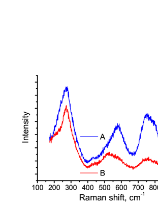
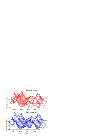
In order to confirm this conjecture, we have investigated the nature of these stripes by polarized Raman spectroscopy. In general, it is known that Raman spectra of these materials change only slightly at ferroelectric phase transitions. In fact, the positions and shapes of the principal Raman bands in our experiments were very similar to those reported in previous investigations of PMN-PT with a comparable compositionSlod10 ; Mar98 ; Kam03 ; Cha04 ; She05 ; Lim09 ; Slo08 ; Ohw01 ; Siny99 . Fortunately, at least the relative intensities of Raman spectra taken in dark and light stripes are different. In the following, we will report Raman spectra in the cross-polarized (HV) geometry, for which the differences were most apparent.
Spectra from selected dark and light stripes are shown in Fig. 3a. The difference in intensity is most obvious when comparing the shape and overall intensity of the phonon bands near 600 cm-1 or 800 cm-1. The difference can be quantified using ratios of intensities between different bands. For example, the ratio of the cross-polarized Raman intensity detected at 570 cm-1 with respect to that detected at 510 cm-1 varies by more than 50 percent when comparing dark and light stripes, and similar contrast is obtained when considering the ratio of intensities at 780 cm-1 and 270 cm-1. Note that both these ratios were previously used in Ref. Yang10, for distinguish two types of coexisting structural ”microregions” of irregular shape in PMN-0.33PT, assigned to the (i.e. rhombohedral-like phase) and (i.e. tetragonal-like phase). We used the latter ratio to map the stripes and observed that in contrast to results of Ref. Yang10, , in our sample the so-obtained relative intensity map reveals clear, about -micron sized stripes, which clearly match the stripes observed optically (Fig. 3b).
To clarify the origin of the Raman contrast associated with the stripes observed, we have recorded the variation of the Raman spectra in a given stripe as a function of the angle between the polarizer and pseudocubic crystal axes, parallel to the edges of the sample. Two sets of spectra collected from laser spot focused within dark and light stripe, respectively, are shown in Fig. 4. The angular dependence of Raman intensity recorded in the dark area is typical for the rhombohedral-like (relaxor) phase of PMN-PT crystals (with maxima at ), and it also agrees with the measurements of pure PMN, documented for example in Ref. Tani11, . In contrast, the angular dependence of Raman intensity recorded in the brighter areas shows the opposite behavior - positions of the intensity maxima are shifted by about 45 degrees with respect to that of the dark areas.
Such angular shift cannot be simply explained by measurement in a different domain of the same rhombohedral phase: all possible Raman tensors of inequivalent rhombohedral ferroelastic domain states can be obtained by rotation around the fourfold axis parallel to the [001] direction. Also, the angular dependence of Raman intensity in elastically compatible tetragonal domain pair is not compatible with our observation. Similar situation can be expected for monoclinic phases that are close to either tetragonal or rhombohedral phase only. On the contrary, projection of the optical axes of one rhombohedral and one tetragonal domain are mutually at 45 degrees. Therefore, the observed angular shift corroborates well the anticipated two-phase picture.
The available experimental setup did not allow a precise in-situ determination of the observed interphases, but the optical observations hint that the interfaces are close to the crystallographic planes. The repeating sequence of nearly parallel interfaces suggests that these are coherent interfaces similar to twin boundaries and that their orientation allows to minimize the interface stresses. Coexistence of different phases is rather frequent phenomenon in case of first-order phase transformations and a planar macroscopic phase front can be stabilized for example with the aid of temperature or concentration gradient.Born99 Macroscopic phase boundary between different ferroelectric phases has been previously observed in PMN-PT crystal with a linear gradient of Ti concentrationShu05 . However, in that work, the phase boundary seems to simply delimit Ti-poorer regions from Ti-richer regions, without any obvious relation to the crystallographic orientation of the sample. Much closer to our observation is the figure 7 of Ref. Fuj98, , showing coexistence of rhombohedral and tetragonal phase in a single crystal of Pb(Zn1/3Nb2/3)1-xTixO3 (PZN-PT). There also the stripes of the tetragonal-like phase are intercalated within the rhombohedral phase and oriented at about 45 degrees with respect to [001]pc direction.comment2 Thus, the phenomenon observed here seems to be common to both PMN-PT and PZN-PT systems.
In fact, the angular dependence recorded in the dark and light areas here are also quite analogous to that of ”G microregions” and ”R microregions”, respectively, discovered in PMN-0.33PT single crystals by authors of the Ref. Yang10, and then also reinvestigated in the temperature study of Ref. Yan11, . In contrast with our observations, the borders of the previously observed ”G microregions” and ”R microregions” have an irregular, spongy character. In the light of the present observations it seems quite likely that these microregions do correspond to the rhombohedral and tetragonal-like areas, even though the geometry of the interfaces is somewhat unexpected. Different morphology is perhaps related to some frozen defects or concentration fluctuations. We also note that their PMN-0.33PT sample was studied ”as-grown”, whereas our observations were made with PMN-0.32PT sample and the macroscopic interfaces were only observed after poling of the sample.
At the microscopic level, coexistence of the rhombohedral-like and tetragonal like phases in relaxor materials with composition close to the phase boundary has been reported in a number of recent works, and it is possible that such interfaces could play some key role in their high piezoelectric propertiesWu12 ; Sato11 ; Schm10 ; Ma12 ; Iwa12 , even though for example the microscopic mechanisms responsible for the piezoelectricity in Na1/2Bi1/2TiO3 based materials are very different from that of lead-based materials.GuenUP For example, it is possible that the phenomenon of overpoling of lead-based relaxor ferroelectric materials is related to the presence of macroscopic (percolated) tetragonal-like areas, while microscopic phase coexistence could still favor the piezoelectric response.Raja07 In either case, we believe that further studies of the macroscopic phase interfaces could help to understand the functional properties of this family of materials.
Our experimental observations clearly indicate that a slow zero-field heating of previously field-cooled PMN-0.32PT specimens is a protocol suitable for the stabilization of macroscopic interfaces between tetragonal and rhombohedral-like (possibly monoclinic) variants of PMN-PT single crystals. Formation of tetragonal and rhombohedral-like stripes occurs in near the transition.
Because of their macroscopic (millimetric) size, these ”morphotropic interfaces” separating adjacent stripes can be easily observed in an optical microscope. In fact, the existence of macroscopic interfaces allowed us also to employ spatially resolved polarized Raman scattering techniques to confirm that the stripe pattern is due to the coexistence of the phases attached to the opposite sides of the morphotropic phase boundary in the temperature-composition phase diagram. However, we believe that the existence of such macroscopic interfaces can be advantageous for other investigations of the coexistence and compatibility of tetragonal and rhombohedral-like (possibly monoclinic) variants MPB systems. Our results also suggest that MPB materials have a clear potential to sustain different interfaces than just ”ordinary” ferroelectric domain walls separating two domains with the same ferroelectric phase. This might considerably enrich the domain-wall engineering opportunities available for this family of MPB crystals.
Acknowledgment. This work was supported by the Czech Science Foundation (Projects Project GACR P204/10/0616 and 202/09/H0041). In addition, the contribution of Ph.D. student I. Rafalovskyi has been supported by Czech Ministry of Education (project SVV-2013-267303).
References
- (1) S. E. Park and T. R. Shrout, J. Appl. Phys. 82, 1804 (1997).
- (2) D. Damjanovic, M. Budimir, M. Davis and N. Setter, Appl. Phys. Lett. 83, 527 (2003).
- (3) S. Zhang and F. Li, J. Appl. Phys. 111, 031301 (2012).
- (4) Z. Feng, X. Zhao, and H. Luo, Solid State Commun. 130, 591 (2004).
- (5) Y. M. Jin, Y. U. Wang, A. G. Khachaturyan, J. F. Li, and D. Viehland, Phys. Rev. Lett. 91, 197601 (2003).
- (6) L. S. Kamzina, I. P. Raevskii, and E. V. Snetkova, Technical Physics Letters 32, 908 (2006).
- (7) R. R. Chien, C.-S. Tu, V. H. Schmidt, and F.-T. Wang, J. Phys.: Condens. Matter 18, 8337 (2006).
- (8) J. Han and W. Cao, Phys. Rev. B 68, 134102 (2003).
- (9) In fact, number of authors report monoclinic symmetry and fine domain pattern in the tetragonal-like phase in the vicinity of the morhotropic phase, too.
- (10) J. Hlinka, P. Ondrejkovic, and P. Marton, Nanotechnology 20, 105709 (2009).
- (11) High quality crystals purchased from APC International, Ltd.
- (12) A. A. Tagantsev, L. E. Cross, and J. Fousek, Domains in Ferroic Crystals and Thin Films (Springer, New York, 2010).
- (13) J. Peräntie, J. Hagberg, A. Uusimäki, J. Tian, and P. Han, J. Appl. Phys. 112, 034117 (2012).
- (14) E. A. McLaughlin, T. Liu, and C. S. Lynch, Acta Materialia 53, 4001 (2005).
- (15) A. Slodczyk and P. Colomban, Materials 3, 5007 (2010).
- (16) M. E. Marssi, R. Farhi, and Y. I. Yuzyuk, J. Phys.: Condens. Matter 10, 9161 (1998).
- (17) S. Kamba, E. Buixaderas, J. Petzelt, J. Fousek, J. Nosek, and P. Bridenbaugh, J. Appl. Phys. 93, 933 (2003).
- (18) B. Chaabane, J. Kreisel, P. Bouvier, G. Lucazeau, and B. Dkhil, Phys. Rev. B 70, 134114 (2004).
- (19) J. A. Lima, W. Paraguassu, P. T. C. Freire, A. G. Souza Filho, C. W. A. Paschoal, J. Mendes Filho, A. L. Zanin, M. H. Lente, D. Garcia, and J. A. Eiras, J. Raman Spectrosc. 40, 1144 (2009).
- (20) A. Slodczyk, P. Daniel, and A. Kania, Phys. Rev. B 77, 184114 (2008).
- (21) H. Ohwa, M. Iwata, H. Orihara, N. Yasuda, and Y. Ishibashi, J. Phys. Soc. Jpn. 70, 3149 (2001).
- (22) I. G. Siny, S. G. Lushnikov, R. S. Katiyar, and V. H. Schmidt, Ferroelectrics 226, 191 (1999).
- (23) M. Shen, G. G. Siu, Z. K. Xu and W. Cao, Appl. Phys. Lett. 86, 252903 (2005).
- (24) Y. Yang, Y. L. Liu, L. Y. Zhang, K. Zhu, S. Y. Ma, G. G. Siu, Z. K. Xu, and H. S. Luo, J. Raman Spectrosc. 41, 1735 (2010).
- (25) H. Taniguchi, M. Itoh, and D. Fu, J. Raman Spectrosc. 42, 706 (2011).
- (26) J. Bornarel and R. Cach, Phys. Rev. B 60, 3806 (1999).
- (27) V. A. Shuvaeva, A. M. Glazer, and D. Zekria, J. Phys.: Condens. Matter 17, 5709 (2005).
- (28) K. Fujishiro, R. Vlokh, Y. Uesu, Y. Yamada, J.-M. Kiat, B. Dkhil, and Y. Yamashita, Jpn. J. Appl. Phys 37, 5246 (1998).
- (29) According to the indicated extinction condition, it seems that the investigated PZN-PT sample was viewed along (001)pc direction.
- (30) Y. Yang, L. Y. Zhang, K. Zhu, and Y. L. Liu, J. Appl. Phys. 109, 083517 (2011).
- (31) C. Ma, H. Guo, S. P. Beckman, and X. Tan, Phys. Rev. Lett. 109, 107602 (2012).
- (32) Y. Sato, T. Hirayama, and Y. Ikuhara, Phys. Rev. Lett. 107, 187601 (2011).
- (33) L. A. Schmitt and H.-J. Kleebe, Func. Mater. Lett. 3, 55 (2010).
- (34) H. Wu, D. Xue, D. Lv, J. Gao, S. Guo, Y. Zhou, X. Ding, C. Zhou, S. Yang, Y. Yang, and X. Ren, J. Appl. Phys. 112, 052004 (2012).
- (35) M. Iwata and Y. Ishibashi, Jpn. J. Appl. Phys. 51, 09LE03 (2012).
- (36) M. Guennou, M. Savinov, J. Drahokoupil, H. Luo, and J. Hlinka, arXiv preprint arXiv:1212.0366, (2012).
- (37) K. K. Rajan, M. Shanthi, W. S. Chang, J. Jin, and L. C. Lim, Sensors and Actuators A 133, 110 (2007).