On the Saturation of the Nonlinear Refractive Index in Atomic Gases
Abstract
Motivated by the ongoing controversy on the origin of the nonlinear index saturation and subsequent intensity clamping in femtosecond filaments, we study the atomic nonlinear polarization induced by a high-intensity and ultrashort laser pulse in hydrogen by numerically solving the time dependent Schrödinger equation. Special emphasis is given to the efficient modeling of the nonlinear polarization at central laser frequency corresponding to 800 nm wavelength. Here, the recently proposed model of the Higher-Order Kerr Effect (HOKE) and two versions of the Standard model for femtosecond filamentation, including either a multi-photon or tunnel ionization rate, are compared. We find that around the clamping intensity the instantaneous HOKE model does not reproduce the temporal structure of the nonlinear response obtained from the quantum mechanical results. In contrast, the non-instantaneous charge contributions included in the Standard models ensure a reasonable quantitative agreement. Therefore, the physical origin for the observed saturation of the overall electron response is confirmed to mainly result from contributions of free or nearly free electrons.
pacs:
32.80.-t,51.50.+vI Introduction
Accurate modeling of high intensity laser pulse propagation in a medium requires the accurate knowledge of its optical response. For laser intensities well below the atomic ionization threshold this response is given by the induced polarization involving bound electrons, usually modeled by a Taylor expansion in terms of the driving electric field. Restriction of the expansion to the third order nonlinear term for isotropic media leads to the well known intensity-dependent Kerr refractive index change Shen (1984). This simplest approach for the polarization already provides all key ingredients to describe fundamental nonlinear phenomena like modulational instability, self-focusing and self-phase modulation Boyd (1992), which manifest in soliton formation, pulse compression or self-similar collapse. The latter effect, however, demonstrates the inherent incompleteness of this simplest approach as it predicts an unphysical blow-up of the beam intensity, which is intimately linked to the onset of laser filamentation.
A theoretical description of laser filamentation therefore requires an accurate modeling of the saturation of the optical nonlinearity, i.e., the induced variation of the nonlinear refractive index with respect to the laser intensity. Originally achieved by accounting for photo-ionization and subsequent interaction of the laser pulse with a free electron plasma Bergé et al. (2007); Bergé and Skupin (2008); Couairon and Berge (2000); Couairon and Mysyrowicz (2007); Chin et al. (2012); Mlejnek et al. (1998); Brodeur et al. (1997), this so-called Standard model for nonlinear refractive index saturation was recently challenged by the measurement of higher order Kerr coefficients Loriot et al. (2009, 2010). Being negatively valued, these coefficients could give rise to an alternative explanation for the saturation of the optical nonlinearity. This strikingly simple approach, based on higher order contributions in the Taylor expansion of the polarization in terms of the driving electric field, is called the Higher-Order Kerr Effect (HOKE) model. Besides the apparent differences in the model equations, there is a fundamental physical difference between HOKE and Standard model. While in the latter model a free electron plasma is responsible for the arrest of the self-focusing, HOKE terms originate exclusively from the response of bound electrons. Thus, the HOKE model questions the very nature of the previous understanding of femtosecond filamentation as a dynamical balance between optical Kerr effect and self-generated electron plasma Béjot et al. (2010, 2012); Kolesik et al. (2010a); Wang et al. (2010); Brée et al. (2011); Wahlstrand and Milchberg (2011); Nurhuda et al. (2008); Teleki et al. (2010); Volkova et al. (2011). From the experimental side, numerous attempts have been undertaken to decide in favor for either bound or ionized electron contributions Polynkin et al. (2011); Chen et al. (2010); Wahlstrand et al. (2011, 2012); Kolesik et al. (2010b); Kosareva et al. (2011); Béjot et al. (2011, 2011) without common agreement. Recent progress was achieved by direct numerical solution of the time dependent Schrödinger equation (TDSE), which showed the excitation of atoms into highly excited Rydberg and Kramers-Henneberger states. Although bound, these states lie at the frontier above which excited electrons may be fully freed, and they behave very similarly to states in the continuum Volkova et al. (2011); Nurhuda et al. (2008); Béjot et al. (2012).
Here, we propose a direct confrontation of both HOKE and Standard models with rigorous quantum mechanical calculations. To this end, we investigate the saturation of the nonlinear polarization by numerically solving the TDSE for an atomic hydrogen model, for the sake of simplicity first in one dimension (1D), and later in full three dimensions (3D) for confirmation. It turns out that a consistent definition of the HOKE coefficients is difficult, because the validity of the Taylor expansion of the nonlinear response in terms of the electric field breaks down at intensities well below the clamping one. Moreover, the instantaneous character of the HOKE model contradicts the quantum mechanical results. In contrast, we achieve reasonable agreement when applying the Standard model with multi-photon or tunnel ionization rate. Although the exact definition of bound and ionized electrons remains ambiguous for atoms in strong laser fields, the saturating contribution to the nonlinear refractive index is still well described by a simple Drude plasma model assuming continuum electrons as encountered once the pulse has passed the interaction region.
The paper is organized as follows. In Sec. II, we introduce our TDSE modeling of the nonlinear polarization as well as the phenomenological models currently discussed in the literature. For sake of simplicity, we focus on 1D TDSE description of atomic hydrogen. Then, we confront the phenomenological models (HOKE and Standard model) with rigorous TDSE results in Sec. III. Finally, we confirm that our findings hold for more realistic 3D TDSE calculations in Sec. IV.
II Modeling Nonlinear Polarization
II.1 1D Quantum Mechanical Treatment
Let us start with 1D quantum mechanical calculations. We assume that the single non-relativistic electron of the 1D hydrogen model in an external laser is described by its wave function , governed by the TDSE
| (1) |
Here with electron mass , and the interaction with the external laser field in velocity gauge is formulated in the dipole approximation, i.e. involving the electron charge and the magnetic potential . We use a soft-core potential adjusted to match the ionization energy of hydrogen, where is the Bohr radius.
The macroscopic polarization is then given by , where is the atomic density at ambient pressure.
As proposed in Nurhuda et al. (2008), we distinguish between bound and continuum electrons by projecting the electronic wavefunction on bound eigenfunctions of the field free Hamiltonian , such that
| (2) |
where we used states up to principal quantum number . Then, the macroscopic polarization can be decomposed into bound, bound-continuum and continuum part, , according to Eq. (2). For example,
| (3) |
To observe the dependence of on the laser pulse intensity, we numerically integrate Eq. (1) for Gaussian pulses
| (4) |
with central frequency ( nm, i.e. cycle period of fs), and peak intensities varying from TW/cm2 to TW/cm2 for pulse durations fs, fs and fs. This intensity range ensures partial ionization by multi-photon and/or tunnel processes as complete ionization for hydrogen appears at TW/cm2 by over-the-barrier ionization Bandrauk et al. (2011).
In order to access the macroscopic nonlinear polarization , we first extract the linear response , where we neglect linear dispersion and absorption, from low intensity calculations TW/cm2 yielding . Then, the nonlinear part of the polarization can be obtained with . For the choice of the carrier envelope phase offset in our test pulses [Eq. (4)], is responsible for self-focusing () or self-defocusing () action, i.e., a positive (resp. negative) nonlinear induced refractive index change. describes nonlinear absorption, which is weak and will be neglected throughout this paper.
II.2 Phenomenological Models
Let us now investigate the possibility to mimic the behavior of the nonlinear polarization from TDSE calculations by means of approximate models. Such phenomenological description of the nonlinear response is extremely important for the efficient numerical modeling of high intense laser pulse propagation. As already mentioned above, we will approach the TDSE results in terms of two controversial models, currently used in the context of femtosecond filamentation. These models involve only a single set of few parameters for all pulse durations and intensities. First, the HOKE model
| (5) |
is fully determined by the nonlinear coefficients . Second, the Standard model
| (6) |
requires an additional equation to describe the free electron density
| (7) |
Here, we will either apply a multi-photon ionization (MPI) rate Popov (2004)
| (8) |
with photon number and ionization cross section or a field-dependent tunnel ionization rate Landau and Lifshitz (1977); Yudin and Ivanov (2001)
| (9) |
involving two parameters , .
II.3 Extraction of Phenomenological Model Parameters from 1D TDSE Results
In order to confront the phenomenological models with TDSE results, we first extract the susceptibilities for the HOKE model and additionally the MPI cross section resp. the tunnel ionization parameters , for the two Standard models from the quantum mechanical data.
For the extraction of the we use the assumption that different orders of the nonlinear polarization become important subsequently for increasing field strength. Additionally, we assume that the characteristic peak at the th harmonic in is mainly caused by . Then, one can deduce consecutively the using
| (10) |
valid for the Gaussian pulses of Eq. (4). Alternatively, we can evaluate at different peak intensities and infer the values of by solving a system of linear equations
| (11) |
where denotes the Fourier transform. Note, that the former approach evaluates the susceptibilities at different harmonic frequencies , whereas the latter one gives access to all directly at . For purely instantaneous HOKE terms of Eq. (5), both approaches should yield the same .
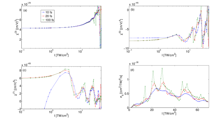
Let us first evaluate at the harmonic frequencies . Results are presented in Figs. 1 (a)-(c), where , and are plotted for different pulse durations vs. peak intensity. We can observe, how difficult it is to extract meaningful coefficients being independent of intensity and pulse duration in the range where becomes relevant, i.e. TW/cm2. The fact that it is not possible to describe the nonlinear polarization in terms of a Taylor expansion at intensities above 10 TW/cm2 already indicates significant changes in the electronic configuration of the atomic system. This involves, e.g., the enhanced population of Stark shifted Rydberg states, staying close to the ionization threshold due to a comparable increase of the ponderomotive potential Agostini et al. (1989). Moreover, the observed dependency of the higher order on the pulse duration reflects strong nonlinear dispersion, i.e., a noninstantaneous character of the nonlinear response at higher intensities. Due to this nonlinear dispersion, and because we are searching for a description of at , we resort to the latter approach of Eqs. (11) in the following. By doing so, we can only extract a minimum set of consistent parameters, namely
| (12) | ||||
| (13) |
all higher order being discarded, since all of them strongly fluctuate along the intensity range. We will further refer to this simple model as HOKE_35. Note that we will later introduce another set of parameters, HOKE_FIT, which includes nonlinear coefficients up to , chosen to obtain a more accurate description of the nonlinear saturation.
For the Standard model with MPI rate, in addition to , the photon number and the cross section has to be determined from the TDSE simulation data. For pulses with wavelength nm and the ionization potential of hydrogen of eV one obtains a minimum photon number of . However, we find the least dependence of on the peak intensity when employing a reduced photon number of . This is plausible, since for the here employed intensities the Keldysh parameter is in the order of one, indicating a transient regime between MPI () and tunnel ionization (), which leads to a reduced exponent in the intensity dependence of the ionization probability. In addition, present atomic resonances connecting the ground and first excited state are of similar order (five and seven photon resonance in 1D and 3D, respectively) Bian and Bandrauk (2011, 2010). Because, we are only interested in the best (phenomenological) description of TDSE results, we will employ the MPI rate with in the following. To calculate , we match the free electron density at the end of the pulse () obtained with the MPI rate with the TDSE one:
| (14) |
where is the continuum part of the wavefunction. Interestingly, the results for all three pulse durations agree, up to strong modulations of at increasing intensity [Fig. 1 (d)]. We attribute these modulations to resonant excitation of high lying Rydberg states, where the energy of an integer number of photons matches the energy gap between the ground and a (Stark shifted) Rydberg state Volkova et al. (2011). We thus select an average value for the Standard model,
| (15) |
matching at TW/cm2. Our choice offers a reasonably good fit of the nonlinear polarization close to the clamping intensity, where changes sign. Below this intensity charge contributions are negligible, thus deviating do not matter. For higher intensities we will overestimate the charge, however, at least in the context of femtosecond filamentation, this regime is expected to be barely relevant as well. In any case, complete ionization of the hydrogen atom is expected at W/cm2 by over-the-barrier ionization Bandrauk et al. (2011).
To extract the tunnel ionization parameters we calculate the free electron density at the end of the pulse according to the tunnel ionization rate for all peak intensity values employed in the TDSE simulations and compare to the TDSE data for fs. Then, and are optimized such that the sum of the squared differences from each comparison is minimized. Performing this least squares fit, we extract
| (16) | ||||
| (17) |
for the parameters entering Eq. (9). These values are used for all pulse durations in the following.
III Confrontation of phenomenological models with 1D TDSE results
III.1 Behavior in Fourier Domain
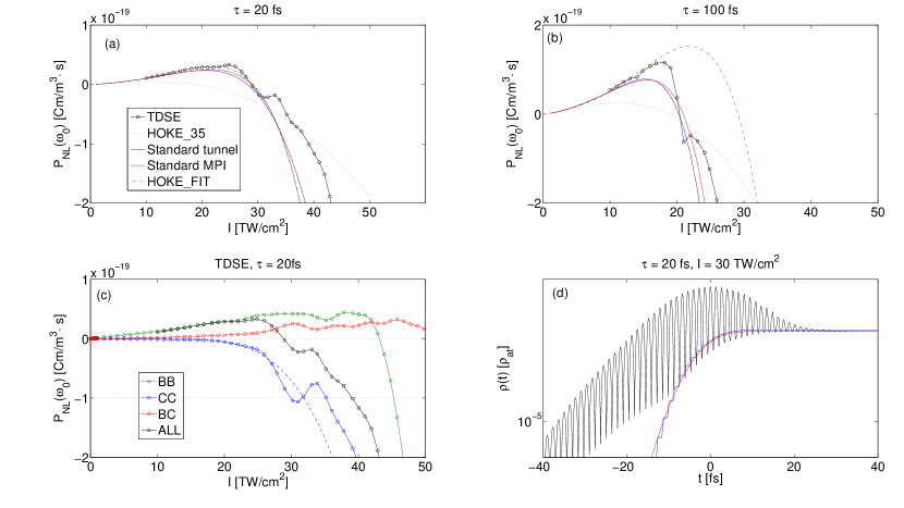
Having all necessary parameters at hand, let us now confront the HOKE and plasma-based (Standard) description of the nonlinear polarization with TDSE results. In Figs. 2 (a) and (b), we compare the nonlinear polarization at center frequency of the laser obtained from TDSE simulations with the approximate models for fs and fs, respectively. Shorter pulses ( fs) yield similar behavior (not shown). For the TDSE results with fs (solid black line) is positive and increases (focusing action) with peak intensity up to TW/cm2. Then, saturation and final change of sign occur at TW/cm2, signaling a qualitative change from focusing to defocusing behavior. This generic behavior also holds for fs with decreased [ TW/cm2, Fig. 2 (b)] and fs with increased [ TW/cm2, not shown] saturation intensity values. Moreover, we observe oscillatory structures of at TW/cm2 ( fs) and TW/cm2 ( fs), which can be attributed to decreased ionization probabilities due to channel closure Kopold et al. (2002). The HOKE_35 model, defined by extracted and coefficients [see Eqs. (12), (13)] (green dotted line) yields significantly lower intensity thresholds for saturation and sign inversion of than observed in the TDSE calculations. Because this discrepancy renders the HOKE_35 unsuitable for practical applications, we suggest a different (purely phenomenological) set of parameters HOKE_FIT
| (18) | ||||
| (19) | ||||
| (20) | ||||
| (21) |
entering Eq. (5) which satisfactorily reproduces the TDSE data for fs. However, changing the pulse duration destroys this agreement [see results for HOKE_FIT, green dash-dotted line in Fig. 2 (a), (b)]. In contrast, comparison to the Standard models with either ionization rate (red line MPI rate, blue tunnel rate) shows reasonable agreement for the physically extracted model parameters [see Eqs. (12) and (15)-(17)] for all pulse durations.
An analysis of the physical mechanism responsible for the observed nonlinear refractive index saturation in the TDSE simulations is detailed in Fig. 2 (c). Here, the resulting contributions to the nonlinear polarization from the bound (’BB’ green line), continuum (’CC’ blue line) and bound-continuum (’BC’ red line) electronic wave function [see Eq. (3)] are shown for fs. The bound (BB) and bound-continuum (BC) contributions to are both positively valued (focusing action) up to 45 TW/cm2, well beyond the threshold intensity where the overall nonlinear polarization (solid black line) changes sign. In contrast, the continuum contribution (CC) is close to zero for small intensities TW/cm2, because no ionization occurs, and yields a negative (defocusing) contribution when free charge generation kicks in. The value of this negative contribution (CC) yields significantly lower intensity thresholds than both the positive contributions of BB and BC. Thus, the observed sign flip in the nonlinear polarization, i.e., the saturation of the nonlinear induced refractive index, can be clearly attributed to the contribution of free electrons according to Eq. (3). In this context it is worth mentioning that, strictly speaking, only the overall polarization is gauge invariant. The projected contributions (BB, BC, CC) may differ for a different gauge in the TDSE equation. However, in particular the CC contribution preserves its qualitative focusing properties using different gauges, as shown in Béjot et al. (2012).
To quantify the impact of the free electrons, ionization is considered in Fig. 2 (d). The free electron density obtained from TDSE simulations via [shown for TW/cm2 and fs; solid black line] develops strong oscillations at times where the pulse interacts with the atom. This is due to the fact that our definition using Eq. (2) for the free electron density is only applicable for the field free situation after the driving pulse has passed. In the presence of the laser field the eigenstates of do no longer represent the bound states of the laser dressed atom. Therefore, spurious oscillations occur due to contributions from electrons in high lying bound (Rydberg/Kramers-Henneberger) states close to the bulge of the bended atomic potential and from truly free electrons. However, in the Standard models we apply ionization rates that describe the truly ionized fraction after the laser pulse only, and the corresponding charge contributions do not feature these oscillations [red line MPI rate, blue line tunnel rate in Fig. 2 (d)]. Importantly, the truly ionized fraction according to the Standard models [dashed blue line for tunnel rate in 2 (c)] reproduces the CC contributions from TDSE simulations for all intensities and pulse durations. Minor deviations for TW/cm2 are due to previously discussed resonant occupation of high lying Rydberg states Volkova et al. (2011), whose description is beyond the scope of simple MPI/tunnel ionization models.
III.2 Behavior in Temporal Domain
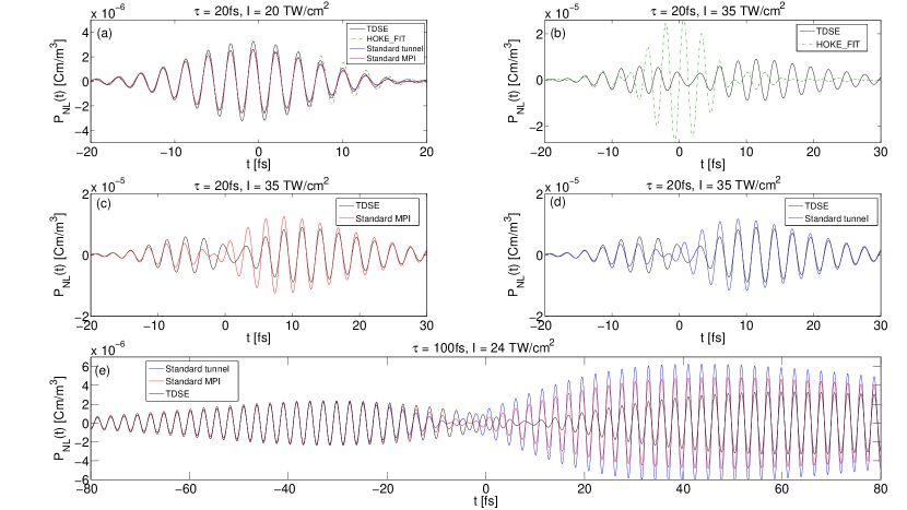
Let us continue our comparison of TDSE results for with approximate models in time domain. In Fig. 3 (a) the time evolution of from TDSE simulations and the approximate HOKE_FIT and Standard models are shown for fs and TW/cm2. For that intensity, charge contributions are small and all three models reproduce accurately the response of the bound electrons, instantaneously following the electric field. For TW/cm2 in (b) charge contributions become important, resulting in a delayed response of opposite sign kicking in at fs, according to TDSE simulations (black solid line). The HOKE_FIT model (dash-dotted green line), although yielding a comparable value of , cannot describe that delayed contribution due to its instantaneous nature and produces much higher polarization amplitudes than observed in the TDSE simulations. In contrast, both Standard models in (c), (d) qualitatively capture this delayed response and they develop only limited differences in amplitude. A similar situation is encountered in (e) for fs and TW/cm2, where also the limits of the Standard models are illustrated more clearly: The qualitative temporal character is described correctly over long evolution times (oscillation in phase with TDSE results). However, due to a slightly overestimated charge contribution, the Standard models yield larger amplitudes for the delayed response coming along with an inaccurate onset of the latter.
IV Comparing to 3D TDSE Calculations
Finally, let us confirm the results obtained from the 1D models by performing the same analysis in 3D. Here, we numerically solve
| (22) |
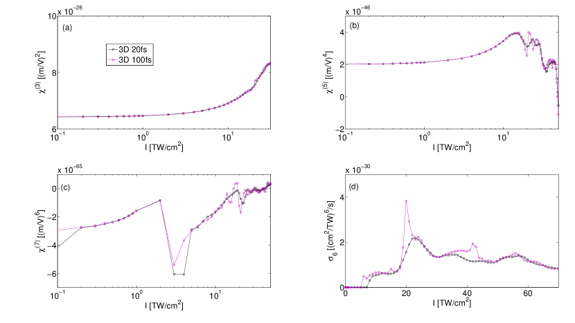
with including with . The interaction with the -linearly polarized external laser field in length gauge is computed in dipole approximation Cormier and Lambropoulos (1997). We followed the same procedure to extract the parameters of the approximate models as in the 1D case (see Sec. II.3).
From low intensity calculations we obtain a linear susceptibility of . The nonlinear susceptibilities evaluated at harmonic frequencies [see Eq. (10)] are presented in Figs. 4 (a)-(c) for pulse durations of fs (solid black line) and fs (solid magenta line). The deviations for different are similar to those observed in the 1D case, indicating similar nonlinear dispersion. Because we again meet difficulties in extracting a consistent set of s from Eqs. (11), we omit a repeated comparison to the HOKE_35 model. Thus we resort to comparisons involving the HOKE_FIT model, defined through
| (23) | ||||
| (24) | ||||
| (25) | ||||
| (26) |
and the Standard models employing
| (27) |
together with ()
| (28) |
for the MPI rate or
| (29) | ||||
| (30) |
for the tunnel ionization rate. Naturally, the values of the model parameters in 3D differ from the ones obtained from 1D TDSE data. Nevertheless, our 3D calculations yield the same order of magnitude for the ’s as observed in 1D. It is interesting that and exhibit opposite signs compared to the 1D case.
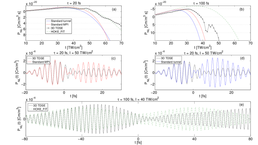
Let us continue with the comparison of 3D TDSE data with HOKE_FIT and Standard model results. In Fig. 5 (a) for fs and (b) for fs the nonlinear polarization inferred from 3D simulations (solid black line) is compared to the HOKE_FIT model (dash-dotted green line) and to the Standard models with a MPI rate (solid red line) or a tunnel ionization rate (solid blue line). As in the 1D case, the HOKE_FIT model exhibits strong deviations for different pulse durations . In contrast, the Standard models with physically extracted parameters reasonably approximate simulation results for both pulse durations . However, for intensities TW/cm2 for the fs case and TW/cm2 for fs the overestimation of charge contributions seems more pronounced than in 1D. In Fig. 5 (c) and (d) this is evident when comparing the temporal evolution of from 3D TDSE simulations (solid black line) to Standard models with (c) MPI (red solid line) and (d) tunnel ionization rate (blue solid line). Here, we show data for fs and TW/cm2, which ensures a considerable charge contribution. Both Standard models qualitatively reproduce simulation results by capturing the instantaneous and delayed property. However, deviations in the amplitude of the delayed contribution are larger than in similar 1D situations [Fig. 3 (c), (d)]. The HOKE_FIT model (dash-dotted green line) in Fig. 5 (e) for fs and TW/cm2 not even qualitatively captures the delayed temporal property as it gives out of phase oscillations with an opposite sign for the field amplitude for fs.
V Conclusion
In conclusion, numerical solution of the TDSE for atomic hydrogen in 1D and 3D reveals the saturation and change of sign of the nonlinear polarization at central laser frequency for rising intensity of the driving pulse. As in Béjot et al. (2012), we identify a time retarded contribution responsible for the transition from the focusing to defocusing regime. This time retarded contribution cannot be modeled by means of an instantaneous HOKE model, as we neither can physically extract consistent HOKE terms nor (even qualitatively) reproduce temporal field structures from simulations with a phenomenologically fitted HOKE_FIT model. On the other hand, applying Standard models with either a MPI or tunnel ionization rate, clearly attributes the delayed response to the generation of free electrons. With both standard models we achieve reasonable agreement with TDSE simulations for the nonlinear polarization at central laser frequency as well as for the temporal field structures. We propose a unique set of parameters, valid for all applied pulse durations and intensities, to model the nonlinear polarization. This simple approach opens the possibility to efficiently simulate laser pulse propagation with reasonable accuracy. However, due to their inherently approximate nature, possibly important details observed in TDSE simulations, e.g. the non-monotonous behavior of the ionization probability due to channel closure and resonant excitation, are not included in the Standard models. Thus, in order to accurately describe laser pulse propagation, a coupling of TDSE calculations to propagation equations as in Lorin et al. (2012) seems mandatory.
We gratefully thank M. Ivanov, M. Kolesik, A.M. Popov and E. Cormier for fruitful and inspiring discussions on the bound electrons in the continuum.
References
- Shen (1984) Y. R. Shen, The Principles of Nonlinear Optics (John Wiley & Sons, New-York, 1984).
- Boyd (1992) R. Boyd, Nonlinear Optics (Academic, New York, 1992).
- Bergé et al. (2007) L. Bergé, S. Skupin, R. Nuter, J. Kasparian, and J.-P. Wolf, Reports on Progress in Physics 70, 1633 (2007).
- Bergé and Skupin (2008) L. Bergé and S. Skupin, Discrete and continuous dynamical systems. Series A 23, 1099 (2008).
- Couairon and Berge (2000) A. Couairon and L. Berge, Physics of Plasmas 7, 193 (2000).
- Couairon and Mysyrowicz (2007) A. Couairon and A. Mysyrowicz, Physics Reports 441, 47 (2007).
- Chin et al. (2012) S. Chin, T. Wang, C. Marceau, J. Wu, J. Liu, O. Kosareva, N. Panov, Y. Chen, J. Daigle, S. Yuan, et al., Laser Physics 22, 1 (2012), 10.1134/S1054660X11190054.
- Mlejnek et al. (1998) M. Mlejnek, E. M. Wright, and J. V. Moloney, Opt. Lett. 23, 382 (1998).
- Brodeur et al. (1997) A. Brodeur, C. Y. Chien, F. A. Ilkov, S. L. Chin, O. G. Kosareva, and V. P. Kandidov, Opt. Lett. 22, 304 (1997).
- Loriot et al. (2009) V. Loriot, E. Hertz, O. Faucher, and B. Lavorel, Opt. Express 17, 13429 (2009).
- Loriot et al. (2010) V. Loriot, E. Hertz, O. Faucher, and B. Lavorel, Opt. Express 18, 3011 (2010).
- Béjot et al. (2010) P. Béjot, J. Kasparian, S. Henin, V. Loriot, T. Vieillard, E. Hertz, O. Faucher, B. Lavorel, and J.-P. Wolf, Phys. Rev. Lett. 104, 103903 (2010).
- Béjot et al. (2012) P. Béjot, E. Cormier, E. Hertz, B. Lavorel, J. Kasparian, J.-P. Wolf, and O. Faucher (2012), URL http://arxiv.org/abs/1206.4906v3.
- Kolesik et al. (2010a) M. Kolesik, E. M. Wright, and J. V. Moloney, Opt. Lett. 35, 2550 (2010a).
- Wang et al. (2010) H. Wang, C. Fan, P. Zhang, C. Qiao, J. Zhang, and H. Ma, Opt. Express 18, 24301 (2010).
- Brée et al. (2011) C. Brée, A. Demircan, and G. Steinmeyer, Phys. Rev. Lett. 106, 183902 (2011).
- Wahlstrand and Milchberg (2011) J. K. Wahlstrand and H. M. Milchberg, Opt. Lett. 36, 3822 (2011).
- Nurhuda et al. (2008) M. Nurhuda, A. Suda, and K. Midorikawa, New Journal of Physics 10, 053006 (2008).
- Teleki et al. (2010) A. Teleki, E. M. Wright, and M. Kolesik, Phys. Rev. A 82, 065801 (2010).
- Volkova et al. (2011) E. Volkova, A. Popov, and O. Tikhonova, JETP Letters 94, 519 (2011), 10.1134/S0021364011190180, URL http://dx.doi.org/10.1134/S0021364011190180.
- Polynkin et al. (2011) P. Polynkin, M. Kolesik, E. M. Wright, and J. V. Moloney, Phys. Rev. Lett. 106, 153902 (2011).
- Chen et al. (2010) Y.-H. Chen, S. Varma, T. M. Antonsen, and H. M. Milchberg, Phys. Rev. Lett. 105, 215005 (2010).
- Wahlstrand et al. (2011) J. K. Wahlstrand, Y.-H. Cheng, Y.-H. Chen, and H. M. Milchberg, Phys. Rev. Lett. 107, 103901 (2011).
- Wahlstrand et al. (2012) J. Wahlstrand, Y.-H. Chen, Y.-H. Cheng, S. Varma, and H. Milchberg, Quantum Electronics, IEEE Journal of 48, 760 (2012).
- Kolesik et al. (2010b) M. Kolesik, D. Mirell, J.-C. Diels, and J. V. Moloney, Opt. Lett. 35, 3685 (2010b).
- Kosareva et al. (2011) O. Kosareva, J.-F. Daigle, N. Panov, T. Wang, S. Hosseini, S. Yuan, G. Roy, V. Makarov, and S. L. Chin, Opt. Lett. 36, 1035 (2011).
- Béjot et al. (2011) P. Béjot, E. Hertz, J. Kasparian, B. Lavorel, J. P. Wolf, and O. Faucher, Phys. Rev. Lett. 106, 243902 (2011).
- Béjot et al. (2011) P. Béjot, E. Hertz, B. Lavorel, J. Kasparian, J.-P. Wolf, and O. Faucher, Opt. Lett. 36, 828 (2011).
- Bandrauk et al. (2011) A. Bandrauk, S. Chelkoowski, and K. Yuan, Int. Rev. Atom. Molec. Phys. 2, 1 (2011).
- Popov (2004) V. S. Popov, Physics-Uspekhi 47, 855 (2004), URL http://stacks.iop.org/1063-7869/47/i=9/a=R01.
- Landau and Lifshitz (1977) L. D. Landau and E. M. Lifshitz, Quantum mechanics: non-relativistic theory, vol. 3 (Pergamon Press, 1977), 3rd ed.
- Yudin and Ivanov (2001) G. L. Yudin and M. Y. Ivanov, Phys. Rev. A 64, 013409 (2001).
- Agostini et al. (1989) P. Agostini, P. Breger, A. L’Huillier, H. G. Muller, G. Petite, A. Antonetti, and A. Migus, Phys. Rev. Lett. 63, 2208 (1989).
- Bian and Bandrauk (2011) X.-B. Bian and A. D. Bandrauk, Phys. Rev. A 83, 041403 (2011).
- Bian and Bandrauk (2010) X.-B. Bian and A. D. Bandrauk, Phys. Rev. Lett. 105, 093903 (2010).
- Kopold et al. (2002) R. Kopold, W. Becker, M. Kleber, and G. G. Paulus, Journal of Physics B: Atomic, Molecular and Optical Physics 35, 217 (2002).
- Cormier and Lambropoulos (1997) E. Cormier and P. Lambropoulos, Journal of Physics B: Atomic, Molecular and Optical Physics 30, 77 (1997).
- Lorin et al. (2012) E. Lorin, S. Chelkowski, E. Zaoui, and A. Bandrauk, Physica D: Nonlinear Phenomena 241, 1059 (2012).