Impact of vibrational entropy on the stability of unsolvated peptide helices with increasing length
Abstract
Helices are a key folding motif in protein structure. The question which factors determine helix stability for a given polypeptide or protein is an ongoing challenge. Here we use van der Waals corrected density-functional theory to address a part of this question in a bottom-up approach. We show how intrinsic helical structure is stabilized with length and temperature for a series of experimentally well studied unsolvated alanine based polypeptides, Ac-Alan-LysH+. By exploring extensively the conformational space of these molecules, we find that helices emerge as the preferred structure in the length range =4-8 not just due to enthalpic factors (hydrogen bonds and their cooperativity, van der Waals dispersion interactions, electrostatics), but importantly also by a vibrational entropic stabilization over competing conformers at room temperature. The stabilization is shown to be due to softer low-frequency vibrational modes in helical conformers than in more compact ones. This observation is corroborated by including anharmonic effects explicitly through ab initio molecular dynamics, and generalized by testing different terminations and considering larger helical peptide models.
I Introduction
Polypeptide helices are a key secondary structure motif in a wide range of proteins Pauling1951_1 ; pdb ; Creighton1987 . It is well known that some amino acids (e.g., alanine) exhibit a stronger helix propensity than others PaceScholtz1998 ; ScholtzBaldwin1991 ; HorovitzFersht1992 ; CreamerRose1992 ; Kinnear:2001p4453 ; MillerKemp2002 ; ScottFersht2007 ; Moreau2009 , but the fact that the helical structure is so abundant in proteins is still intriguing. From a thermodynamic point of view, there are at least two possible limits in which helices compete with other structure prototypes. Towards high temperature, one expects the transition to a random coil ZimmBragg1959 , which should become entropically favored as the temperature increases. Towards low temperature, however, helices may themselves compete with other enthalpically stable conformations. However, in the most interesting regime, namely intermediate, physiological temperatures, stability may be determined by a delicate balance between enthalpy and entropy Kinnear:2002p4458 . We here unravel this balance quantitatively for the emergence of helical structure in a particularly well studied series of unsolvated polyalanine based peptides Ac-Alan-LysH+, =4-8 HudginsJarrold1998 ; KohtaniJarroldWater2004 . We consider explicitly not just the helical part of conformational space, but actually the much larger, general low-energy conformational space of the peptides, of which helices are a part. In this paper we show: (i) a comprehensive search of the conformational space for Ac-Alan-LysH+, =4-8; (ii) harmonic free energy calculations for several structural candidates; (iii) outlook on the role of anharmonicities in the potential energy surface; and (iv) a theoretical comparison for longer model peptides, considering only helical motifs, with a different termination, in order to clarify the impact of Lys on the soft vibrational modes. Our key finding is that there is a significant vibrational entropic stabilization of helices compared to other, more compact conformers, a contribution that should indeed make a difference in actual proteins as well. This contribution is intrinsic to the helix and should therefore act largely independently, not entangled with environment-dependent terms such as a solvent entropy. Interestingly, an essential role of low-frequency modes is also actively debated in other areas of protein science, related to their function TournierSmith2003 ; BrooksKarplus1985 ; NatChemFocus ; HayScrutton2012 ; GlowackiHarveyMulholland2012 .
Beginning with the terms that shape the potential-energy surface (PES), known reasons for helix stability include ScholtzBaldwin1992 ; Baldwin-review (i) their efficient hydrogen-bond (H-bond) network and increasing H-bond cooperativity with helix length GuoKarplus1994 ; Baldwin2003 ; IretaGalvan2003 ; dannenbergccoperativity2 ; TkatchenkoRossi2011 , (ii) suitably bonded and/or electrostatically favorable termination, for instance the LysH+ termination considered here BlagdonGoodman1975 ; PrestaRose1988 ; SerranoFersht1989 ; TakahashiOoi1989 ; HudginsJarrold1998 ; MarquseeBaldwin1989 ; DugourdJarrold2005 ; WilliamsKemp1998 , and (iii) remarkably, a rather specific favorable contribution of van der Waals (vdW) interactions for -helices TkatchenkoRossi2011 ; ShuguiShuhua2011 . Clearly, the peptide chain length plays a role: Too short chains have too few and too weak hydrogen bonds for helices to compensate the cost of strain in the backbone MarquseeBaldwin1989 ; KohtaniJarroldWater2004 ; LiuBowers2004 ; JobKemp2006 ; ElstnerSuhai2000 . In practice, environment effects will necessarily influence helix stability ElstnerSuhai2000 ; SalvadorDannenberg2007 ; Garcia2002 ; TiradoRives-Jorgensen2000 . In an aqueous medium, the hydrophobic effect will be prominent Kauzmann1959 . In water-poor conditions, like membranes, helices are frequently observed MacKenzieEngelman1997 ; Vitaly2003 . Finally, in vacuum conditions, the longer members (8) of the polyalanine based peptide series studied here assume helical structure in experiment HudginsJarrold1998 ; JarroldReview2007 ; Rossi2010 . These helices are stable in vacuo even up to extreme temperatures (not expected in solution) TkatchenkoRossi2011 ; KohtaniHighT:2004 , or after soft landing on a surface WangLaskin2010 .
The potential energy surface also shapes entropy, and thus the effect of temperature . With increasing , the conformational entropy of the backbone will favor an unfolded state ZimmBragg1959 ; Baldwin-review ; BrooksKarplusPettitt ; PoulainCalvoDugourd2007 (so-called “random coil”), while at low helices may also compete with other, enthalpically more stable conformers. For instance, gas-phase ion mobility spectrometry (IMS) by Jarrold and coworkers Kinnear:2002p4458 showed that the Ac-Ala4-Gly7-Ala4H+ polypeptide is helical at =400 K but globular at room temperature. A similar structural change was observed in experiments involving multiply protonated polyalanine in the gas-phase in Ref. CountermanClemmer2003 . Empirical force-field based simulations by Ma and coworkers MaTsaiNussinov2000 of more than 60 small peptides indicate that the vibrational entropy (harmonic approximation) could stabilize -helices or -hairpins over competing low-temperature conformers. Very recently, Plowright and coworkers PlowrightMons2011 used density-functional theory (DFT) including dispersion contributions (the B97-D Grimme2006 exchange-correlation functional) to suggest that, for a small neutral four-residue peptide, -sheets and conformers containing 310 helical loops are stabilized by the harmonic vibrational entropy at finite temperatures.
In the present work, we provide independent, unambiguous, and quantitative computational evidence that the vibrational entropy acts to stabilize helical conformers with increasing temperature over more compact, enthalpically competitive structures. The reason, in short, is traced to the softer low-frequency modes of helices, which are also reflected in the dynamics (anharmonic case). We focus on polyalanine-based peptides, since alanine is known to have a high helix propensity both in solution ScholtzBaldwin1992 ; ChakBaldwin1995 and in vacuo Kinnear:2001p4453 . For Ac-Alan-LysH+ (=4-20) in the gas phase, IMS KohtaniJarroldWater2004 and first-principles calculations compared to experimental vibrational spectroscopy at room temperature Rossi2010 suggest a cross-over from non-helical to helical preferred conformers as a function of polyalanine chain length. For =5 there is a competition between different conformers, while the =10 and =15 conformers are found to be firmly in the helical range Rossi2010 . In a previous publication TkatchenkoRossi2011 we have quantified, from first principles, the contributions from electrostatics, H-bond cooperativity, and van der Waals interactions on the stability of unsolvated polylalanine-based helices against unfolding. This class of systems is thus an ideal testing ground to clarify the structural competition of non-helical (compact) and helical conformers as a function of chain length also toward the opposite temperature limit (low temperature, folded state). In the following, we address conformational preference of Ac-Alan-LysH+ in vacuo for =4-8, i.e., the length range in which the helical preference at room temperature develops.
Our work is based on an exhaustive prediction of low-energy conformers using DFT and the PBE PBE exchange-correlation functional, corrected to account for long-range vdW interactions tkatchenko-scheffler2009 (here called PBE+vdW). This level of theory treats accurately, and without system-specific empirical parameters, critical length-dependent contributions such as H-bond cooperativity GuoKarplus1994 ; IretaGalvan2003 ; dannenbergccoperativity2 ; TkatchenkoRossi2011 and vdW interactions TkatchenkoRossi2011 , including their effect on vibrational frequencies and in ab initio molecular dynamics.
II Methods
Our conformational search strategy, used to find structure candidates of Ac-AlanLysH+, =4-8, consists of two steps. For a detailed discussion of the search strategy we use here, we refer the reader to Ref. mythesis . Both the Lys residue and the C-terminal COOH are considered protonated throughout, as is known to be the gas-phase preference when the N-terminus is capped.HudginsJarrold1998 ; mclean2010 ; WieczorekDannenberg2004
In the first step, we begin by an extensive, unconstrained force field (OPLS-AA KaminskiJorgensenoplsaa2001 ) basin-hopping search (using the TINKER package tinker ), aiming to simply “list” as many different conformers as possible. The same strategy was employed in Ref. Rossi2010 , enumerating a huge number of candidate structures: at least 105 conformers for each of the molecules in question. Our particular choice of the OPLS-AA force field is not motivated by any other reason than that an input structure “generator” for DFT was needed. In particular, care was taken that small changes to the parameters in the OPLS-AA part of the search do not affect the structure manifold considered in the DFT step below.mythesis
In the second step, a wide range of conformers suggested by the force field is fully relaxed using DFT with the PBE+vdW exchange-correlation functional in all-electron total energy calculations (FHI-aims program package aims-Blum:2009 ). We employ essentially converged numeric atom-centered basis sets and other numerical settings aims-Blum:2009 ; Havu2009 for the relaxations. In detail, the PBE+vdW part of our searches covers 1068, 1000, 800, 800, and 820 conformers for =4, 5, 6, 7, 8, respectively. For =6, 7, and 8, these numbers are chosen to ensure that at least all conformers identified in the lowest 7 kcal/mol ( 0.3 eV) of the force field step are included in the PBE+vdW relaxations. For =4 and 5, we explored the limits of our search strategy mythesis . When comparing locally relaxed minima in PBE+vdW that were obtained by starting from an OPLS-AA local PES minimum structure, we observe a correlation of the relative energy hierarchies, but with a large scatter (max. around 50 meV per residue). Therefore, a large number of conformers must be considered for post-relaxation in PBE+vdW. The comparison also reveals structure-specific force field errors that might otherwise go unnoticed. For example, we observe that OPLS-AA systematically overestimates the energy of 310-helical structures in vacuo. To exclude any unwanted impact of the overestimation of 310-helices, we performed additional constrained basin-hopping searches in which we forced certain H-bonds of the molecule to remain 310-helical, again followed by individual, unconstrained PBE+vdW relaxations.
The PBE+vdW relaxed conformers were sorted into “families” according to their H-bond pattern. We define an H-bond to be present when an O acceptor atom is closer than 2.5 Å to a H donor atom. Within each H-bond family thus defined, small conformational variations are still possible, e.g., by slight bends of the backbone atoms or different rotamers of the LysH+ side chain. Typically, the lowest-energy PBE+vdW conformer is found among the family members arising within 5 kcal/mol (0.2 eV) of the lowest-energy force field conformer.
Harmonic vibrational frequencies and intensities were computed from finite differences. The accuracy of the vibrational frequencies calculated in this manner is estimated by analyzing the rotational and translational modes. These modes should be at zero frequency, and thus the deviation observed gives a limit for the accuracy of the rest of the frequencies. We have not observed deviations of more than 2 cm-1 in any calculations. Harmonic free energies were calculated in the harmonic oscillator/rigid body approximation. Vibrational density of states beyond the harmonic approximation were calculated from the Fourier transform of the velocity time autocorrelation function, taken from ab initio molecular dynamics trajectories.
For all molecules containing ¿8 alanine residues and for the Li+ terminated model structures discussed in this work, no extensive conformational search was performed. These peptides are structure models used specifically for a computer experiment to determine the development of low-frequency vibrational modes with increasing helix length for two different terminations. Their geometries are fully relaxed PBE+vdW structures.
For additional details about these calculations we refer the reader to the Supporting Information.
III Results and Discussion
III.1 Conformational energy hierarchy
Fig. 1 summarizes the energetic ordering of the lowest-energy (PBE+vdW) H-bond families for =4–8, found employing our search strategy. Only the energy of the lowest energy structure belonging to each family is reported, and families are included up to 0.12 eV ( 3 kcal/mol) of the lowest identified minimum of the PBE+vdW PES. -helical conformers are highlighted in red. The 310-helical conformer for =4 is highlighted in blue. We define purely - (or purely 310-) helical conformers as those where, counting from the N-terminus, all the backbone CO groups at residues are either connected to NH groups at residues (or ) or to the LysH+ side chain (usually the final three or four CO groups at the C terminus). Coordinates and a more detailed analysis of all the geometries shown in Fig. 1 are given in the Supporting Information.
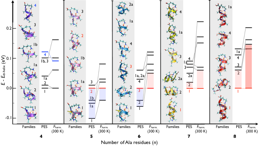
The conformer associated with the lowest-energy PES minimum for =4 is rather small, connecting almost all its backbone CO groups to the LysH+ termination. The remaining H-bond at the N-terminus is bifurcated, comprising an - and a 310-helical H-bond. This conformer could therefore be classified as the smallest possible -helical prototype in this series. In contrast, the lowest-energy PES minima for =5 and 6 are not simple helices. Each contains an “inverted” H-bond where one CO group points to the N-terminus and its connecting NH group points to the C-terminus, producing more compact structures. In Fig. 1, these bonds are highlighted in yellow and pointed to by an arrow. For =5, we have previously denoted this conformer as “g-1” Rossi2010 . For =7 and 8, the lowest-energy PES minima correspond to -helices. In each case, they are closely followed by a conformer that we characterize as compact/globular (Families 2 of =7 and 8, with an energy separation of 20 and 60 meV respectively). Thus, we already observe a cross-over with peptide length to -helical lowest-energy minima of the PES at =7. However, based on the energy hierarchies of the structures of the local PES minima alone, one would not expect a purely helical ensemble of conformers at room temperature at =7 or 8. Simple Boltzmann factors would indicate a mix of structure candidates. Yet, the experimental work by Kohtani and Jarrold KohtaniJarroldWater2004 does indicate a complete room-temperature structural cross-over at =8 at the latest, albeit based on a completely different line of reasoning (water adsorption behavior of “helical” versus “globular” conformers).
For the low-energy conformers in Fig. 1, we also compute and show in the same figure the
impact of the vibrational free
energy at room temperature (=300 K) in the harmonic approximation
111The free energy contributions from
rigid-body rotations are also included, but
differences between these energies for different conformers were
found to be of the order of only a few meV, with the maximum
difference amounting to 8 meV..
Remarkably, the relative stability of the
-helices is systematically enhanced by the
vibrational free energy contribution for all .
For =5 and 6 the -helical conformers move down in energy with
respect to the (non-helical) lowest energy PES minimum. For =7 and 8
the -helices now become the isolated minima. In detail, we observe:
- For =8, the energetic interval between the -helical
lowest energy conformer and the nearest globular one (red shaded areas) now amounts to
0.14 eV. The additional Family 1a at 0.13 eV is another -helix
with a slightly modified terminating H-bond network. With the vibrational
free energy included, the energy hierarchy is thus consistent with the
experimental claim that -helices
dominate over all other possible conformers in gas-phase experiments
for =8 KohtaniJarroldWater2004 ; WangLaskin2010 .
- For =7, the same qualitative picture emerges. Here, the next
remaining conformer (Family 3) at 300 K is a
mixed 310/-helix. The competing compact conformers (representatives of Family 2 and 2a) are
significantly destabilized by ().
- For =6, the -helix emerges as the room-temperature
minimum free energy conformer, but the competing non-helical PES
minimum, Family 1, remains close (50 meV).
- For =5, the difference between the -helix and Family 1 (g-1) decreases by
60 meV at 300 K, compared to the PES minimum, but Family 1 remains
overall more favorable.
In essence, there is an uniform stabilization of helical over more
compact (globular) structures as the temperature increases. The
stabilization tendency increases with peptide length, confirming
quantitatively and systematically from first principles the related
observations in
Refs. Kinnear:2002p4458 ; MaTsaiNussinov2000 ; PlowrightMons2011 .
We next demonstrate that it is indeed the
vibrational entropy term which is critical, and then pinpoint
the physical reason among the low-frequency vibrational modes.
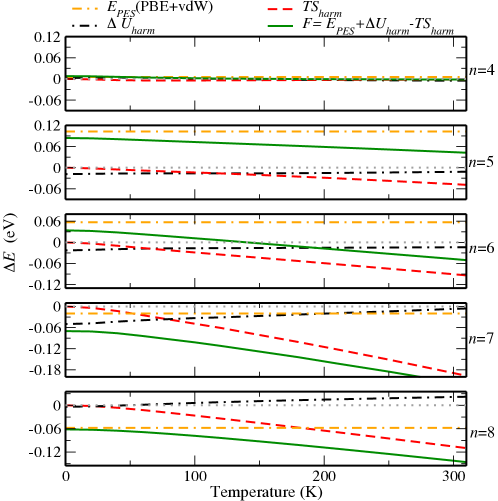
III.2 Origin of the entropic stabilization
In Fig. 2 we analyze the individual quantities composing the vibrational free energy differences between: Families 1 and 2 of =4, Families 1 and the -helical conformers of =5 and 6, as well as Families 1 (-helical) and 2 for =7 and 8. The energy terms plotted are the PBE+vdW PES minimum energy, the harmonic internal energy (containing the zero-point energy) , and the entropy term . They are reported in Fig. 2 as energy differences, taking the non-helical conformer of each as the reference, such that negative slopes correspond to a stabilization of the -helices. Upon inspection of Fig. 2, we observe a monotonic stabilization of all helical conformers with increasing . While for the shortest molecule (=4) there is hardly any observable effect, the stabilization trend is enhanced with increasing length. For =6, we predict a cross-over of the lowest-energy structures at 150 K. It is clear that among the individual contributions to the vibrational part of the free energy , the entropy term is indeed always the most important helix-favoring term. The zero-point-energy ( at =0), is also favorable, but on a smaller scale.
The observed stabilization of the -helical conformers can be understood by analyzing the lowest-energy vibrational modes of different conformations. For simple helices, these modes have been estimated and analyzed before ItohNishi2011 . The important point here is that we have available a direct comparison against many other, non-helical structure candidates. These modes dominate in the harmonic free energy expression at room , so that conformers with lower-frequency modes will effectively be stabilized over others. In Table 1, the frequencies (in cm-1) corresponding to the first normal modes of all conformers shown in Fig. 1 are reported. The lowest-energy -helices, marked in red in Table 1, show first vibrational normal modes between 8 cm-1 and 13 cm-1. The same is true for families 1a of =7 and 8, which only differ in details of the termination. In contrast, the conformers that have first vibrational modes of at least 20 cm-1 are all compact non-helical, for example: 28 cm-1 for Family 2 of =7, and 22 cm-1 for Family 1 (g-1 motif) of =5. The g-1 motif has the first vibrational mode lying around 20 cm-1 for =5, 6, and 7 (marked with a * symbol in Table 1).
Of course, the energy destabilization of the compact conformers with respect to the helices will depend not only on the first vibrational mode but also on the overall distribution of low-frequency modes. To illustrate this point, we show the frequencies of vibration lying between 0 and 50 cm-1 for Families 1 () and 2 (globular) of =7 and 8 in Fig. 3(a) and (b). The -helices have lower first vibrational normal modes, and have slightly higher (one or two modes) densities of modes in this region than the globular conformers.
| length/conformer | 4 | 5 | 6 | 7 | 8 |
|---|---|---|---|---|---|
| Family 1 | 23 | 22* | 20* | 12 | 11 |
| Family 1a | 27 | 23 | 21 | 10 | 11 |
| Family 1b | 25 | 26 | |||
| Family 2 | 17 | 13 | 20 | 28 | 20 |
| Family 2a | 23 | 22 | |||
| Family 3 | 27* | 20 | 8 | 15 | 16 |
| Family 4 | 17 | 18 | 20* | 15 |
Here, a comment regarding structures that are more elongated than -helices is in order. It is customary to compare the stability of -helices with 310 helices and -sheets or fully extended structures (e.g., in references IretaGalvan2003 ; dannenbergccoperativity2 ; IsmerIretaNeugebauer2008 ; ImprotaScuseria2001 ; Topol2001 ; WuZhao2001 ; ShiKallenbach2002 ; bourkubelkakeiderling2002 ; peneviretashea2008 and many others). Following our rationale above, the more extended the structure is, the softer the low vibrational modes will be. This indeed happens for the most extreme case, the fully extended structure (FES), where we find the first vibrational mode to lie around only 2 cm-1 (calculated for =8 and 15). In fact, entropically stabilized -sheets in neutral polyalanine in the gas-phase have been suggested in Ref. DugourdJarrold2005 . For 310 helices, in our own structure searches for =4-8, we find the first vibrational mode to lie always very close to their -helical counterpart. Accordingly, it is the enthalpic energy difference that favors -helices specifically over 310 for all molecules studied. For infinite periodic structures, calculation of phonons and vibrational free energies for -, 310-, and -helices, and the FES in Ref. IsmerIretaNeugebauer2008 corroborate our results. There, all helices are destabilized with respect to the FES at 300 K. The -helix, which is the most compact among the helices studied in Ref. IsmerIretaNeugebauer2008 , is most destabilized by the vibrational entropy term.
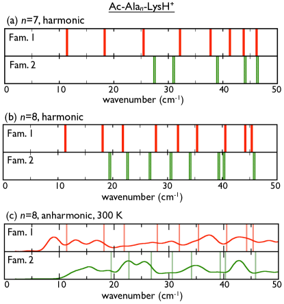
III.2.1 Beyond the harmonic approximation: Ab initio molecular dynamics
We next show that the observations discussed in the last paragraph should hold also in a fully anharmonic picture. Even at relatively low temperature, where the structure still stays generally constant, we expect first the inherent anharmonicity of the local, nearly harmonic PES, then the lowest-barrier transitions between basins (side chain rotations), and then transitions between locally different backbone conformations and H-bond networks TournierSmith2003 ; HongSmith2011 to contribute. Unfortunately, a direct calculation of these terms (e.g., by thermodynamic integration) is computationally prohibitive in DFT. We can, however, use explicit ab initio molecular dynamics simulations to gain some insights. In Fig. 3(c), we compare the Fourier-transformed velocity autocorrelation functions of the Family 1 (helical) and Family 2 (compact non-helical) conformers of =8, extracted from explicit microcanonical ab initio molecular dynamics simulations (21 ps total time, 1 fs time step, initially thermalized to approximately room temperature). The Fourier transform of the velocity autocorrelation function corresponds to a vibrational density of states (VDOS). At =0 and for classical nuclei, the autocorrelation function should reflect only the harmonic eigenmodes of Fig. 3(b). Compared to these modes, the onset of the calculated VDOS at 300 K is noticeably shifted towards lower frequencies for both conformers, but the onset frequency for the helix is still significantly lower than for the non-helical structure. Thus, the lower vibrational frequencies of the helix are also carried over to the full (anharmonic) motion of the conformers. In addition, the integral over the VDOS up to 50 cm-1 is 4% larger for the -helical Family 1 than for the compact Family 2, i.e., the general downshift of frequencies in this region is also preserved.
Regarding the overall local structure stability during the AIMD simulation, we observe that the H-bond pattern of Family 2 (compact non-helical) stays essentially the same throughout the entire simulation (see Figure S.1 in the Supporting Information). In contrast, Family 1 displays local structural fluctuations in the helical part, occasionally forming short-lived 310-like H-bond connections. Similar fluctuations also occur in simulations of longer helices (e.g., Ac-Ala15-LysH+ in Ref. Rossi2010 ). It seems plausible that the overall greater “floppiness” of the helix compared to the more compact non-helical H-bond network adds another favorable entropic contribution at room temperature. This direct observation that we make has been guessed as the motive for the loss in entropy observed in -helical formation, compared to a PPII helical structure in Ref. ShiKallenbach2002 . According to our reasoning, the PPII structure, being less compact than the -helices should indeed show more fluctuations, just like -helices do if compared to more compact conformers.
III.3 The role of the termination
Finally, we show that, especially for the short peptides considered here,
there are two independent aspects to the stabilization of helical
conformers: length and termination. To isolate their role, we examine
the character of the lowest-frequency modes as a function of
peptide length (also for longer helices) for two different
terminations. The first is the LysH+-terminated series, which is
the main subject of this work. We contrast this series with
Li+-terminated polyalanine molecules Alan-Li+, a much more
rigid termination as we shall see. For the latter, we assume
-helices for all (fully relaxed in DFT-PBE+vdW), which are at least
locally stable when Li+ is placed in contact with the last three
residues at the C terminus.
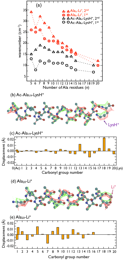
In Fig. 4(a), we compare the position of the first and
second vibrational modes of these -helical
geometries of Alan-Li+, =5-15, and 20, with the -helical
geometries of Ac-Alan-LysH+, =5-15, and 19.
For both peptide series, there is a monotonic decrease of
the first and second vibrational frequencies for 7, but the starting point
is much higher for the Li+ termination (26 cm-1 at =7)
than for the LysH+ termination. A softening trend of the respective
modes in neutral polyalanine helices with increasing length has also been observed in Ref. ItohNishi2011 .
In order to characterize this vibrational mode in more detail, Fig. 4(b)
visualizes the displacement of the backbone atoms of Ac-Ala19-LysH+ when
deforming this molecule along the first vibrational mode. Fig. 4(c) shows the relative
length changes of the hydrogen bonds in the structure upon deformation
along this mode. Subfigures (d) and (e) show the equivalent data for Ala20-Li+.
For both molecules, the vibration spans the helical part of the structures.
For the LysH+ terminated molecule, we show
that the actual LysH+ termination (connected to the last
four CO residues) is clearly involved in the vibration. In contrast, the
hypothetical Li+ charged termination
constrains especially the C terminus to be much more rigid, as
evidenced by the almost zero change of all Li-O
distances. Here, the N-terminus is much more involved in the
lowest-frequency modes. The same trends are observed for the smaller
molecules in both series.
Movies containing 3D visualizations of the first vibrational modes for helical
and compact structures
are contained in the Supporting Information.
We thus conclude two points:
(i) Helices are entropically favored by allowing delocalized, soft
low-frequency modes that we do not observe in competing, more compact
conformers of the same LysH+ termination (evidenced by
Table 1).
(ii) For short conformers, the already
electrostatically favorable LysH+ termination PrestaRose1988 ; MarquseeBaldwin1989 is additionally helpful by allowing soft,
delocalized modes to include the termination also for short
conformers, in contrast to the hypothetical, much harder
charged termination by Li+. For long enough helices these softer low frequency modes
should exist regardless of the termination.
IV Conclusions
In summary, we show from first principles, quantitatively, and for a particularly well studied series of polyalanine peptides how helices emerge with length and temperature as the leading structural pattern from a vast array of possible competing conformers. The crossover to helical stability with length is already apparent based on local structural minima of the potential-energy surface alone, due to the critical role of H-bond networks including their cooperativity GuoKarplus1994 ; Baldwin2003 ; IretaGalvan2003 ; dannenbergccoperativity2 ; TkatchenkoRossi2011 as well as that of vdW terms TkatchenkoRossi2011 . In addition, the contribution from softer low-frequency vibrational modes acts to stabilize helices with increasing temperature over their more compact competition. The specific experimental claim KohtaniJarroldWater2004 of exclusively helical conformers at and above =8 is thus explained by both enthalpic and entropic effects acting together at finite temperature. Ab initio molecular dynamics simulations corresponding to approximately room temperature suggest that these trends are further strengthened by anharmonic effects.
The emergence of room-temperature helix stability with length in Ac-Alan-LysH+ is thus the result of a subtle balance of enthalpic and entropic terms. In a non-vacuum environment, further terms would obviously contribute, but we expect the fact that helices, in general, allow locally softer vibrational modes to hold. Regarding the overall ubiquity of the helical motif in folded peptides and proteins, at least, we here show that low-frequency modes will be a significant quantitative contribution.
V Acknowledgement
The authors would like to acknowledge Dr. Carsten Baldauf for numerous helpful discussions and suggestions about the figures and paper in general, including the schematic backbone vibration visualization in Fig. 4.
VI Supplemental Information
Additional computational details, XYZ geometries of conformers discussed in this paper, detailed information about the structures discussed in this paper, and movies illustrating the first vibrational modes of helices and compact conformers.
References
- (1) Pauling, L.; Corey, R.; Branson, H. Proc. Natl. Acad. Sci. USA 1951, 37, 205–211.
- (2) Berman, H.M.; et al. Nucl. Acids Res. 2000, 28, 235–242. http://www.pdb.org.
- (3) Creighton, T.E. Nature 1987, 326, 547–548.
- (4) Pace, N.; Scholtz, J.M. Biophys. J. 1998, 75, 422–427.
- (5) Scholtz, J.M.; York, E.J.; Stewart, J.M.; Baldwin, R.L. J. Am. Chem. Soc. 1991, 113, 5102–5104.
- (6) Horovitz, A.; Matthews, J.M.; Fersht, A.R. J. Mol. Biol. 1992, 227, 560–568.
- (7) Creamer, T.P.; Rose, G.D. Proc. Natl. Acad. Sci. 1992, 89, 5937–5941.
- (8) Kinnear, B.; Jarrold, M. J. Am. Chem. Soc. 2001, 123, 7907–7908.
- (9) Miller, J.; Kennedy, R.; Kemp, D. J. Am. Chem. Soc. 2002, 124, 945–962.
- (10) Scott, K.A.; Alonso, D.V.; Sato, S.; Fersht, A.R.; Daggett, V. Proc. Nat. Acad. Sci. 2007, 104, 2661–2666.
- (11) Moreau, R.; et. al. J. Am. Chem. Soc. 2009, 131, 13107–13116.
- (12) Zimm, B.H.; Bragg, J.K. J. Chem. Phys. 1959, 31, 526–535.
- (13) Kinnear, B.; Hartings, M.; Jarrold, M. J Am Chem Soc 2002, 124, 4422–4431.
- (14) Hudgins, R.R.; Ratner, M.A.; Jarrold, M.F. J. Am. Chem. Soc. 1998, 120, 12974–12975.
- (15) Kohtani, M.; Jarrold, M. J. Am. Chem. Soc. 2004, 126, 8454–8458.
- (16) Tournier, A.L.; Smith, J.C. Phys. Rev. Lett. 2003, 91, 208106.
- (17) Brooks, B.; Karplus, M. Proc. Natl. Acad. Soc. 1985, 82, 4995–4999
- (18) Editorial, Nature Chemistry Focus issue Of polemics and progress, Nature Chem. 2012, 4, 141.
- (19) Hay, S.; Scrutton, N.S. Nature Chem. 2012, 4, 161–168.
- (20) Glowacki, D.R.; Harvey, J.N.; Mulholland, A.J. Nature Chem. 2012, 4, 169–176.
- (21) Scholtz, J.M.; Baldwin, R.L. Annu. Rev. Biophys. Biomol. Struct. 1992, 21, 95–118, and references therein.
- (22) Baldwin, R.L. J. Mol. Biol. 2007, 371, 283–301.
- (23) Guo, H.; Karplus, M. J. Phys. Chem. 1994, 98, 7104–7105.
- (24) Baldwin, R.L. J. Biol. Chem. 2003, 278, 17581–17588.
- (25) Ireta, J.; Neugebauer, J.; Scheffler, M.; Rojo, A.; Galván, M. J. Phys. Chem. B 2003, 107, 1432–1437.
- (26) Wieczorek, R.; Dannenberg, J.J. J. Am. Chem. Soc. 2004, 126, 14198–14205.
- (27) Tkatchenko, A.; Rossi, M.; Blum, V.; Ireta, J.; Scheffler, M. Phys. Rev. Lett. 2011, 106, 118102.
- (28) Blagdon, D.E.; Goodman, M. Biopolymers 1975, 14, 241–245.
- (29) Presta, L.G.; Rose, G.D. Science 1988, 240, 1632–1641.
- (30) Serrano, L.; Fersht, A.R. Nature 1989, 342, 296–299.
- (31) Takahashi, S.; Kim, E.H.; Hibino, T.; Ooi, T. Biopolymers 1989, 28, 995–1009.
- (32) Marqusee, S.; Robbins, V.; Baldwin, R. Proc. Natl. Acad. Sci. 1989, 86, 5286–5290.
- (33) Dugourd, P.; Antoine, R.; Breaux, G.; Broyer, M.; Jarrold, M. J. Am. Chem. Soc. 2005, 127, 4675–4679.
- (34) Williams, L.; Kristian, K.; Kemp, D. J. Am. Chem. Soc. 1998, 120, 11033–11043.
- (35) Hua, S.; Xu, L.; Li, W.; Li, S. J. Phys. Chem. B 2011, 115, 11462–11469.
- (36) Liu, D.; Wyttenbach, T.; Bowers, M. Int. J. Mass Spectrom. 2004, 236, 81–90.
- (37) Job, G.E.; et al. J. Am. Chem. Soc. 2006, 128, 8227–8233.
- (38) Elstner, M.; Jalkanen, K.J.; Knapp-Mohammady, M.; Frauenheim, T.; Suhai, S. Chem. Phys. 2000, 256, 15–27.
- (39) Salvador, P.; Asensio, A.; Dannenberg, J.J. J. Phys. Chem. B 2007, 111, 7462–7466.
- (40) García A.E.; Sanbonmatsu, K.Y. Proc. Natl. Acad. Sci. 2002, 99, 2782–2787.
- (41) Tirado-Rives, J.; Maxwell, D.S.; Jorgensen, W.L. J. Am. Chem. Soc. 1993, 115, 11590–11593.
- (42) Kauzmann, W. Adv. Protein Chem. 1959, 14, 1–63.
- (43) MacKenzie, K.R.; Prestegard, J.H.; Engelman, D.M. Science 1997, 276, 131–133.
- (44) Khutorsky, V. Biochem. Biophys. Res. Commun. 2003, 301, 31–34.
- (45) Jarrold, M. Phys. Chem. Chem. Phys. 2007, 9, 1659–1671, and references therein.
- (46) Rossi, M.; et al. J. Phys. Chem. Lett. 2010, 1, 3465–3470.
- (47) Kohtani, M.; Jones, T.; Schneider, J.; Jarrold, M. J. Am. Chem. Soc. 2004, 24, 7420–7421.
- (48) Hu, Q.; Wang, P.; Laskin, J. Phys. Chem. Chem. Phys. 2010, 12, 12802–12810.
- (49) Brooks, C.; Karplus, M.; Pettitt, M. Proteins, Advances in Chemical Physics (Wiley-VCH) 1988 1st edition, 71, 180–183.
- (50) Poulain, P.; Calvo F.; Antoine R.; Broyer M.; Dugourd Ph. Europhys Lett 2007 79, 66003.
- (51) Counterman, A.; Clemmer, D. J. Phys. Chem. B 2003 107, 2111-2117.
- (52) Ma, B.; Tsai, C.J.; Nussinov, R. Biophys J 2000, 79, 2739–2753.
- (53) Plowright, R.J.; Gloaguen, E.; Mons, M. ChemPhysChem 2011, 12, 1889–1899.
- (54) Grimme, S. J. Comput. Chem. 2006, 27, 1787–1799.
- (55) Chakrabartty, A.; Baldwin, R.L. Adv. Protein Chem. 1995, 46, 141–176.
- (56) Perdew, J.P.; Burke, K.; Ernzerhof, M. Phys. Rev. Lett. 1996, 77, 3865–3868.
- (57) Tkatchenko, A.; Scheffler, M. Phys. Rev. Lett. 2009, 102, 073005.
- (58) Rossi, M. Ab initio study of alanine-based polypeptide secondary-structure motifs in the gas phase, Ph.D. thesis (Fritz-Haber-Institut der Max-Planck-Gesellschaft and Technische Universität Berlin), 2011, http://opus.kobv.de/tuberlin/volltexte/2012/3429/.
- (59) McLean, J.; et. al. J. Phys. Chem. B 2010, 114, 809–816
- (60) Wieczorek, R.; Dannenberg, J. J. Am. Chem. Soc. 2004, 126, 12278–12279
- (61) Kaminski, G.A.; Friesner, R.A.; Tirado-Rives, J.; Jorgensen, W.L. J. Phys. Chem. B 2001, 105, 6474–6487.
- (62) Ponder, J. Tinker - software tools for molecular design. In this work we used versions 4.2 and 5.1 of the program and the force-fields’ versions distributed within the package.
- (63) Blum, V.; et al. Comp. Phys. Comm. 2009, 180, 2175–2196.
- (64) Havu, V.; Blum, V.; Havu, P.; Scheffler, M. J. Comput. Phys. 2009, 228, 8367–8379.
- (65) Itoh, K.; Ikeda, A.; Iwamoto, T.; Nishizawa, S. J. Molec. Struct. 2011, 1006, 52–58.
- (66) Ismer, L.; Ireta, J.; Neugebauer, J. J. Phys. Chem. B 2008, 112, 4109–4112.
- (67) Improta, R.; Barone, V.; Kudin, K.; Scuseria, G. J. Am. Chem. Soc. 2001, 123, 3311–3322.
- (68) Topol, I.; et. al., J. Am. Chem. Soc. 2001, 123, 6054–6060.
- (69) Wu, Y.-D.; Zhao, Y.-L. J. Am. Chem. Soc. 2001, 123, 5313–5319
- (70) Shi, Z.; Olson, C.; Rose, G.; Baldwin, R.; Kallenbach, N. Proc. Nat. Acad. Sci. 2002, 99, 9190–9195.
- (71) Bour, P.; Kubelka, J.; Keiderling, T. Biopolymers 2002, 65, 45–59.
- (72) Penev, E.; Ireta, J.; Shea J-E. J. Phys. Chem. B 2008, 112, 6872–6877.
- (73) Hong, L.; Smolin, N.; Lindner, B.; Sokolov, A.P.; Smith, J.C. Phys. Rev. Lett. 2011, 107, 148102.