Is ion channel selectivity mediated by confined water?
Abstract
Ion channels form pores across the lipid bilayer, selectively allowing inorganic ions to cross the membrane down their electrochemical gradient. While the study of ion desolvation free-energies have attracted much attention, the role of water inside the pore is less clear. Here, molecular dynamics simulations of a reduced model of the KcsA selectivity filter indicate that the equilibrium position of Na+, but not of K+, is strongly influenced by confined water. The latter forms a stable complex with Na+, moving the equilibrium position of the ion to the plane of the backbone carbonyls. Almost at the centre of the binding site, the water molecule is trapped by favorable electrostatic interactions and backbone hydrogen-bonds. In the absence of confined water the equilibrium position of both Na+ and K+ is identical. Our observations strongly suggest a previously unnoticed active role of confined water in the selectivity mechanism of ion channels.
FRIAS] School of Soft Matter, Freiburg Institute for Advanced Studies, Freiburg, Germany
1 Introduction
Neurons enable us to think, act and remember 1. At the fundamental level, an important role is played by ion channels. Forming potassium-selective pores that span the cell membrane, potassium channels are the most widely distributed channels in nature 1, 2. The breakthrough in the structure determination came from the identification of the bacterial homolog from Streptomyces lividans (KcsA) 3. This channel is characterized by a tetrameric structure in which four identical protein subunits associate around a central ion conducting pore. At the extracellular side, the selectivity filter is formed by a highly conserved sequence of five residues (TVGYG) 4, 3.
Originally, the high complementary of the selectivity filter to K+ was thought to be the reason for the selectivity 5. But it is now clear that the interplay between structure and dynamics of the selectivity filter is at the origin of the mechanism 6, 7, 8, 9. Recently, atomic models of the selectivity filter were used to elucidate the role of dynamics in the process 10, 11, 12, 13. In one of these models, the filter is reduced to the most selective binding site of KcsA, called S2 14. This is done by harmonically constraining the four backbone segments defining S2 to the experimental conformation 10, 12. Solvation free-energy calculations illustrated that the model is selective, allowing a statistical mechanics treatment of the limiting cases of rigid and very flexible binding sites 12. Moreover, calculations on larger models of the filter showed that backbone fluctuations are influenced by the presence of Na+ or K+ at position S2 11. These results support the view that several aspects of selectivity can be elucidated by analyzing ion binding to simplified models of the filter.
Another important player in selectivity is water. Simulation results showed that confined water appears together with cations in the conduction pore 15, 16, 17. Notwithstanding, it is not clear yet if water actively mediates selectivity or not.
Here, the role of confined water is investigated by molecular dynamics simulations of a S2 binding site model, providing evidence that the equilibrium position of Na+ within the binding site is displaced by the presence of a water molecule. Our calculations are in agreement with a recent crystallographic study 18 and multi-ion free-energy calculations 18, 19. These concepts support the idea that KcsA can bind both Na+ and K+ with similar strength but different mechanism.
2 Methods
The S2 model. A reduced model of the S2 binding site of the KcsA channel (PDB code: 1K4C, 1) was built with four diglycine peptides as done in Ref. 12. The heavy atoms of the reduced model were constrained with an harmonic potential of force constant kJ/mol/nm2 ( Å2). The coordinates were translated with the vector , taking the axial coordinate parallel to (1,0,0). A pdb file of the reduced model is provided as Supplementary Information (SI).
Molecular dynamics simulations. All calculations were performed with the GROMACS program 20, 21 and the AMBER-03 force field 22, 23. A cubic box of initial length of 4 nm solvated with TIP4P-Ew water was used 24. Simulations were integrated with the Langevin equations () at 300 K coupled with a Berendsen barostat ( ps) 25. Long range electrostatics was computed with PME 26 with a 1.0 nm cut-off for all non-bonded interactions. After 10 ns of equilibration, each ion was simulated by a 100 ns long trajectory. For both Na+ and K+, the starting configuration was taken as the center of the binding site (, see 1). To check that there was no influence on the starting position, 20 runs of 5 ns each were further performed (Figure S1 in SI). Calculations performed with a TIP3P water model are in agreement with the present analysis (see Figure S2 in SI).
Potential of mean force The potential of mean force for Na+ and K+ was computed with GROMACS 20, 21 along the pore axis from (the bulk) to (position ). Umbrella sampling calculations were spaced by 0.1 Å along the axial coordinate with the ion restrained in the normal plane with an harmonic potential (=1000 kJ/mol/nm2). An additional harmonic potential of force constant and 20000 kJ/mol/nm2 was applied in the and range, respectively. After 1 ns of equilibration, trajectories were run for 1 ns. The weighted histogram method was used to reconstruct the potential of mean force 27.
Ion interaction energy in vacuo. To calculate the ion interaction energy , the cation was harmonically restrained ( kJ/mol/nm2) at 0.25 Å spaced positions along the axial coordinate from -5.0 to 0.0 Å. After 1 ns of equilibration, each run was performed for 10 ns at 300 K and constant volume.
3 Results
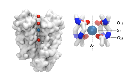
The KcsA channel is shown in 1. In our simulation study, a reduced model of the protein selectivity filter was used. The S2 binding site was modeled by four peptides constrained to the experimental structure (right panel, see Methods for details) 10, 12. Within this model, the pore axis centered along the channel is labeled as , while the origin of the axis is taken as the experimental position of the K+ ion. This position is conventionally called . Similarly, the positions of the carbonyl oxygens defining the binding site at the extra and intra-cellular sides of the model are denoted as and , respectively (1).
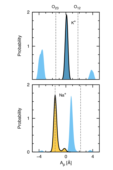
The behavior of K+ and Na+ inside the S2 binding site was studied by molecular dynamics simulations in explicit water (see Methods for details). In both cases, the starting position of the ion was . 2 shows the probability distribution of the ion position on the axis. As expected, K+ was found at position (black countoured blue area) between and (dashed lines), coordinating with the eight carbonyl oxygens of the binding site 14, 10.
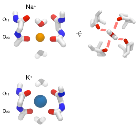
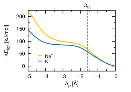
This is not the case for Na+. After few ns of simulation (see Figure S1 in SI), the ion hopped from to , assuming a configuration perfectly in plane with the 4 carbonyl oxygens (black countoured orange area in bottom panel of 2). This position is very stable, representing the 93.6% of the total simulation time, with a high barrier to hop back to (roughly four transitions in 100 ns). Analysis of the closest water molecules to the ion provided a mechanism for the configuration shift. Contrary to the case of K+ where the solvent is at the outside of the pore, one water molecule enters the channel effectively shifting the position of Na+ to . This is shown by the probability distribution of the closest water to the ion represented as a light blue area in the figure. A structural representation of the confined water is illustrated in 3. Per se, there is no energetic preference in shifting Na+ to the position as shown by the average interaction energy between the ion and the pore in vacuo (4). In the absence of water, is the most stable position for both K+ and Na+, strongly indicating that the binding site shift is due to the presence of a confined water.
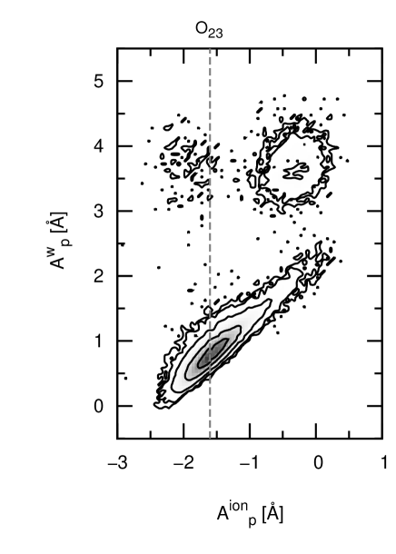
The confined water at position is stabilized by multiple strong contacts (3). The water oxygen makes electrostatic interactions with Na+, while hydrogen bonds with two of the four carbonyls at position are formed (3, top right panel). These hydrogen bonds are also entropically stabilized, being the confined water capable of binding to all four carbonyls of by rotating itself around the axis (bond lifetime around 2 ps, see Figure S3 in SI). This molecule is extremely stable, never exchanging with the bulk. At the outside of the channel another water molecule was found at the other side of Na+, forming favorable electrostatic interactions with the ion (ghost water in the top panel of 3). The position of the confined water correlates with the fluctuations of Na+ around , indicating that the ion-water configuration behaves as a complex (5). In the rare occasions when Na+ hops back to , the water molecule is expelled and the complex broken ( and , 5).
On the other hand, for the case of K+ water was only found at the outside of the pore (ghost molecules at the bottom of 3). Those waters are stabilized by the formation of hydrogen bonds with the carbonyls of the binding site. But the interaction with the ion is much weaker in this case, being the water oxygens facing the bulk.
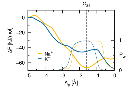
To complement the simulation study, free-energy calculations were performed. In 6 the potential of mean force (PMF, see Methods) along the axial coordinate is shown (thick lines). The most stable configurations for Na+ and K+ were respectively found at positions and , confirming our analysis. For both cases, position is coupled with the presence of a water molecule inside the filter ( around , thin lines). Though energetically unfavorable, K+ at this position is in complex with a confined water as observed for Na+. Interestingly, the free-energy difference between the outside of the pore () and the most stable binding position is remarkably similar for the two ions, indicating no strong preference towards K+ binding. These results strongly support the idea that the filter has the ability to bind both ions with different mechanisms but similar strength as already suggested in Ref. 18, 19.
4 Discussion
It has been known, for more than a decade now, that the selectivity filter of KcsA presents several binding sites. Each of them forms a cage of optimal coordination for K+ by means of eight backbone carbonyls. Most of the calculations on KcsA selectivity were based on the relative stability of Na+ over K+ inside the pore with respect to the bulk 10, 12. That is, the solvation free-energy to move an ion from bulk water to a specific binding site inside the selectivity filter. These works suggested the S2 binding site as the most selective portion of the filter, being the free-energy difference in the position much more favorable for K+ compared to Na+. But this approach is not without problems if is not a stable configuration for Na+. In fact, Na+ and K+ might be characterized by distinct equilibrium positions inside the pore. X-ray crystallography 18 and multi-ion free-energy calculations18, 19 supported this idea, showing that Na+ preferentially adopts a configuration in-plane with backbone carbonyls. The observation of distinct binding positions has several consequences to our understanding of ion selectivity. Previous free-energy calculations using position restrains to the center of the binding sites need to be extended taking into account the correct equilibrium configurations.
Our calculations on a minimalistic model of the filter provided a mechanism for the position shift. The presence of a confined water molecule in complex with Na+ effectively modifies the equilibrium configuration. The confined water is stabilized by favorable electrostatic interactions with the ion as well as multiple hydrogen-bonds with the backbone carbonyls. The binding position shift disappears in the absence of confined water, making the latter an essential ingredient for the preferential position of Na+. In-plane binding always implies the presence of confined water even in the unfavorable case of K+.
In the past, water was mostly considered in terms of solvation free-energies and its screening effects without much attention to the molecular mechanism. Our results reinforce the idea that biological water is an active player at the molecular level 28.
5 Acknowledgments
This work is supported by the Excellence Initiative of the German Federal and State Governments.
References
- Kandel et al. 2000 Kandel, E. R.; Schwartz, J. H.; Jessell, T. M. Principles of Neural Science, 4th ed.; McGraw-Hill Medical, 2000
- Hille 2001 Hille, B. Ionic Channels of Excitable Membranes, 3rd ed.; Sinauer Associates, 2001; Chapter 5, pp 131–168
- Doyle et al. 1998 Doyle, D. A.; Morais Cabral, J.; Pfuetzner, R. A.; Kuo, A.; Gulbis, J. M.; Cohen, S. L.; Chait, B. T.; MacKinnon, R. Science 1998, 280, 69–77
- Heginbotham et al. 1994 Heginbotham, L.; Lu, Z.; Abramson, T.; MacKinnon, R. Biophys J 1994, 66, 1061–1067
- Zhou et al. 2001 Zhou, Y.; Morais-Cabral, J. H.; Kaufman, A.; MacKinnon, R. Nature 2001, 414, 43–48
- Fowler et al. 2008 Fowler, P.; Tai, K.; Sansom, M. Biophys J 2008, 95, 5062–5072
- Roux et al. 2011 Roux, B.; Bernèche, S.; Egwolf, B.; Lev, B.; Noskov, S.; Rowley, C.; Yu, H. J Gen Phys 2011, 137, 415–426
- Dixit and Asthagiri 2011 Dixit, P.; Asthagiri, D. J Gen Phys 2011, 137, 427–433
- Varma et al. 2011 Varma, S.; Rogers, D.; Pratt, L.; Rempe, S. J Gen Phys 2011, 137, 479–488
- Noskov et al. 2004 Noskov, S. Y.; Berneche, S.; Roux, B. Nature 2004, 431, 830–834
- Asthagiri et al. 2006 Asthagiri, D.; Pratt, L. R.; Paulaitis, M. E. J Chem Phys 2006, 125, 024701+
- Yu et al. 2010 Yu, H.; Noskov, S. Y.; Roux, B. Proc Natl Acad Sci U S A 2010, 107, 20329–20334
- Kast et al. 2011 Kast, S.; Kloss, T.; Tayefeh, S.; Thiel, G. J Gen Phys 2011, 138, 371–373
- Berneche and Roux 2001 Berneche, S.; Roux, B. Nature 2001, 414, 73–77
- Berneche and Roux 2000 Berneche, S.; Roux, B. Biophys J 2000, 78, 2900–2917
- Guidoni et al. 2000 Guidoni, L.; Torre, V.; Carloni, P. FEBS Lett 2000, 477, 37–42
- Domene and Sansom 2003 Domene, C.; Sansom, M. Biophys J 2003, 85, 2787–2800
- Thompson et al. 2009 Thompson, A. N.; Kim, I.; Panosian, T. D.; Iverson, T. M.; Allen, T. W.; Nimigean, C. M. Nat Struct Mol Biol 2009, 16, 1317–1324
- Kim and Allen 2011 Kim, I.; Allen, T. W. Proc Natl Acad Sci U S A 2011, 108, 17963–17968
- Van Der Spoel et al. 2005 Van Der Spoel, D.; Lindahl, E.; Hess, B.; Groenhof, G.; Mark, A. E.; Berendsen, H. J. C. J Comput Chem 2005, 26, 1701–1718
- Hess et al. 2008 Hess, B.; Kutzner, C.; van der Spoel, D.; Lindahl, E. J Chem Theory Comput 2008, 4, 435–447
- Duan et al. 2003 Duan, Y.; Wu, C.; Chowdhury, S.; Lee, M. C.; Xiong, G.; Zhang, W.; Yang, R.; Cieplak, P.; Luo, R.; Lee, T.; Caldwell, J.; Wang, J.; Kollman, P. J Comput Chem 2003, 24, 1999–2012
- Sorin and Pande 2005 Sorin, E. J.; Pande, V. S. Biophys J 2005, 88, 2472–2493
- Horn et al. 2004 Horn, H. W.; Swope, W. C.; Pitera, J. W.; Madura, J. D.; Dick, T. J.; Hura, G. L.; Gordon, T. H. J Chem Phys 2004, 120, 9665–9678
- Berendsen et al. 1984 Berendsen, H. J. C.; Postma, J. P. M.; van Gunsteren, W. F.; DiNola, A.; Haak, J. R. J. Chem. Phys. 1984, 81, 3684–3690
- Darden et al. 1993 Darden, T.; York, D.; Pedersen, L. J Chem Phys 1993, 98, 10089–10092
- Kumar et al. 1992 Kumar, S.; Rosenberg, J.; Bouzida, D.; Swendsen, R.; Kollman, P. J Comp Chem 1992, 13, 1011–1021
- Ball 2011 Ball, P. Nature 2011, 478, 467–468