Surface patterning of carbon nanotubes can enhance their penetration through a phospholipid bilayer
Abstract
Nanotube patterning may occur naturally upon the spontaneous self-assembly of biomolecules onto the surface of single-walled carbon nanotubes (SWNTs). It results in periodically alternating bands of surface properties, ranging from relatively hydrophilic to hydrophobic, along the axis of the nanotube. Single Chain Mean Field (SCMF) theory has been used to estimate the free energy of systems in which a surface patterned nanotube penetrates a phospholipid bilayer. In contrast to un-patterned nanotubes with uniform surface properties, certain patterned nanotubes have been identified that display a relatively low and approximately constant system free energy ( kT) as the nanotube traverses through the bilayer. These observations support the hypothesis that the spontaneous self-assembly of bio-molecules on the surface of SWNTs may facilitate nanotube transduction through cell membranes.
Journal link:
Understanding the mechanism of transduction of carbon nanotubes through cell membranes is a challenging undertaking from many perspectives. Apart from fundamental interest, details of the mechanism may answer questions about the cytotoxicity of nanotubes 1, 2, 3, 4 and their potential use as delivery vehicles 5, 6, 7, 8, 9, 10 or to probe bilayer properties 11, 12. However, despite considerable effort devoted to this question, a consensus on the mechanism has not yet been reached and direct evidence of spontaneous translocation of nanotubes through the membranes of cells is still lacking.
In a recent paper, Pogodin and Baulin 13 considered the thermodynamics of a system in which an uncharged nanotube with uniform surface properties penetrates a phospholipid bilayer. A coarse grained model of the phospholipid molecule was adopted, which had been previously shown to adequately characterise the key thermodynamic properties of a phospholipid bilayer in a fluid phase 14. Estimates were made of the free energies of the equilibrium states for the system as the nanotube was progressively moved perpendicularly into the bilayer using a numerical implementation of the Single Chain Mean Field (SCMF) theory 14. The SCMF methodology and three models of the phospholipid bilayer have been discussed in details in Ref. 14. The simplest 3-beads model of phospholipids has been proved to be successful in describing the thermodynamic properties of the bilayer. In essence it involves a coarse grained description of the lipid molecule where the monomers are grouped into two types of beads, one hydrophilic representing the polar heads and two hydrophobic representing the tails of the lipid. The size and the interaction parameters of the beads we adjusted to reproduce the essential thermodynamic properties of a fluid phase of the phospholipid bilayer such as the thicknesses of the layer and the hydrophobic core, the equilibrium area per lipid and the compressibility constant 14.
The objective of these calculations was to determine whether nanotubes of various diameters and surface properties might penetrate a bilayer as a consequence of their thermal motion. Thus, nanotubes of 1, 2.43 and 4.86 nm diameter were considered, characterised by an energy per contact with the coarse grained phospholipid tail, , ranging from 0 kT (representing steric repulsion) to kT, which corresponds to strong hydrophobic attraction. The perpendicular orientation was chosen since this represents the minimum contact area between nanotube and phospholipids per unit depth of penetration and hence the minimum free energy of interaction. An output of the SCMF calculations was the equilibrium free energies and the spatial mean field concentration distribution of the phospholipid heads and tails in the bilayer, which varied as a consequence of nanotube penetration. The model thus demonstrated the structural rearrangement of phospholipids at the molecular level that was induced by insertion of the nanotube, and the equilibrium free energy change of the system for each position of the nanotube.
In summary, the calculations showed that the free energy change of the systems for kT rose monotonically with increasing nanotube penetration and were substantial at full penetration (e.g. for the 2.43 nm diameter nanotube the free energy of the system at full penetration was about 100 kT). For kT, and particularly for kT, the initial penetration of the nanotube resulted in a significant fall in the system free energy (e.g. to ca. kT for penetration of a 2.43 nm diameter nanotube with kT to the centre of the bilayer). Further penetration of these nanotubes then led to a steep rise in the system free energy. Overall, the calculations showed that hydrophilic and weakly hydrophobic nanotubes face a substantial energy barrier to penetration, whilst intermediate and strongly hydrophobic nanotubes penetrate little or become entrapped in a free energy well within the bilayer.
Inferences can be drawn from these calculations for the transduction of cylindrical nano-objects such as single-walled carbon nanotubes (SWNTs) through the outer membrane of cells. Untreated SWNTs are significantly hydrophobic and their thermal motion might lead to their accumulation within the core of the cell membrane, but they are unlikely to translocate across the membrane in view of the steep energy barrier that they face to pass out of the membrane core. Hydrophilically functionalised SWNTs, for example pegylated SWNTs 15 face a substantial energy barrier in even penetrating the membrane to its core. These thermodynamic calculations create a conundrum since numerous experimental reports exist that show the accumulation of SWNTs within the cytoplasm of cells16, 17. How may this be?
Unlike the model system that is envisaged in Ref. 13, the environment around a cell is complex and comprises of diverse biomolecular species that might interact with SWNTs. Indeed, a number of publications have reported the ordered self-assembly of polar lipids 18, 19, single stranded DNA 20, 21, 22, polysaccharides 23, 24, amphiphilic proteins 25, 26, 27 and even vitamins 28 onto nanotubes. The electrostatics also give rise to the most general patterns on the nanotubes 29. This self-assembled patterning occurs spontaneously upon mixing the nanotubes with the patterning agent in aqueous solution. The resulting molecular structures commonly take the form of discrete hydrophilic rings along the axis of the nanotube in the case of polar lipids, to helices for polysaccharides and DNA. Thus, in practice, in cell culture systems nanotubes may not have homogeneous surface properties but may display a distinct regular patterning. Furthermore, it is not evident that a nanotube that has been naturally patterned in this way interacts with phospholipid bilayers in a similar manner to a naked nanotube or to a nanotube with a homogeneous adsorption layer. Our point of view is supported by experimental evidence that ordered arrangements of hydrophilic and hydrophobic surface functional groups can alter the penetration of spherical nanoparticles through the cell membranes 30.

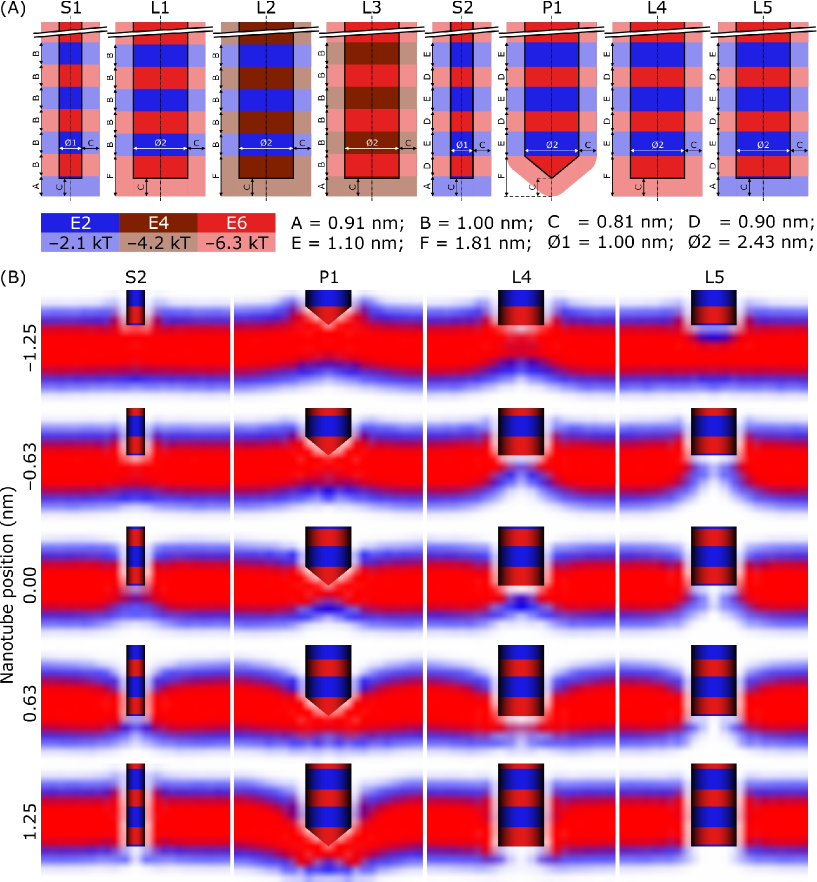
Consider then a patterned hydrophobic nanotube. Along the nanotube the self-assembly of polar lipids (or other biomolecular species) leads to equi-spaced rings of different relative hydrophobicity to the naked nanotube. Moving along the axis of the nanotube, the surface characteristics alternate periodically as indicated in Figure 1(A). The spatially segregated surface characteristics of such a nanotube is thus substantially different from the uniform surface character assumed in the previous SCMF calculations 13, and this may influence the free energy change of the system when the nanotube penetrates a phospholipid bilayer. Note, that the surface patterning has bigger effect on the translocation through a bilayer than the shape or geometry of nanoparticles31, especially for small particles.
Comparison of the energy curves of uniform nanotubes at different positions13 suggest that alternation of stripes with certain interaction can, in principle, reduce considerably the energy barrier of translocation. For example, two stripes with opposite energies placed together may cancel the contribution of each other. Since the thickness of the hydrophobic core is about 2 nm, the width of alternating stripes in this case should be of order 1 nm so that the core is in contact simultaneously with two opposite stripes. Two distinct patterns were considered for a 1 nm diameter nanotube (Figure 1(A), S1 and S2) that differ only in the relative widths of the two sets of rings (i.e. whereas B=1.00 nm for S1, D=0.90 nm and E=1.10 nm for S2). Both S1 and S2 are characterised by alternating rings of interaction energy and kT respectively. A larger nanotube of diameter 2.43 nm was also considered. In this case five different patterns were considered (Figure 1(A), L1, L2, L3, L4 and L5). The effect of the bottom of the nanotube was also investigated, L4 and L5 only differ in the interaction of the bottom part, while P1 has an edge with the same interaction parameter as L4. The corresponding concentration profiles of nanotubes with similar patterns, S2, P1, L4 and L5, are shown in Figure 1(B). We emphasise that these patterns are purely representative and their dimensions and energies are not those for any specific biomolecular system.
The concentration profiles for the patterns L1, L2, L3 look identical, while the energy of penetration of these patterns is quite different. This is because the same positions of the lipids around the nanotube may lead to different enthalpic contributions. Thus, the snapshots usually provided by MD simulations may not be sufficient to distinguish between different scenarios, while the equilibrium energy of insertion may be crucial for understanding the mechanisms of insertion into phospholipid bilayers.
| S1 | S2 | P1 | L1 | L2 | L3 | L4 | L5 | |
|---|---|---|---|---|---|---|---|---|
| (kT) | 29 | 36 | 68 | 22 | 19 | |||
| (kT) | 37 | |||||||
| (nm) | 1.88 | 1.25 | ||||||
| Max. force (pN) | 56 | 51 | 275 | 286 | 184 | 68 | 241 | 78 |
| Max. pressure (MPa) | 72 | 64 | 59 | 62 | 40 | 15 | 52 | 17 |
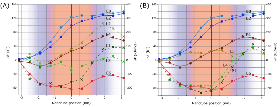
The free energy of penetration of these variously patterned nanotubes into a coarse grained representation of a phospholipid bilayer was estimated using a numerical implementation of SCMF, in the manner described by Pogodin and Baulin 13. The resulting free energies for the small nanotubes are shown in Figure 2. For the nanotube pattern S1 with equi-sized rings (Figure 1(A), S1) the system free energy always lies within the range 0 to kT. Neglecting the end of the nanotube, the mean interaction energy along the length of S1 is kT. Comparing S1 with the free energy for penetration of a nanotube with a uniform surface of interaction energy E4, kT (Figure 2, E4) it is evident that no steep energy barrier is encountered by the patterned nanotube. Variation of the relative sizes of the rings (to give Figure 1(A), S2) results in the free energy of the system being always within the range 0 kT, allowing relatively unimpeded passage of the nanotube through the bilayer. Note, that the oscillations of the energy curve is due to abrupt passage from one stripe to another. However, one can expect that a helical pattern would allow for smooth transition from one minimum to another similar to screwdriving. Since the error bar of these curves is about few kT, this pattern implies a zero energy cost of translocation.
A similar behaviour was observed for the larger diameter nanotube (Figure 3(A), L1, L2 and L3). Judicious choice of patterning (L3) resulted in free energies for penetration within the range to kT with no steep energy barrier. Injudicious patterning (L1 and L2) resulted in free energy profiles that varied significantly with depth of penetration and which both presented very significant energy barriers to penetration. The result for pattern L1 suggests a critical dependence upon the interaction energy of the end face of the nanotube since the pattern L1 differs from that of a smaller nanotube S1 only insofar as the end face has been changed to the highest hydrophobicity (c.f. kT in L1 and kT in S1). The free energy profile for L1 is thus similar in characteristic to the uniformly hydrophobic nanotube, though presents a significantly steeper energy barrier to full penetration. For L2 the effect is opposite, the lower hydrophobicity of the end face and rings causes the free energy profile to resemble that for a nanotube of uniformly low hydrophobicity and again presents a steep energy barrier at a relatively low extent of penetration. Finally, the error bars on the point at 0 nm for curve L3 in Figure 3(A) are included to show the standard error of six separate SCMF calculations for this arrangement and extent of penetration (approximately 10 kT). The characteristic values of the energies, forces and pressures for different patterns are summarized in Table 1.
The effect of the end face was investigated in Figure 3(B). The pattern of L4, L5 and P1 is similar to a successful patterning of the small nanotube S2, only the end face is different. The larger nanotube L5 has the same end face as S2 and shows no steep energy barrier for penetration, although the energy is slightly shifted to positive values. The same pattern, but different end face, L4, results in serious changes in the penetration energy in the beginning of insertion. In fact, the energy curve for L4 follows the curve for homogeneous nanotube E6 which has the same interaction parameter as the end face. In turn, when the nanotubes are fully inserted, the end face does not influence and the curves L4 and L5 coincide. Modification of the shape of the end of the nanotube does not lead to serious changes. Since our calculations consider equilibrium insertion, only the area of contact of different stripes matters and the nanotube with sharp tip P1 behaves similar to L4. However, one can expect that the shape of the tip may be important for dynamics of the insertion.
In the calculations reported here, no attempt has been made to determine the optimum surface patterning along the axes of the nanotubes that results in the most uniform system free energy throughout bilayer penetration that we leave for future work. Nor have we sought to determine the optimal sets of ’s. Rather, calculations have been conducted for discrete patterns of combination of previously reported ’s for homogeneous nanotubes 13. Nevertheless, for the systems studied the effect of nanotube patterning had a significant effect upon the system free energy change during bilayer penetration and some patterns were shown to result in a relatively uniform free energy throughout penetration. In the most preferable case, S2, any free energy barrier to penetration is kT, which is comparable in magnitude to the standard error in the SCMF free energy calculation. Extrapolating these observations to the practical case of cell membrane penetration by SWNTs, it is tempting to speculate that patterning of the tubes by one or other of the biomolecules that are commonly present in cell culture supernatants may significantly enhance the possibility of their transduction through cell membranes.
Acknowledgements.
The authors acknowledge the UK Royal Society International Joint Project with Cambridge University and Spanish Ministry of education MICINN project CTQ2008-06469/PPQ.References
- Cheng et al. 2009 Cheng, C.; Muller, K. H.; Koziol, K. K. K.; Skepper, J. N.; Midgley, P. A.; Welland, M. E.; Porter, A. E. Toxicity and Imaging of Multi-Walled Carbon Nanotubes in Human Macrophage Cells. Biomaterials 2009, 30, 4152–4160.
- Davoren et al. 2007 Davoren, M.; Herzog, E.; Casey, A.; Cottineau, B.; Chambers, G.; Byrne, H. J.; Lyng, F. M. In Vitro Toxicity Evaluation of Single Walled Carbon Nanotubes on Human A549 Lung Cells. Toxicology in Vitro 2007, 21, 438–448.
- Liu and Hopfinger 2008 Liu, J.; Hopfinger, A. J. Identification of Possible Sources of Nanotoxicity from Carbon Nanotubes Inserted into Membrane Bilayers Using Membrane Interaction Quantitative Structure-Activity Relationship Analysis. Chem. Res. Toxicol. 2008, 21, 459–466.
- Smart et al. 2006 Smart, S.; Cassady, A.; Lu, G.; Martin, D. The Biocompatibility of Carbon Nanotubes. Carbon 2006, 44, 1034–1047.
- Kam and Dai 2005 Kam, N. W. S.; Dai, H. Carbon Nanotubes As Intracellular Protein Transporters: Generality and Biological Functionality. J. Am. Chem. Soc. 2005, 127, 6021–6026.
- Kam et al. 2006 Kam, N. W. S.; Liu, Z.; Dai, H. Carbon Nanotubes As Intracellular Transporters for Proteins and DNA: An Investigation of the Uptake Mechanism and Pathway. Angew. Chem. Int. Ed. 2006, 45, 577–581.
- Pantarotto et al. 2004 Pantarotto, D.; Briand, J.-P.; Prato, M.; Bianco, A. Translocation of Bioactive Peptides across Cell Membranes by Carbon Nanotubes. Chem. Commun. 2004, 1, 16–17.
- Pantarotto et al. 2004 Pantarotto, D.; Singh, R.; McCarthy, D.; Erhardt, M.; Briand, J.-P.; Prato, M.; Kostarelos, K.; Bianco, A. Functionalized Carbon Nanotubes for Plasmid DNA Gene Delivery. Angew. Chem. Int. Ed. 2004, 43, 5242–5246.
- Klumpp et al. 2006 Klumpp, C.; Kostarelos, K.; Prato, M.; Bianco, A. Functionalized Carbon Nanotubes as Emerging Nanovectors for the Delivery of Therapeutics. Biochim. et Biophys. Acta 2006, 1758, 404–412.
- Liu et al. 2009 Liu, Q.; Chen, B.; Wang, Q.; Shi, X.; Xiao, Z.; Lin, J.; Fang, X. Carbon Nanotubes as Molecular Transporters for Walled Plant Cells. Nano Letters 2009, 9, 1007–1010.
- Balasubramanian and Burghard 2006 Balasubramanian, K.; Burghard, M. Biosensors Based on Carbon Nanotubes. Anal. Bioanal. Chem. 2006, 385, 452–468.
- Kouklin et al. 2005 Kouklin, N. A.; Kim, W. E.; Lazareck, A. D.; Xu, J. M. Carbon Nanotube Probes for Single-Cell Experimentation and Assays. Applied Phys. Lett. 2005, 87, 173901.
- Pogodin and Baulin 2010 Pogodin, S.; Baulin, V. A. Can a Carbon Nanotube Pierce through a Phospholipid Bilayer? ACS Nano 2010, 4, 5293–5300.
- Pogodin and Baulin 2010 Pogodin, S.; Baulin, V. A. Coarse-Grained Models of Phospholipid Membranes within the Single Chain Mean Field Theory. Soft Matter 2010, 6, 2216–2226.
- Chattopadhyay et al. 2006 Chattopadhyay, J.; de Jesus Cortez, F.; Chakraborty, S.; Slater, N.; Billups, W. Synthesis of Water-Soluble PEGylated Single-Walled Carbon Nanotubes. Chem. Mater. 2006, 18, 5864–5868.
- Porter et al. 2007 Porter, A. E.; Gass, M.; Muller, K.; Skepper, J. N.; Midgley, P. A.; Welland, M. Direct Imaging of Single-Walled Carbon Nanotubes in Cells. Nature Nanotech. 2007, 2, 713–717.
- Porter et al. 2009 Porter, A. E.; Gass, M.; Bendall, J. S.; Muller, K.; Goode, A.; Skepper, J. N.; Midgley, P. A.; Welland, M. Uptake of Noncytotoxic Acid-Treated Single-Walled Carbon Nanotubes into the Cytoplasm of Human Macrophage Cells. ACSNano 2009, 3, 1485–1492.
- Richard et al. 2003 Richard, C.; Balavoine, F.; Schultz, P.; Ebbesen, T. W.; Mioskowski, C. Supramolecular Self-Assembly of Lipid Derivatives on Carbon Nanotubes. Science 2003, 300, 775–778.
- Thauvin et al. 2008 Thauvin, C.; Rickling, S.; Schultz, P.; Celia, H.; Meunier, S.; Mioskowski, C. Carbon Nanotubes as Templates for Polymerized Lipid Assemblies. Nature Nanotech. 2008, 3, 743–748.
- Gigliotti et al. 2006 Gigliotti, B.; Sakizzie, B.; Bethune, D. S.; Shelby, R. M.; Cha, J. N. Sequence-Independent Helical Wrapping of Single-Walled Carbon Nanotubes by Long Genomic DNA. Nano Lett. 2006, 6, 159–164.
- Tu et al. 2009 Tu, X.; Manohar, S.; Jagota, A.; Zheng, M. DNA Sequence Motifs for Structure-Specific Recognition and Separation of Carbon Nanotubes. Nature 2009, 460, 250–253.
- Campbell et al. 2008 Campbell, J. F.; Tessmer, I.; Thorp, H. H.; Erie, D. A. Atomic Force Microscopy Studies of DNA-Wrapped Carbon Nanotube Structure and Binding to Quantum Dots. J. Am. Chem. Soc. 2008, 130, 10648–10655.
- Zhang et al. 2009 Zhang, X.; Meng, L.; Lu, Q. Cell Behaviors on Polysaccharide-Wrapped Single-Wall Carbon Nanotubes: A Quantitative Study of the Surface Properties of Biomimetic Nanofibrous Scaffolds. ACS Nano 2009, 3, 3200–3206.
- Numata et al. 2005 Numata, M.; Asai, M.; Kaneko, K.; Bae, A.-H.; Hasegawa, T.; Sakurai, K.; Shinkai, S. Inclusion of Cut and As-Grown Single-Walled Carbon Nanotubes in the Helical Superstructure of Schizophyllan and Curdlan (betta-1,3-Glucans). J. Am. Chem. Soc. 2005, 127, 5875–5884.
- Dieckmann et al. 2003 Dieckmann, G. R.; Dalton, A. B.; Johnson, P. A.; Razal, J.; Chen, J.; Giordano, G. M.; Munoz, E.; Musselman, I. H.; Baughman, R. H.; Draper, R. K. Controlled Assembly of Carbon Nanotubes by Designed Amphiphilic Peptide Helices. J. Am. Chem. Soc. 2003, 125, 1770–1777.
- Dalton et al. 2004 Dalton, A. B.; Ortiz-Acevedo, A.; Zorbas, V.; Brunner, E.; Sampson, W. M.; Collins, S.; Razal, J. M.; Yoshida, M. M.; Baughman, R. H.; Drapper, R. K. et al. Hierarchical Self-Assembly of Peptide-Coated Carbon Nanotubes. Adv. Funct. Mat. 2004, 14, 1147–1151.
- Zorbas et al. 2004 Zorbas, V.; Ortiz-Acevedo, A.; Dalton, A. B.; Yoshida, M. M.; Dieckmann, G. R.; Draper, R. K.; Baughman, R. H.; Jose-Yacaman, M.; Musselman, I. H. Preparation and Characterization of Individual Peptide-Wrapped Single-Walled Carbon Nanotubes. J. Am. Chem. Soc. 2004, 126, 7222–7227.
- Ju et al. 2008 Ju, S.-Y.; Doll, J.; Sharma, I.; Papadimitrakopoulos, F. Selection of Carbon Nanotubes with Specific Chiralities Using Helical Assemblies of Flavin Mononucleotide. Nature Nanotech. 2008, 3, 356–362.
- Vernizzi et al. 2009 Vernizzi, G.; Kohlstedt, K. L.; de la Cruz, M. O. The Electrostatic Origin of Chiral Patterns on Nanofibers. Soft Matter 2009, 5, 736–739.
- Verma et al. 2008 Verma, A.; Uzun, O.; Hu, Y.; Hu, Y.; Han, H.-S.; Watson, N.; Chen, S.; Irvine, D. J.; Stellacci, F. Surface-Structure-Regulated Cell-Membrane Penetration by Monolayer-Protected Nanoparticles. Nature Materials 2008, 7, 588–595.
- Yang and Ma 2010 Yang, K.; Ma, Y.-Q. Computer Simulation of the Translocation of Nanoparticles with Different Shapes across a Lipid Bilayer. Nature Nanotech. 2010, 5, 579–583.