kxl014 \gridframeN\cropmarkN
Chain Length Determines the Folding Rates of RNA
Abstract
We show that the folding rates (s) of RNA are determined by , the number of nucleotides. By assuming that the distribution of free energy barriers separating the folded and the unfolded states is Gaussian, which follows from central limit theorem arguments and polymer physics concepts, we show that . Remarkably, the theory fits the experimental rates spanning over seven orders of magnitude with . An immediate consequence of our finding is that the speed limit of RNA folding is about one microsecond just as it is in the folding of globular proteins. Received for publication Dec 2011 and in final form x xxxx xxxx.Address reprint requests and inquiries to C. Hyeon or D. Thirumalai, E-mail: hyeoncb@kias.re.kr, thirum@umd.edu
doi:
doi: 10.1016/bpj.xxxxxxxxxRNA molecules are evolved biopolymers whose folding has attracted a great deal of attention [1, 2, 3] because of the crucial role they play in a number of cellular functions. The slightly branched polymeric nature of RNA implies that the shapes, relaxation dynamics, and even their folding rates must depend on . In support of this assertion it has been shown that the radius of gyration of the folded states, using data available in Protein Data Bank (PDB), scales Å, where the Flory exponent varies from 0.33 to 0.40 [4, 5, 6].
Although this result is expected from the perspective of polymer physics
it is surprising from the viewpoint of structural biology because it might be argued that sequence and the complexity of secondary and tertiary structure organization could lead to substantial deviations from the predictions based on Flory-like theory. Here, we show that folding rates, s, of RNA are also primarily determined by , thus adding to the growing evidence that it is possible to understand folding of RNA using polymer physics principles.
Theoretical Considerations. Theoretical arguments, with genesis in the dynamics of activated transitions in disordered systems, suggest that
| (1) |
where should be 0.5 [7]. The rationale for this finding hinges on the observation that favorable base-pairing interactions and the hydrophobic nature of the bases tend to collapse RNA whereas the charged phosphate residues are better accommodated by extended structures. Thus, the distribution of activation free energy, , between the folded and unfolded states is a sum of favorable and unfavorable terms. We expect from central limit theorem that the distribution of should be roughly Gaussian with dispersion . Thus, with .
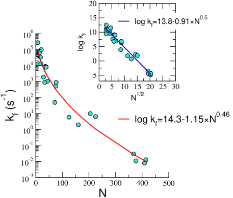
We analyzed the available experimental data (see the Table for a list of RNA molecules) on RNA folding rates by assuming that grows as with as a free parameter. The theoretical value for is 0.5. The folding rates of RNA spanning over seven orders of magnitude is well fit using with correlation coefficient of 0.98 (Fig.1). The fit yields , and . In the inset we show the fit obtained by fixing . Apart from the moderate differences in the values the theoretical prediction and the numerical fits are in agreement, which demonstrates that the major determining factor in determining RNA folding rates is .
It is known that RNA, such as Tetrahymena ribozyme, folds by multiple pathways that is succinctly described by the kinetic partitioning mechanism (KPM) [8].
According to KPM a fraction, , of molecules reaches the native states rapidly whereas the remaining fraction is trapped in an ensemble of misfolded intermediates. For Tetrahymena ribozyme [9]. The -dependence given by Eq. (1) holds for the majority of molecules that fold to the native state from the compact intermediates, which form rapidly under folding conditions [10].
Conclusions. Implications of our findings are: (i) The inverse of the prefactor, , is almost six orders of magnitude larger than the transition state theory estimate of . The value of , which coincides with the typical base pairing time [11], is the speed limit for RNA folding. Interestingly, general arguments based on the kinetics of loop formation have been used to predict that the speed limit for protein folding is also about one [12, 13, 14]. It remains to be ascertained if the common folding speed limit for proteins and RNA is due to evolutionary pressure on the folding of evolved sequences. \doiline
(ii) It is worth pointing out that Dill [15] has recently shown that the dependence of rates and stabilities of protein folding depend only the number of amino acids, which in turn places strict constraints on their functions in the cellular context. Taken together these studies show that despite the complexity of protein and RNA folding only a few variables might determine their global properties, which suggests that there may be simple principles that determine biological functions.
Acknowledgements. This was supported by a grant from the National Science Foundation through grant number CHE 09-14033.
References
- [1] Thirumalai, D., and C. Hyeon, 2005. RNA and Protein folding: Common Themes and Variations. Biochemistry 44:4957–4970.
- [2] Treiber, D. K., and J. R. Williamson, 1999. Exposing the kinetic traps in RNA folding. Curr. Opin. Struct. Biol. 9:339–345.
- [3] Woodson, S., 2010. Compact intermediates in RNA folding. Annu. Rev. Biophys. 39:61–77.
- [4] Hyeon, C., R. I. Dima, and D. Thirumalai, 2006. Size, shape and flexibility of RNA structures. J. Chem. Phys. 125:194905.
- [5] Yoffe, A., P. Prinsen, A. Gopal, C. Knobler, W. Gelbart, and A. Ben-Shaul, 2008. Predicting the sizes of large RNA molecules. Proc. Natl. Acad. Sci. USA 105:16153.
- [6] Hajdin, C. E., F. Ding, N. V. Dokholyan, and K. M. Weeks, 2010. On the significance of an RNA tertiary structure prediction. RNA 16:1340–1349.
- [7] Thirumalai, D., 1995. From Minimal Models to Real Proteins: Time Scales for Protein Folding Kinetics. J. Phys. I (Fr.) 5:1457–1467.
- [8] Guo, Z., and D. Thirumalai, 1995. Kinetics of Protein Folding: Nucleation Mechanism, Time Scales, and Pathways. Biopolymers 36:83–102.
- [9] Pan, J., D. Thirumalai, and S. A. Woodson, 1997. Folding of RNA involves parallel pathways. J. Mol. Biol. 273:7–13.
- [10] Thirumalai, D., N. Lee, S. A. Woodson, and D. K. Klimov, 2001. Early Events in RNA Folding. Annu. Rev. Phys. Chem. 52:751–762.
- [11] Pörschke, D., O. Uhlenbeck, and F. Martin, 1973. Thermodynamics and kinetics of the helix-coil transition of oligomers containing GC base pairs. Biopolymers 12:1313–1335.
- [12] Hagen, S. J., J. Hofrichter, A. Szabo, and W. A. Eaton, 1996. Diffusion-limited contact formation in unfolded cytochrome c: Estimating the maximum rate of protein folding. Proc. Natl. Acad. Sci. USA 93:11615–11617.
- [13] Yang, W. Y., and M. Gruebele, 2003. Folding at the speed limit. Nature 423:193 – 197.
- [14] Kubelka, J., J. Hofrichter, and W. A. Eaton, 2004. The protein folding ’speed limit’. Curr. Opin. Struct. Biol. 14:76–88.
- [15] Dill, K., K. Ghosh, and J. Schmit, 2011. Physical limits of cells and proteomes. Proc. Natl. Acad. Sci. USA 108:17876–17882.
- [16] Proctor, D. J., H. Ma, E. Kierzek, M. Gruebele, and P. C. Bevilacqua, 2004. Folding thermodynamics and kinetics of YNMG RNA hairpins: Specific incorporation of 8-bromoguanosine leads to stabilization by enhancement of the folding rate. Biochemistry 43:14004–14014.
- [17] Porschke, D., 1974. Thermodynamic and kinetic parameters of an oligonucleotide hairpin helix. Biophys. Chem. 1:381–386.
- [18] Riesner, D., and R. Römer, 1973. Physico-Chemical Properties of Nucleic Acids, Academic Press, London, volume 2, chapter Thermodynamics and Kinetics of Conformational Transitions in Oligonucleotides and tRNA, 237–318.
- [19] Serebrov, V., R. Clarke, H. Gross, and L. Kisselev, 2001. Mg2+-induced tRNA folding. Biochemistry 40:6688–6698.
- [20] Kuznetsov, S., C. Ren, S. Woodson, and A. Ansari, 2008. Loop dependence of the stability and dynamics of nucleic acid hairpins. Nucleic Acids Research 36:1098.
- [21] Narayanan, R., Y. Velmurugu, S. Kuznetsov, and A. Ansari, 2011. Fast folding of RNA pseudoknots initiated by laser temperature-jump. J. Am. Chem. Soc. 133:18767–18774.
- [22] Tan, E., T. J. Wilson, M. K. Nahas, R. M. Clegg, D. M. J. Lilley, and T. Ha, 2003. A four-way junction accelerates hairpin ribozyme folding via a discrete intermediate. Proc. Natl. Acad. Sci. USA 100:9308–9313.
- [23] Sosnick, T. R., and T. Pan, 2004. Reduced Contact Order and RNA Folding Rates. J. Mol. Biol. 342:1359–1365.
- [24] Deras, M. L., M. Brenowitz, C. Y. Ralston, M. R. Chance, and S. A. Woodson, 2000. Folding Mechanism of the Tetrahymena Ribozyme P4-P6 Domain. Biochemistry 39:10975–10985.
- [25] Rangan, P., B. Masquida, E. Westhof, and S. A. Woodson, 2003. Assembly of core helices and rapid tertiary folding of a small bacterial group I ribozyme. Proc. Natl. Acad. Sci. 100:1574–1579.
- [26] Fang, X., T. Pan, and T. R. Sosnick, 1999. Mg-dependent folding of a large ribozyme without kinetic traps. Nature Struct. Biol. 12:1091–1095.
- [27] Xiao, M., M. J. Leibowitz, and Y. Zhang, 2003. Concerted folding of a Candida ribozyme into the catalytically active structure posterior to a rapid RNA compaction. Nucleic Acids Res. 31:3901–3908.
- [28] Zarrinkar, P. P., J. Wang, and J. R. Williamson, 1996. Slow folding kinetics of RNase P RNA. RNA 2:564–573.
- [29] Sclavi, B., M. Sullivan, M. R. Chance, M. Brenowitz, and S. A. Woodson, 1998. RNA Folding at Millisecond Intervals by Synchrotron Hydroxyl Radical Footprinting. Science 279:1940–1943.
| RNA | N | ||
|---|---|---|---|
| GCUUCGGC [16] | 8 | tetraloop hairpin | |
| GCUUCGGC [16] | 8 | tetraloop hairpin | |
| GGUUCGCC [16] | 8 | tetraloop hairpin | |
| GGUUCGCC [16] | 8 | tetraloop hairpin | |
| GGACUUUUGUCC [16] | 12 | tetraloop hairpin | |
| GGACUUCGGUCC [16] | 12 | tetraloop hairpin | |
| A6C6U6 [17] | 18 | tetraloop hairpin | |
| Extra-arm of (yeast) [18] | 21 | tRNA | |
| pG-half of (yeast) [18] | 36 | tRNA | |
| CCA-half of (yeast) [18] | 39 | tRNA | |
| CCA-half of (wheat) [18] | 39 | tRNA | |
| (yeast) [19] | 76 | tRNA | |
| (yeast) [18] | 77 | tRNA | |
| -hairpin [20] | 14 | hairpin (52+4) | |
| -hairpin [20] | 19 | hairpin (52+9) | |
| -hairpin [20] | 29 | hairpin (52+19) | |
| -hairpin [20] | 44 | hairpin (52+34) | |
| VPK pseudoknot [21] | 34 | pseudoknot | |
| Hairpin ribozyme (4-way junction) [22, 23] | 125 | natural form of hairpin ribozyme | |
| P5abc [24] | 72 | Group I intron . ribozyme | |
| P4-P6 domain(Tetrahymena ribozyme) [24] | 160 | Group I intron . ribozyme | |
| Azoarcus [25, 23] | 205 | ||
| B.subtilis RNase P RNA catalytic domain [26] | 225 | ||
| Ca.L-11 ribozyme [27] | 368 | ||
| E.coli RNase P RNA [28] | 377 | ||
| B.subtilis RNase P RNA [28] | 409 | ||
| Tetrahymena ribozyme [29, 23] | 414 | Group I intron . ribozyme |