Fluid membranes can drive linear aggregation of adsorbed spherical nanoparticles
Abstract
Using computer simulations we show that lipid membranes can mediate linear aggregation of spherical nanoparticles binding to it for a wide range of biologically relevant bending rigidities. This result is in net contrast with the isotropic aggregation of nanoparticles on fluid interfaces or the expected clustering of isotropic insertions in biological membranes. We present a phase diagram indicating where linear aggregation is expected, and compute explicitly the free energy barriers associated with linear and isotropic aggregation. Finally, we provide simple scaling arguments to explain this phenomenology.
Lipid membranes have unique mechanical properties that are crucial for many biological processes, including cellular recognition, signal transduction, inter- and intracellular transport, and cell adhesion. Most of these processes require interactions of a lipid-bilayer with a variety of nano- and micro-size objects, such as proteins, DNA, viruses and other biomacromolecules. Along with its fundamental importance, understanding the interactions of fluid membranes with nano-objects is a crucial component in targeted drug-delivery design and in nanotoxicity studies. It also has intriguing implications for medical imaging kostarelos and for the development of biosensors and functional biomimetic materials costanzo ; webb .
Lipid membranes are typically very flexible and under thermal perturbations they undergo surface deformations that are significantly larger than their thickness. Because of such a flexibility, they can easily be deformed when interacting with nano-particles that can be either adsorbed on the membrane surface or embedded in the lipid bilayer. The resulting membrane deformations may in turn mediate interactions between the membrane-bound objects. This phenomenon has been extensively studied over the past two decades, both experimentally and theoretically. Most of the previous studies focused on the interactions between embedded inclusions. A bending-mediated Casimir-like isotropic interaction was initially proposed as a possible mean of driving protein aggregation on a lipid bilayer safran ; pincus . Later papers have shown that a more accurate accounting of the local constraints imposed by non-isotropic inclusions on the membrane can lead to additional complex terms whose sign and functional form is very much dependent on how the objects anchor to the surface; see for instance netz ; fournier ; chou ; deserno ; chen and references therein. Hydrophobic mismatch killian , difference in curvature between the membrane and the embedded objects leibler ; lipowsky ; pep or line tension between the lipids and the inclusions baumgart can also induce domain formations.
Adsorption or inclusion of objects comparable in size to the membrane thickness ( 5nm) greatly perturbs the local packing of the lipids leading to quite complex phenomena dependent on the molecular details of the membrane-object interactions. When considering larger objects, on the contrary, it becomes feasible to describe the membrane as a continuous surface and coarse-grain its interactions with the nanoscopic objects with generic binding potentials. Here we are interested in membrane driven interaction between adsorbing colloidal particles that are more than one order of magnitude larger than the membrane thickness. Despite their structural complexity, for sufficiently large scales the behavior of lipid membranes can be described by a small number of elastic parameters that capture their response to deformation; a bending rigidity of the order of , and a small surface tension are the most important ones. Both can be altered either by dispersing within the bilayer additional molecular components, or by changing the lateral forces/osmotic pressure applied on the membrane.
In this paper we show that spherical nanoparticles adhering to fluid membranes can self-assemble into a variety of two-dimensional aggregates. Significantly, for intermediate and biologically relevant values of the bending rigidity we find that particles preferentially arrange into linear/flexible aggregates. This result is in striking contrast with most of the theoretical studies on membrane inclusions that predict isotropic aggregation when the embedding object imposes an isotropic deformation on the surface. Linear aggregation is expected only for sufficiently anisotropic wedge-like local deformations fournier , and this is clearly not the case for spherical nanoparticles. We find that the key to understand the stability of linear versus isotropic aggregates resides in the interplay between bending and binding energies of the nanoparticles. The latter term, usually and correctly neglected when dealing with embedded nano-components, does indeed play a major role in the structural morphology of the aggregates formed by non-embedded adhering components.
It should be stressed that string-like formations very similar to those we present here have been observed experimentally in several systems. For instance, colloidal particles bound to giant phospholipid vesicles (GUV) via streptividin-biotin bonds or by electrostatic physisorption form one-dimensional ring-like assembles safinya . Similarly, the cationic lipid-DNA complexes of low net charge assemble into linear colloidal aggregates when adsorbed to the cell membrane safinya1 . However, to the best of our knowledge, this phenomenon has never been explained, nor studied in detail. In this paper we use a combination of numerical simulations and scaling arguments to detail the physical origin behind it.
We performed Monte Carlo simulations of planar and spherical fluid membranes interacting with adsorbing nanoparticles. The membrane is modeled using a simple one particle-thick solvent-free model, and consists of hard spherical beads, of diameter , connected by flexible links to form a triangulated network ho ; nelson ; gompper whose connectivity is dynamically rearranged to simulate the fluidity of the membrane. The membrane bending energy acts on neighboring triangles, and has the form
| (1) |
where is the bending rigidity, and the and are the normals of two triangles and sharing a common edge. The cost associated with area changes is included via the energy term , where is the tension of the surface and is its total area. A nanoparticle is modeled as a sphere of diameter , with or . Excluded volume between any two spheres in the system (nanoparticles and surface beads) is enforced with a hard-sphere potential. Finally, the nanoparticle-to-surface adhesion is modeled via a generic power-law potential between the nanoparticles and the surface beads defined as
| (2) |
with , and cutoff at . Following smit , simulations of the planar membrane were carried out in the ensemble, while the ensemble was used for the spherical membrane. In each simulation the number of nanoparticles is held constant, and the surface tension is set to . For we have nanoparticles of diameter and surface tensions .
We begin by computing the phase behavior of the system for different values of the surface bending rigidity and nanoparticles’ adhesive energy . The results are summarized in the left panel of Fig. 1, and report the structure of the aggregates observed for each pair in the case of the planar geometry.
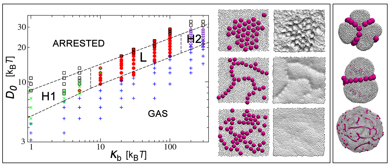
A gas phase occurs when is too weak for the particles to deform the membrane. In this phase particles are just lightly bound to the surface, they are highly mobile, and have a certain probability of detaching from it. An arrested phase occurs for large values of . In this case particles bind very strongly to the membrane resulting in large local deformations that heavily limit their mobility over the surface. This typically leads to configurations that are kinetically trapped or even to nanoparticle engulfment. Three ordered phases occur for moderate values of . Each of the three phases spans a range of values. For small values of the bending rigidity, particles create well defined deep-spherical imprints in the membrane and organize into ordered hexagonal arrays (H1). Low cost in bending energy and high gain in surface binding allows for these deep deformations. In this phase the nanoparticles are not in direct contact with each other, but are separated by the pinched parts of the membrane. Close-packing maximizes sharing of the pinched regions between neighboring nanoparticles, thus maximizing the surface-to-nanoparticle contact area. An identical result is obtained when repeating the simulations on the spherical membrane, and is reminiscent of the experimentally observed two-dimensional hexagonal crystal formed by negatively charged particles on positively-charged surfactant vesicles reported in weitz . Even in this case the colloids are extensively wrapped by the membrane and are not in direct contact with each other.
For biologically relevant values of , our nanoparticles create smooth channel-like distortions on the membrane and self-assemble into linear aggregates (L) non unlike those predicted for anisotropic membrane inclusions fournier . Although we have not computed a structural phase diagram for our vesicle model, we find that the simulations on the vesicle performed at different nanoparticle concentrations and vesicle radii lead to analogous results. Here particles form sinuous linear patterns that tend to follow the equatorial lines of the vesicle. Snapshots from our simulations are shown in the right panel of Fig. 1. This phase strikingly resembles the linear aggregates of colloidal particles on Giant Phospholipid Vesicles (GUV) obtained in safinya .
For very large values of the nanoparticles re-organize into the familiar hexagonal lattice, however, unlike what happens for the small aggregates, the membrane now remains almost completely flat and the nanoparticles are in contact with each other (H2). Because of its high stiffness, particles can only weekly deform the membrane to gain in binding energy, as a result the binding energy is minimized by recruiting the largest number of membrane beads in the vicinity of the nanoparticles. This effectively drives the crystallization of the region of the membrane that directly interacts with the nanoparticles, creating a line tension between crystalline and fluid membrane regions that is minimized when isotropic aggregation takes place lipowsky ; pep .
As mentioned before, the formation of linear aggregates is quite surprising. To ensure that our results are not affected by the triangulation underlying the definition of our membrane model, we repeated our simulations using the coarse-grained, but tether-free model proposed by Zhang et al. li . This model also accounts for possible topological changes in the surface, however the elastic properties of the membrane are not fed to the system in the form of parameters of an elastic energy, but are encoded into the molecular details of the anisotropic pair potentials between the effective building blocks of the membrane, and need to be extracted by analyzing the fluctuations spectrum of the surface li , or by other means. It is comforting to report that no qualitative difference was found on the overall phenomenology of the phase diagram: linear aggregates do indeed form and are not an artifact of our model. We also checked that linear aggregates do no form when limiting the area of the particles’ binding region to enforce a finite (constant) contact angle between particles and membrane. This case is basically equivalent to enforcing isotropic regions with intrinsic curvature, mimicking for instance the local perturbation of a protein, in a lipid bilayer for which isotropic aggregation is expected pep .
To understand why linear aggregates become more favorable for moderate bending rigidities, we placed three nanoparticles, and in linear formation and at a kissing distance over a planar membrane, and calculated the free energy cost required to disrupt the linear arrangement. The idea is to keep in place particles and and force particle to form an angle between the vector connecting particles and and that connecting particles and while keeping the relative particle distance unaltered.
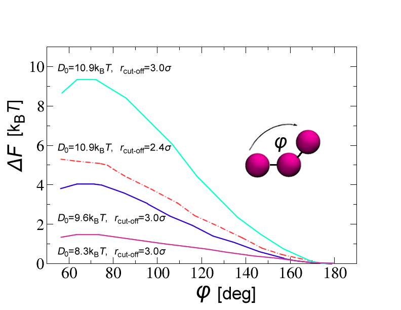
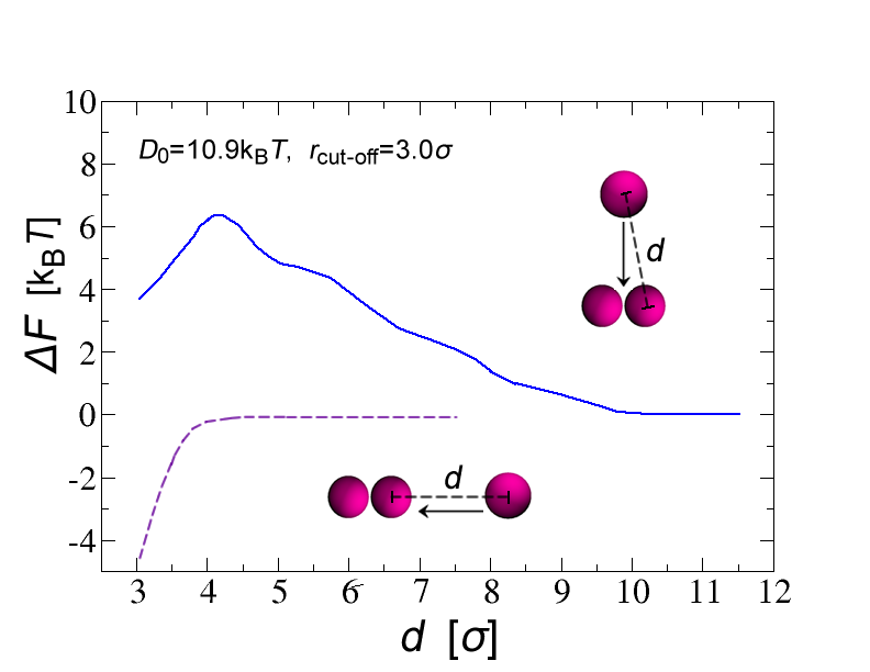
Using the umbrella sampling method valleau , we can reconstruct piece-wise the probability that the trimer arranges according to any of the explored angles, which in turns gives us access to the free energy difference . All simulations were repeated for different values of and two different ranges of the binding potential. The results are shown in Fig. 2, and undoubtedly tells us that in this region of the phase diagram the linear configuration is the most stable one, with the close-packed compatible configuration () sitting in a metastable shallow minimum of the free-energy curve separated from the linear configuration () by a significant barrier. The height of the barrier depends on the exact parameters, but is typically larger than 4 inside the linear region of the phase diagram. Fig. 2 shows the free-energy as a function of particle separation when the third particle approaches the other two from infinity, either in the linear or perpendicular alignment, as depicted in the insets in Fig. 2. When the third particle approaches the dimer to form a linear aggregate, the free-energy (when particles are sufficiently close) decreases monotonically down to a minimum at contact. When the third particle approaches the dimer from a direction that is perpendicular to the dimer’s axis, we observe a repulsive free energy barrier that precedes a shallow minimum at contact. Remarkably the range of the repulsion is felt as far as three nanoparticle diameters (up to 9 times the range of the attractive part), revealing correlations in the three body interactions that are significantly longer than the ones expect from a simple Casimir effect netz ; fournier .
To understand the unexpected stability of the linear aggregates over the close-packed structures in the regime where linear aggregation occurs, we measured the energy of the system associated with linear and hexagonal aggregates. The l.h.s of Fig. 3 shows explicitly how the total energy difference between linear and hexagonal aggregates, computed for the same values of and , as a function of particle number , is partitioned between the bending and the binding contribution. This analysis reveals that despite the smaller bending cost, hexagonal aggregates provide a fairly small gain in binding energy when compared to linear aggregates, and this leads to a net energy balance that favors the latter. It is worth mentioning that we monitored the difference in free energy due to the surface tension between the two configurations, and found it to be indeed negligible. We also checked that linear aggregates form for our largest nanoparticles, .
To rationalize these numerical data we offer the following scaling argument. A quick look at the typical surface deformations in this region of the phase diagram (see snapshots on the r.h.s of Fig. 3) suggests that in either linear or hexagonal configuration the contribution to the system energy can be split in two parts. The first part comes from the overall deformation of the membrane due to the collective arrangement of the particles. The second part comes from the shallow surface indentations (corrugations) produced by each particle on top of the overall deformation. Let’s assume that the energy due to the corrugation is fairly independent of the overall arrangement of the aggregates. We can think of it as a particle self energy that is constant for a given , and . The total self-energy is than .
When particles arrange into linear structures (), they generate a channel-like profile in the membrane with length proportional to the number of the nanoparticles and width proportional to . The bending energy of the channel can be estimated using the standard elastic energy landau with being the area and being the constant curvature of the deformation. Ignoring the energy due to the contribution of the surface tension and subtracting the contribution of the particles’ self energy, we can write the total free energy of the channel as , where is a parameter that accounts for the degree of surface wrapping per nanoparticle, and is related to the overall height of the channel. Close-packed hexagonal () arrangements form a flat, two-dimensional imprint of lateral size proportional to . In this case, apart from a geometrical prefactor, the free energy due to the rim of the imprint scales as . In fact, here the area is proportional to the length of the rim and grows as , and the term accounts for the small bending cost associated with the in-plane curvature of the rim with . Structural stability requires both free energies to be negative ( for large ), which results in , for sufficiently large values of making the linear aggregates more stable than the isotropic ones. In other words, the gain in binding energy overwhelms the larger cost in bending.

In conclusion, we have computed a phase diagram showing the different aggregates formed by nanoparticles adsorbing onto a lipid bilayer as a function of the surface bending rigidity and nanoparticles adhesive energy. Our main result is that for a wide range of bending rigidities , nanoparticles can organize into linear aggregates provided the binding energy is sufficiently large.
Although linear aggregates are expected to form on elastic (polymerized) surfaces due to the global constraints imposed on the surface deformations by the stretching rigidity ( at least in the large limit) cacciuto , for fluid membranes . Our result is therefore quite different than the expected, and usually assumed, isotropic aggregation mediated by either local isotropic deformations of the surface or due to hydrophobic mismatch. The binding energy of the nanoparticles, the missing ingredient in studies of aggregation of membrane inclusions, is the key to rationalize this phenomenology. We hope our work will induce further analysis of membrane-mediated interactions between adhering nanoparticles.
ACKNOWLEDGMENTS
This work was supported by the National Science Foundation under Career Grant No. DMR-0846426.
References
- (1) T. A.-J. Wafa and K. Kostarelos, Nanomedicine 2, 85 (2007).
- (2) P. J. Costanzo, E. Liang, T. E. Patten, S. D. Collins, and R. L. Smith, Lab Chip 5, 606 (2005).
- (3) R. J. Mart, K. P. Liem and S. J. Webb, Pharm. Res. 26, 1701 (2009).
- (4) H. Aranda-Espinoza, A. Berman, N. Dan, P. Pincus and S. Safran, Biophys J. 71, 648 (1996).
- (5) M. Goulian, R. Bruisma and P. Pincus, Europhys. Lett. 22, 145 (1993).
- (6) R.R. Netz, J. Phys. I France 7, 833 (1997).
- (7) P.G. Dommersnes and J.-B. Fournier, Eur. Phys. J. B, 12, 9 (1999).
- (8) T. Chou, K. S. Kim and G. Oster, Biophysical Journal, 80, 10075 (2001).
- (9) C. Yolcu, I. Z. Rothstein, and M. Deserno, Europhys. Lett, 96, 20003 (2011).
- (10) S. Mkrtchyan, C. Ing, and J. Z. Y. Chen, Phys. Rev. E 81, 011904 (2010).
- (11) J. A. Killian, Biochim. Biophys. Acta Rev. Biomembr. 1376, 401 (1998).
- (12) S. Leibler, J. Phys. France 47, 507-516, (1986).
- (13) F. Jülicher and R. Lipowsky, Phys. Rev. Lett. 70, 2964 (1992).
- (14) R.N. Frese, J. C. Pamies, J. D. Olsen, S. Bahatyrova, C.D. Van Der Weij-De Wit, T. J. Aartsma, C. Otto, N. Hunter, D. Frenkel and R. Van Grondelley, Biophys. J. 94, 640 (2008).
- (15) T. Baumgart, S. T. Hess, W. W. Webb, Nature, 425, 821-824, (2003).
- (16) I. Koltover, J. O. Rädler and C. R. Safinya, Phys. Rev. Lett. 82, 1991 (1999).
- (17) I. Koltover, T. Salditt and C. R. Safinya, Biophys J. 77, 915 (1999).
- (18) J.-S. Ho and A. Baumgärtner, Europhys. Lett. 12, 295 (1990); A. Baumgärtner and J.-S. Ho, Phys. Rev. A 41, 5747 (1990).
- (19) Y. Kantor, M. Kardar, and D. R. Nelson, Phys. Rev. Lett. 57, 791 (1986).
- (20) G. Gompper and D. M. Kroll, J. Phys.: Condens. Matter 9, 8795 (1997).
- (21) M. Kranenburg, M. Venturoli and B. Smit, Phys. Rev. E 67, 060901 (2003).
- (22) H. Yuan1, C. Huang, J. Li, G. Lykotrafitis and Sulin Zhang, Phys. Rev. E 82, 011905 (2010).
- (23) G. M. Torrie, J. P. Valleau, J. Comp. Phys. 23, 187 (1977)
- (24) L. Ramos, T. C. Lubensky, N. Dan, P. Nelson and D. A. Weitz, Science, 286, 2325 (1999).
- (25) L. D. Landau and E. M. Lifshitz, Theory of Elasticity, Pergamon: New York, (1970).
- (26) A. Šarić and A. Cacciuto, Soft Matter, 7, 8324 (2011).; A. Šarić and A. Cacciuto, Soft Matter, 7, 1784 (2011).