Rigidity sensing explained by active matter theory
Abstract
The magnitude of traction forces exerted by living animal cells on their environment is a monotonically increasing and approximately sigmoidal function of the stiffness of the external medium. This observation is rationalized using active matter theory: adaptation to substrate rigidity results from an interplay between passive elasticity and active contractility.
Living cells respond to mechanical as well as biochemical cues. Rigidity sensing designates the web of complex mechanisms whereby a cell will adapt, as a function of the elastic modulus of its environment, aspects of its phenotype as diverse as its motility, gene expression, proliferation, or fate after differentiation Discher2005 ; Janmey2009 .
The traction forces that a cell exerts on a flat, elastic plate depend in a non-trivial way on the extracellular stiffness Saez2005 : roughly proportional to the elastic modulus in a softer environment, they saturate to a finite value for stiffer substrates Ghibaudo2008 . Similar force-rigidity data are obtained whether forces are measured locally Ghibaudo2008 or globally Mitrossilis2009 ; Mitrossilis2010 ; Webster2011 , for integrin-mediated Saez2005 ; Ghibaudo2008 as well as cadherin-mediated adhesion Ganz2006 ; Ladoux2010 , and even when the traction forces are exerted by assemblies of cells in a monolayer epithelium Saez2010 . Single-cell rheology assays Mitrossilis2010 ; Webster2011 show that the response of a cell to a sudden change of substrate rigidity is faster than the data acquisition rate allows to detect. These observations call for a simple, generic explanation, valid on short time scales where cell signaling cannot operate.
In the context of adhesion-dependent mechanosensing, a simple ’two-spring model’ was introduced in Schwarz2006 , predicting that stiffer environments lead to stronger traction forces. A ’three-spring model’ was later proposed in order to explain the stiffness-dependent orientation of stress fibers in adherent cells Zemel2010 , where contractility modulates cytoskeletal stiffness via a phenomenological polarizability coefficient. In this Letter, we formulate and solve a simpler model derived from active matter theory, a generic description of living matter where the mechano-chemical transduction due to molecular motors (activity) plays a central role Kruse2005 ; Juelicher2007 . We obtain a (static) force-rigidity relationship that agrees well with experimental data. We give an expression of the (dynamic) loading rate, expected to be valid on time scales too short for cytoskeletal remodeling and protein recruitment to occur ( s Icard-Arcizet2008 ; Allioux-Guerin2009 ).
MODEL
Active matter theory formulates constitutive equations that take into account viscoelasticity, activity, the polar nature of cytoskeletal biopolymers Kruse2005 , and respect the principles of linear irreversible thermodynamics. In one spatial dimension, we write the stress of an homogeneous, elastic and contractile material as the sum of an elastic and an active contribution: . The active stress is proportional to , the difference of chemical potential between products and reagents of the chemical reaction responsible for mechanochemical transduction (ATP hydrolysis): , where is a material parameter of the cytoskeleton Juelicher2007 . For simplicity, we restrict our description of the cytoskeleton to the linear regime, and include:
-
(i)
elasticity, described by a linear spring of length at time , rest length and spring constant ;
-
(ii)
activity, modeled by an active force across a section of area .
In the nonlinear regime, elastic moduli may also depend on activity, as hypothesized in Zemel2010 . Assuming, as in Kaunas2011 , that the rest length of elastic cytoskeletal structures is time-dependent due to motor activity is also beyond the scope of the linear regime that we consider here.
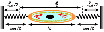
The extra-cellular environment is represented by a linear spring of length at time , rest length , and spring constant (see Fig. 1). The traction force exerted by the cell on its environment reads:
| (1) |
The sign is chosen as for contraction. Under usual experimental conditions, the total length of the system (cell + substrate) is constant: . The force balance equation reads:
| (2) |
STATICS
The amplitude of the traction force at equilibrium reads:
| (3) |
When , the traction force saturates to :
| (4) |
the sum of and of residual stresses . We expect the ensemble average of residual stresses to cancel: . As long as , the traction force is a linear function of : .
Experimentally, a wide range of rigidities can be obtained when the substrate is a dense array of cylindrical elastomer micropillars whose stiffness depends on their radius and height. Depending on the coating protein, traction forces are transmitted through integrin-mediated adhesions (with fibronectin) Saez2005 ; Ghibaudo2008 or through cadherin-mediated adhesions (with N-cadherin) Ganz2006 ; Ladoux2010 . Fig. 2 show that, in both cases, experimental data are well fitted by the force-rigidity relation Eq. (3). Note that the fitted values of saturation forces were not observed: they correspond to values of so large that the deflections of pillars would fall below the experimental spatial resolution.
The data encompasses three cell types, Madin-Darby Canine Kidney (MDCK) cells, 3T3 fibroblasts, and C2 mouse myogenic cells. Cytoskeletal organisation and adhesive properties of the cells vary substantially according to the type of adhesions and the range of substrate rigidity. More diffuse cortical actin dominates when the environment is softer, wheras actomyosin bundles are preferentially formed at higher rigidities Ghibaudo2008 ; Ladoux2010 . In all cases, our simple model captures the essence of the force-rigidity dependence, and sums up biological variation into two quantitative parameters, the asymptotic traction force and the cytoskeletal stiffness . The order of magnitude of the saturation force corresponds to an active stress of the order of Pa. We obtain this value upon neglecting possible residual stresses, and using , where the section of a micropillar is of order . The cytosketal rigidity is of the order of , corresponding to elastic moduli of the order of Pa, a value intermediate between moduli typical of cortical actin Wottawah2005 and of stress fibers Deguchi2006 (we used with ).
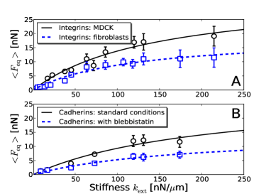
When cells are treated with blebbistatin, an inhibitor of contractility, the value of is halved (Fig. 2 B). Inspection of Eq. (4) suggests that traction forces remain non-zero due to residual activity of myosin motors, as proposed in Ladoux2010 , or to non-zero residual stresses, or to a combination of both effects. We note that the value of is almost unchanged. However, assuming as in Zemel2010 that activity modulates cytoskeletal stiffness via a polarizability coefficient leads to a ratio that depends through on cytoskeletal contractility. Experimental data Ladoux2010 suggests that is independent of the level of contractility, in agreement with our prediction, Eq. (3).
DYNAMICS
The loading rate exerted by T cells immediately after receptor engagement with a model antigen-presenting cell was measured thanks to a biomembrane force set-up where micropipette aspiration controls the external rigidity, and found to be linear in Husson2011 . Motivated by this result, we turn to the dynamics of traction forces, and modify Eq. (2) by taking into account internal protein friction in a linear force-velocity relationship:
| (5) |
where is the stall force, and is a friction coefficient Tawada1991 . Eliminating other variables in Eq. (2), we obtain a differential equation for the traction force:
| (6) |
with a viscoelastic time . Integration from an initial time with initial force gives . For zero initial force, we find that the initial loading rate is proportional to the substrate rigidity:
| (7) |
in agreement with Husson2011 , where the initial time is set when pulling starts so that . We checked that Eq. (7) still holds if we replace the linear force-velocity equation (5) by Hill’s equation Hill1938 . Since is a function of activity, we predict that the loading-rate-rigidity data will be modified upon treatment with contractility agonists and antagonists.
CONCLUSION AND OUTLOOK
At low external rigidity, cell traction forces increase linearly with the stiffness of the substrate. Their constant ratio was first interpreted as a displacement regulated by the cell Saez2005 . We show that regulation is not necessary to explain the force-rigidity relationship. Within the framework of linear irreversible thermodynamics, we propose a minimal model whose consequences are consistent with available experimental data. We predict that both the saturation force , exerted for large stiffness, and the constant displacement , observed at low stiffness, depend upon the contractility level. Our description is relevant for several types of adhesive structures and cytoskeletal organisation. Unlike Schwarz2006 , we ignore the dynamics of adhesive contacts through which force is transmitted to the substrate. Other monotonically increasing functions of stiffness that depend on two parameters also fit the same experimental data: it is our hope that this work will foster further quantitative experiments to confirm – or disprove – our model.
To treat the dynamics of traction forces, we include internal friction, and obtain an initial loading rate proportional to external stiffness, as observed experimentally Husson2011 . In accord with single-cell rheology assays Mitrossilis2010 ; Webster2011 , the loading rate responds instantaneously to variations of .
The merit of our analysis is to show that the simplest equations of active matter dictated by symmetry and conservation laws are sufficient to describe a behavior which might seem to require a more elaborate regulation at first sight. This suggests that other features such as stress fiber diameter and equilibrium with the rest of the actin-myosin system could be described within the general framework of active gels. We hope that extensions of our model will allow to understand quantitatively how more complex cell processes depend on extracellular rigidity. Including membrane elasticity and cortical tension in an appropriate geometry may explain why the initial loading rate exerted by T cells upon receptor engagement saturates for stiffer environements Husson2011 . The dynamics of wetting of the microplate by the cell must be taken into account to describe traction forces exerted during cell spreading Mitrossilis2009 ; Mitrossilis2010 . Finally, biochemical signaling, protein recruitment and remodeling of adhesive and cytoskeletal structures act over longer time scales Icard-Arcizet2008 ; Allioux-Guerin2009 and may enhance the mechanical effects described here.
ACKNOWLEGMENTS
The authors thank A. Asnacios, F. Graner, J.-F. Joanny, J. Husson, B. Ladoux and P. Silberzan for fruitful discussions. This work was supported by JSPS, MAEE and MESR under the Japan-France Integrated Action Program (SAKURA).
References
- (1) Discher, D.S., P. Janmey and Y.-L. Wang. 2005. Tissue cells feel and respond to the stiffness of their substrate. Science 310:1139–1143.
- (2) Janmey, P.A., J.P. Winer, …, Q. Wen. 2009. The hard life of soft cells. Cell Motil. Cytoskel. 66:597–605.
- (3) Saez, A., A. Buguin, …, B. Ladoux. 2005. Is the mechanical activity of epithelial cells controlled by deformations or forces?. Biophys. J. 89:L52–L54.
- (4) Ghibaudo, M., A. Saez, …, B. Ladoux. 2008. Traction forces and rigidity sensing regulate cell functions. Soft Matter 4:1836–1843.
- (5) Mitrossilis, D., J. Fouchard, …, A. Asnacios. 2009. Single-cell response to stiffness exhibits muscle-like behavior. Proc. Natl. Acad. Sci. U.S.A. 106:18243–18248.
- (6) Mitrossilis, D., J. Fouchard, …, A. Asnacios. 2010. Real-time single-cell response to stiffness. Proc. Natl. Acad. Sci. USA. 107:16518–16523.
- (7) Webster, K.D., A. Crow and D.A. Fletcher. 2011. An AFM-based stiffness clamp for dynamic control of rigidity. PLoS One 6:e17807.
- (8) Ganz, A., M. Lambert, …, B. Ladoux. 2006. Traction forces exerted through N-cadherin contacts. Biol. Cell 98:721–730.
- (9) Ladoux, B., E. Anon, …, R.-M. Mège. 2010. Strength dependence of cadherin-mediated adhesions. Biophys. J. 98:534–542.
- (10) Saez, A., E. Anon, …, B. Ladoux. 2010. Traction forces exerted by epithelial cell sheets. J. Phys. Cond. Mat. 22:194119.
- (11) Schwarz, U.S., T. Erdmann and I.B. Bischofs. 2006. Focal adhesions as mechanosensors: the two-spring model. Biosystems 83:225–232.
- (12) Zemel, A., F. Rehfeldt, …, S.A. Safran. 2010. Optimal matrix rigidity for stress-fibre polarization in stem cells. Nat. Phys. 6:468–473.
- (13) Kruse, K., J.-F. Joanny, …, K. Sekimoto. 2005. Generic theory of active polar gels: a paradigm for cytoskeletal dynamics. Eur. Phys. J. E. 16:5–16.
- (14) Jülicher, F., K. Kruse, …, J.-F. Joanny. 2007. Active behavior of the cytoskeleton. Phys. Rep. 49:3–28.
- (15) Icard-Arcizet, D., O. Cardoso, …, S. Hénon. 2008. Cell stiffening in response to external stress is correlated to actin recruitment. Biophys. J. 94:2906–2913.
- (16) Allioux-Guérin, M., D. Icard-Arcizet, …, M. Coppey-Moisan. 2009. Spatio-temporal analysis of cell response to a rigidity gradient: a quantitative study by multiple optical tweezers. Biophys. J. 96:238–247.
- (17) Kaunas, R., H.-J. Hsu and S. Deguchi. 2011. Sarcomeric model of stretch-induced stress fiber reorganization. Cell Health Cytoskel. 3:13–22.
- (18) Wottawah, F., S. Schinkinger, …, J. Käs. 2005. Optical rheology of biological cells. Phys. Rev. Lett. 94:098103.
- (19) Deguchi, S., T. Ohashi and M. Sato. 2006. Tensile properties of single stress fibers isolated from cultured vascular smooth muscle cells. J. Biomech. 39:2603–2610.
- (20) Husson, J., K. Chemin, …, N. Henry. 2011. Force generation upon T cell receptor engagement. PLoS One 6:e19680.
- (21) Tawada, K. and K. Sekimoto. 1991. A physical model of ATP-induced actin-myosin movement in vitro. Biophys. J. 59:343–356.
- (22) Hill, A.V. 1938. The heat of shortening and the dynamic constants of muscle. Proc. Roy. Soc. London B 126:136–195.