Compressive auto-indexing in femtosecond nanocrystallography
Abstract
Ultrafast nanocrystallography has the potential to revolutionize biology by enabling structural elucidation of proteins for which it is possible to grow crystals with 10 or fewer unit cells on the side. The success of nanocrystallography depends on robust orientation-determination procedures that allow us to average diffraction data from multiple nanocrystals to produce a three dimensional (3D) diffraction data volume with a high signal-to-noise ratio. Such a 3D diffraction volume can then be phased using standard crystallographic techniques. “Indexing” algorithms used in crystallography enable orientation determination of diffraction data from a single crystal when a relatively large number of reflections are recorded. Here we show that it is possible to obtain the exact lattice geometry from a smaller number of measurements than standard approaches using a basis pursuit solver.
eprint LBNL-4008E
I Introduction
X-ray crystallography is currently the leading method for atomic resolution imaging of macromolecules. Third generation synchrotron sources permit successful structure solution from crystals 5 microns in size or greater Holton (2009). Obtaining sufficiently large crystals is currently an important stumbling block in structure determination.
The Linac Coherent Light Source (LCLS) recently began operation Emma et al. (2010) at the SLAC National Accelerator Laboratory in Palo Alto, California, using energetic electrons from a linear accelerator to produce coherent x-rays with an instrument called a free electron laser (FEL). Free Electron Laser sources produce pulses of light that are over 9 orders of magnitude brighter than current third generation synchrotron sources Ackermann et al. (2007). Several other x-ray laser sources of this type are being built or planned worldwide Shintake et al. (2008); Altarelli et al. (2006).
The high number of photons incident on a specimen are expected to produce measurable diffraction patterns from nanocrystals perhaps even down to a single period (single molecule) Chapman (2009), enabling high resolution structure elucidation of systems which can only be crystallized into very small crystals that are not suitable for conventional crystallography. Even for larger crystals, the short pulses can circumvent the radiation damage problem Neutze et al. (2000); Chapman et al. (2006) which limits the resolution of many sensitive crystals.
In such an experiment, two-dimensional (2D) diffraction images of randomly oriented nanocrystal of the same type can be captured within an exposure time of a few femtoseconds. These images can then be used to deduce the 3D structure of the molecule. To see the structure in 3-D, one has to merge the data from all these individual nanocrystals, whose orientations are not known.
Femtosecond nanocrystallography brings new challenges to data processing Kirian et al. (2010). One problem is that the orientation of each diffraction image obtained is unknown. Another problem is that a single snapshot of the crystal diffraction pattern may contain very few reflections. In traditional crystallography, a small angular range of integration ensures that many Bragg reflections are recorded while ensuring that overlaps are minimized. This is not possible with ultrafast x-ray pulses. The relentless improvements of these light sources (beam energy, beam divergence and wavelength) will further exacerbate the problem.
These new difficulties make indexing such patterns a hard problem for existing crystallographic software.
II Structure Determination from Crystal Diffraction

The diffracted photon flux (photons/pulse/ pixel) produced by a crystallite is given by
where is the continuous scattering from one unit cell (molecule), is the 3D rotation matrix of the unknown object orientation, a 3D vector that relates the Bragg “reflection” on a two dimensional detector to a point in a 3D Fourier space, is the incident photon flux density (photons/pulse/area), is the electron cross section, is a polarization factor, is the solid angle subtended by a detector pixel at the sample, the integer values are called Miller indices, identify the Bravais lattice characteristic of the crystal periodic structure, and is the shape transform of the crystallite finite dimensions. For large crystals, is simply a Dirac -function. For a crystal made of a few unit cells, is broadened and may introduce an error in the location of the reflection.
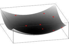
Each set of pixel coordinates on a detector placed at a distance from the sample is and corresponds to a value of in the 3D reciprocal space according to the geometric description of elastic scattering shown in Figure 1.
In this figure, and are the incident and scattered wave vectors that satisfy , where is the wavelength of the x-ray. The direction of and are the same as the direction of the incident beam and the outgoing beam . The coordinates of a lattice point corresponding to satisfy
| (2) | |||||
The end point of the vector lies on a 2D surface called the Ewald sphere. This spherical surface of radius intersects the origin ( when ), and is centered at (while the origin of is at the sample).
In traditional crystallography, the most common data collection method is the rotation method in which the diffraction image is collected while rotating the sample, i.e. varying in Eq. II.
A small angular range of integration ensures that all Bragg reflections are recorded while overlaps are minimized. The strength (measured intensity) and orientation of each Bragg reflection is estimated from the diffraction geometry (including source divergence, bandwidth, pixel size and angular average).
In x-ray crystallography, the term indexing refers to the task of assigning the measured Bragg peaks to the discrete locations of a periodic lattice. Auto-indexing uses the position of these peaks to deduce the shape and orientation () of the lattice, and to identify the lattice coordinates of each measured peak.
It is accomplished in several steps.
-
•
For the purpose of autoindexing, one can simply assign the value of 1 to for every peak above a noise threshold. As a result, one obtains a 3D map in the reciprocal space that contains the values of either 1 or 0. Note that is only affected by the content of a unit cell when is so small that the reflection is not detected. Assuming , Eq. (II) becomes:
(3) -
•
Some type of computational analysis is performed on the 3D map to ascertain the orientation and the unit cell parameters of the crystal (). The analysis typically proceeds by making use of Fourier transform and peak searches. An efficient algorithm that uses many 1D Fourier transform was proposed in Steller et al. (1997); Rossmann and van Beek (1999). It is used in many existing autoindexing software packages such as MOSFLM Leslie (1992). We will provide details of these algorithms in the next section, as this problem will be the focus of our paper.
-
•
Once the lattice vectors and orientation are determined, the lattice coordinates that overlap with the Ewald sphere will provide the index of a reflection. Multiple solutions due to mirror symmetries of the lattice are generated. These solutions can be distinguished using the measured intensities.
Once the orientation and the unit cell parameters associated with a crystal has been determined, one may then proceed to estimate the intensities of the crystal, from the diffraction geometry (including source divergence, bandwidth, pixel size and angular average). Finally, a phase retrieval algorithm is used to recover the phase of the Fourier transform and subsequently the 3D density map of the crystal.
For the purpose of this paper, we will not discuss the issues of structure factor determination or the phase retrieval problem. Instead, we will focus on the second item of the autoindexing problem, how to determine the lattice parameters and orientation.
Multiple solutions due to symmetries of the lattice (but not of the crystal) will still have to be sorted out using measured intensities. In this paper we do not address this problem, which presents another challenge when attempting to merge many thousand of low-signal snapshots.
III Real space autoindexing
Most autoindexing algorithms search for peaks in real space, by applying some form of 3D Fourier transform of the binary reciprocal space map .
If the region of space that was measured is large, its 3D FT will provide the real space lattice. A simple numerical thresholding may reveal the positions of the 3D lattice points in real space. They can subsequently be used to determine the unit cell parameters, crystal orientation and type.
The binary 3D mask that defines the region of space that was measured (=1 if was measured, =0 otherwise) can be viewed as a 3-dimensional optical transfer function. The 3D Fourier transform of the binary mask forms a point spread function which convolves the real space lattice. If the PSF is larger than the lattice spacing, these cannot be resolved.
As we will show in section VI, when the number of measured Bragg peaks is less than 10, the real space lattice points cannot be easily distinguished from the rest of the sampled voxels based on the intensity of the inverse 3D Fourier transform.
The use of a 3D Fourier transform around the origin for indexing a diffraction pattern was suggested over two decades ago Bricogne (1986). A similar approach appears to have been incorporated in the program DENZO, which has been distributed as part of the diffraction-image processing suite HKL Otwinowski and Minor (1997). A 3D FFT has been used to index diffraction images by calculating a Patterson function from a set of reflections which have all been assigned unit intensity Campbell (1998). Efficient software implementations avoid the use of a full 3D Fourier transform, by using the Fourier projection-slice theorem Natterer and Wübbeling (2001), calculating 1D sections of the 3D FTs by a series of projections and 1-dimensional FFTs Rossmann and van Beek (1999) Powell (1999). Indexing software such as MOSFLM Steller et al. (1997), LABELIT Sauter et al. (2004) utilize this approach.
The complexity of the projection-FFT approach is , where is the number of direction vectors that must be generated, and is the number of samples along the projected 1D intensity profile, which is proportional to , where is the total number of sampled voxels contained in the crystal. A typical value of is between 5,000 and 20,000. Clearly, this method will not work well if the number of Bragg points on a diffraction pattern is small.
Although the argument used in Rossmann and van Beek (1999) for abandoning the full 3D FFT is the high cost for performing 3D FFTs of large crystals, this is no longer a serious issue due to the rapid growth in the processing speed and memory capacity of modern multi-core microprocessors. At the time of writing, a 3D FFT takes about 0.15 seconds on a GPU processor, while a takes about 7 minutes on a machine with sufficient memory. Furthermore, there are now algorithms that we may use to take full advantage of the sparsity of the 3D reciprocal space map Ying (2009), i.e., there are a few non-zeros in the 3D map constructed from the Bragg reflections, and reduce the complexity of the 3D FFT calculation from the standard to that of , where is the total number of sampled voxels in the crystal.
IV Recovering Real Space Lattice via L1 Minimization
An alternative technique for retrieving the positions of the real (and reciprocal) space lattice points associated with a crystal is to use the recently developed compressive sensing methodology Donoho (2006); Candès et al. (2006a, b) and formulate the problem as an L1 minimization problem.
Let be a vector representation of the 3D density map of a crystal lattice to be determined in real space. Similar to the 3D inverse Fourier transform approach, we will use the magnitude of each component of to determine whether the 3D voxel associated with that component is a real space lattice point.
The vector is related to the diffraction measurement through the following equation:
| (4) |
where is the th pixel of the binary image corresponding to a sampled 3D reciprocal space voxel that lies on the Ewald sphere, is the total number of voxels on the Ewald sphere, is the matrix representation of a 3D discrete Fourier transform, is the index of the voxel (in ) that lie on the Ewald sphere, and is the th column of the identity matrix. To ensure that is real, Friedel’s conjugate symmetry is imposed on . Therefore, we have equations in (4) even though the number of samples on the Ewald sphere is .
Because the number of sampled voxels on a Ewald sphere is always far fewer than the number of reciprocal lattice points, the linear system defined by (4) is clearly underdetermined. Therefore, cannot be recovered by simply solving (4). However, because the vector to be recovered is expected to be “sparse”, i.e., it is expected to have nonzero values at a subset of real space voxels, it follows from the recently developed compressive sensing theory Candès et al. (2006a, b), that we may be able to recover by solving the following convex minimization problem
| (5) |
where is an sparse “sensing” matrix that contains , , as its rows, is a vector representation of the intensity values (0’s and 1’s) assigned to voxels on the Ewald sphere (and its Friedel symmetric counterpart), denotes the norm of a vector.
The equality constraint in (5) can be relaxed to an inequality constraint of the form
where denotes the -norm of a vector, for some small constant to allow imprecise measurements or noise in the data. The relaxed minimization problem is often known as the basis pursuit denoising (BPDN) problem, and the original L1 minimization problem (5) is also known as the basis pursuit (BP) problem.
V Algorithms for Solving the L1 Minimization Problem
The L1 minimization problem (5) and its BPDN relaxed form can be solved in a number of ways. In the software package SPGL1 Van den Berg and Friedlander (2009), which we use to perform the numerical experiments shown in the next section, the BPDN problem is reduced to a sequence of what is known as the LASSO Tibshirani (1996) problems
| (6) |
where is a parameter that is determined in an iterative process that involves finding the root of nonlinear equation , where , which is the optimal value of the objective function in (6) for a given , is known as the Pareto curve. The LASSO problem is solved by a spectral projected gradient method Birgin et al. (2000, 2005); Dai and Fletcher (2005) in SPGL1.
An alternative approach for solving the BPDN problem is to apply a first-order method developed by Y. Nesterov Nesterov (1983, 2005) to solve (5) directly. A software package based on this approach is called NESTA Becker et al. (2009).
The computational cost of both SPGL1 and NESTA is dominated by the calculation of and , i.e., 3D fast Fourier and inverse Fourier transforms required in each iteration. Therefore, the overall cost of an autoindexing scheme based on L1 minimization formulation of the problem is higher compared to the existing approaches. However, as we will see in the next section, the advantage of the method is that it can recover the real space lattice points reliably even when only a few Bragg peaks can be identified on a diffraction image.
VI Computational Experiment
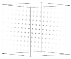
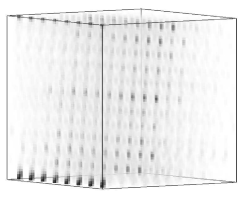
To test the algorithm we created a real space volume which was then populated with a cubic lattice that contains voxels. A rotation was then applied to the lattice (fig. 2(a)) and the result was Fourier transformed to generate the 3D diffraction volume (fig. 2(d)). The size of the problem was chosen for visualization purposes.
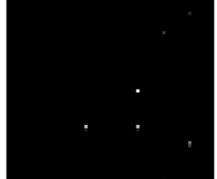
The simulated diffraction data was calculated by selecting those voxels which are crossed by the Ewald sphere (fig. 2(b)) and projecting them onto the detector plane according to the geometry shown in Figure 1. Figure 2(c) shows the simulated 2D diffraction pattern. The detector plane is uniformly sampled with pixels, and we set the distance between the crystal and the detector to 64 pixels.
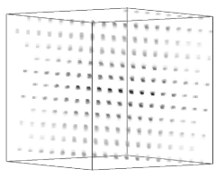
We then tried to recover the real space lattice using two methods. In the first method we simply took the inverse 3D FFT of the diffraction volume in which the voxels that were not “observed” were set to zero. The intensity of the transformed volume is shown in Figure 2(e). In the second approach, we solve the L1 minimization problem (5) discussed in the previous section by using the SPGL1 software provided by the authors of Van den Berg and Friedlander (2009). The amplitude of the reconstructed real space lattice , after 300 iterations of SPGL1 is shown in Figure 2(f). After only 10 iterations it is already possible to see the unit cell positions clearly enough to determine the crystal orientation and unit cell dimensions.
It is clear from Figures 2(e) and 2(f) that the latter approach results in a much sharper image from which the real space lattice points can be easily extracted.
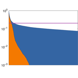
To quantify this difference we normalized recovered real space intensity values of both methods and sorted them in decreasing order. We plotted the sorted values as 1D curves in Figure 3. The curve that separates the orange and blue region of the plot is associated with the solution to the L1 minimization problem. The curve that separates the blue region and the white area above it is associated with the sorted intensities obtained from a direct 3D inverse FFT. Clearly, the intensity associated with the solution to the L1 minimization problem decreases much more rapidly, thereby making it easy to select a threshold (shown as the magenta line in Figure 3) that can be used to identify real space lattice points.
VII Concluding Remarks
We presented a new technique for autoindexing nanocrystal diffraction images. The technique is based on dividing the indexing problem in three steps. We reformulate the critical step of recovering a real space 3D map of the lattice as an L1 minimization (or BP) problem and solving the problem by an efficient and robust numerical algorithm. A simple numerical thresholding reveals the positions of the 3D lattice points in real space. They can subsequently be used to determine the unit cell parameters, crystal orientation and type as currently done in existing crystallographic software. Mirror symmetries of the lattice generate multiple solutions that still need to be sorted out in a final step using the measured intensities.
Although the algorithm is more costly than the existing approach because it is iterative and performs multiple 3D FFTs, it has the advantage of recovering crystal lattice reliably when only a few Bragg peaks can be measured. Greedy algorithms that make use of the sparsity of the solution may avoid this problem and increase speed significantly Mallat (1999); Needell and Vershynin (2009); Needell and Tropp (2009); Donoho et al. (2006); Eldar et al. (2010).
Once the lattice vectors and orientation are determined, the lattice coordinates that overlap with the Ewald sphere will provide the index of a reflection.
We demonstrate the feasibility of the technique with a simple example. More studies are needed to test the efficacy of the method on different types of Bravais lattices and on datasets that may be contaminated with noise. However, we believe that our preliminary results already indicate that compressive sensing based autoindexing is a promising tool for ultrafast nanocrystallography. Moreover, this type of technique allows other constraints to be easily incorporated into L1 minimization formulation to improve the reliability of indexing. It may even be possible to extend this approach to index multiple crystals, powder diffraction or Laue data.
Acknowledgment
We thank J. C. H. Spence for raising the problem, and Y. C. Eldar for discussions. This work was supported by the Laboratory Directed Research and Development Program of Lawrence Berkeley National Laboratory under U.S. Department of Energy Contract No. DE-AC02-05CH11231. Funding for FRNCM was provided by the American Recovery and Reinvestment Act Computational Science and Engineering Petascale Initiative.
References
- Holton (2009) J. M. Holton, A beginner’s guide to radiation damage, Journal of Synchrotron Radiation 16 (2009) 133–142.
- Emma et al. (2010) P. Emma, R. Akre, J. Arthur, R. Bionta, C. Bostedt, J. Bozek, A. Brachmann, P. Bucksbaum, R. Coffee, F. J. Decker, Y. Ding, D. Dowell, S. Edstrom, A. Fisher, J. Frisch, S. Gilevich, J. Hastings, G. Hays, P. Hering, Z. Huang, R. Iverson, H. Loos, M. Messerschmidt, A. Miahnahri, S. Moeller, H. D. Nuhn, G. Pile, D. Ratner, J. Rzepiela, D. Schultz, T. Smith, P. Stefan, H. Tompkins, J. Turner, J. Welch, W. White, J. Wu, G. Yocky, J. Galayda, First lasing and operation of an ångstrom-wavelength free-electron laser, Nature Photonics 4 (2010) 641–647.
- Ackermann et al. (2007) W. Ackermann, G. Asova, V. Ayvazyan, A. Azima, N. Baboi, J. Bahr, V. Balandin, B. Beutner, A. Brandt, A. Bolzmann, R. Brinkmann, O. I. Brovko, M. Castellano, P. Castro, L. Catani, E. Chiadroni, S. Choroba, A. Cianchi, J. T. Costello, D. Cubaynes, J. Dardis, W. Decking, H. Delsim-Hashemi, A. Delserieys, G. Di Pirro, M. Dohlus, S. Dusterer, A. Eckhardt, H. T. Edwards, B. Faatz, J. Feldhaus, K. Flottmann, J. Frisch, L. Frohlich, T. Garvey, U. Gensch, Gerthch, M. Gorler, N. Golubeva, H. J. Grabosch, M. Grecki, O. Grimm, K. Hacker, U. Hahn, J. H. Han, K. Honkavaara, T. Hott, M. Huning, Y. Ivanisenko, E. Jaeschke, W. Jalmuzna, T. Jezynski, R. Kammering, V. Katalev, K. Kavanagh, E. T. Kennedy, S. Khodyachykh, K. Klose, V. Kocharyan, M. Korfer, M. Kollewe, W. Koprek, S. Korepanov, D. Kostin, M. Krassilnikov, G. Kube, M. Kuhlmann, C. L. S. Lewis, L. Lilje, T. Limberg, D. Lipka, F. Lohl, H. Luna, M. Luong, M. Martins, M. Meyer, P. Michelato, V. Miltchev, W. D. Moller, L. Monaco, W. F. O. Muller, O. Napieralski, O. Napoly, P. Nicolosi, D. Nolle, T. Nunez, A. Oppelt, C. Pagani, R. Paparella, N. Pchalek, J. Pedregosa-Gutierrez, B. Petersen, B. Petrosyan, G. Petrosyan, L. Petrosyan, J. Pfluger, E. Plonjes, L. Poletto, K. Pozniak, E. Prat, D. Proch, P. Pucyk, P. Radcliffe, H. Redlin, K. Rehlich, M. Richter, M. Roehrs, J. Roensch, R. Romaniuk, M. Ross, J. Rossbach, V. Rybnikov, M. Sachwitz, E. L. Saldin, W. Sandner, H. Schlarb, B. Schmidt, M. Schmitz, P. Schmuser, J. R. Schneider, E. A. Schneidmiller, S. Schnepp, S. Schreiber, M. Seidel, D. Sertore, A. V. Shabunov, C. Simon, S. Simrock, E. Sombrowski, A. A. Sorokin, P. Spanknebel, R. Spesyvtsev, L. Staykov, B. Steffen, F. Stephan, F. Stulle, H. Thom, K. Tiedtke, M. Tischer, S. Toleikis, R. Treusch, D. Trines, I. Tsakov, E. Vogel, T. Weiland, H. Weise, M. Wellhofer, M. Wendt, I. Will, A. Winter, K. Wittenburg, W. Wurth, P. Yeates, M. V. Yurkov, I. Zagorodnov, K. Zapfe, Operation of a free-electron laser from the extreme ultraviolet to the water window, Nat Photon 1 (2007) 336–342.
- Shintake et al. (2008) T. Shintake, H. Tanaka, T. Hara, T. Tanaka, K. Togawa, M. Yabashi, Y. Otake, Y. Asano, T. Bizen, T. Fukui, S. Goto, A. Higashiya, T. Hirono, N. Hosoda, T. Inagaki, S. Inoue, M. Ishii, Y. Kim, H. Kimura, M. Kitamura, T. Kobayashi, H. Maesaka, T. Masuda, S. Matsui, T. Matsushita, X. Marechal, M. Nagasono, H. Ohashi, T. Ohata, T. Ohshima, K. Onoe, K. Shirasawa, T. Takagi, S. Takahashi, M. Takeuchi, K. Tamasaku, R. Tanaka, Y. Tanaka, T. Tanikawa, T. Togashi, S. Wu, A. Yamashita, K. Yanagida, C. Zhang, H. Kitamura, T. Ishikawa, A compact free-electron laser for generating coherent radiation in the extreme ultraviolet region, Nature Photonics 2 (2008) 555–559.
- Altarelli et al. (2006) M. Altarelli, R. Brinkmann, M. Chergui, W. Decking, B. Dobson, S. Dusterer, G. Grubel, W. Graeff, H. Graafsma, J. Hajdu, J. Marangos, J. Pfluger, H. Redlin, D. Riley, I. Robinson, J. Rossbach, A. Schwarz, K. Tiedtke, T. Tschentscher, I. Vartaniants, H. Wabnitz, H. Weise, R. Wichmann, K. Witte, A. Wolf, M. Wulff, M. Yurkov, XFEL: The European X-Ray Free-Electron Laser : Technical design report, DESY, Hamburg, 2006.
- Chapman (2009) H. N. Chapman, X-ray imaging beyond the limits, Nat Mater 8 (2009) 299–301.
- Neutze et al. (2000) R. Neutze, R. Wouts, D. van der Spoel, E. Weckert, J. Hajdu, Potential for biomolecular imaging with femtosecond x-ray pulses, Nature 406 (2000) 752–757.
- Chapman et al. (2006) H. N. Chapman, A. Barty, M. J. Bogan, S. Boutet, M. Frank, S. P. Hau-Riege, S. Marchesini, B. W. Woods, S. Bajt, H. W. Benner, R. A. London, E. Plonjes, M. Kuhlmann, R. Treusch, S. Dusterer, T. Tschentscher, J. R. Schneider, E. Spiller, T. Moller, C. Bostedt, M. Hoener, D. A. Shapiro, K. O. Hodgson, D. van der Spoel, F. Burmeister, M. Bergh, C. Caleman, G. Huldt, M. M. Seibert, F. R. Maia, R. W. Lee, A. Szoke, N. Timneanu, J. Hajdu, Femtosecond diffractive imaging with a soft-x-ray free-electron laser, Nature Physics 2 (2006) 839–843.
- Kirian et al. (2010) R. A. Kirian, X. Wang, U. Weierstall, K. E. Schmidt, J. C. H. Spence, M. Hunter, P. Fromme, T. White, H. N. Chapman, J. Holton, Femtosecond protein nanocrystallography—data analysis methods, Opt. Express 18 (2010) 5713–5723.
- Steller et al. (1997) I. Steller, R. Bolotovsky, M. G. Rossmann, An algorithm for automatic indexing of oscillation images using Fourier analysis, J. Appl. Cryst. 30 (1997) 1036–1040.
- Rossmann and van Beek (1999) M. G. Rossmann, C. G. van Beek, Data processing, Acta Cryst. D 55 (1999) 1631–1640.
- Leslie (1992) A. G. W. Leslie, Recent changes to the MOSFLM package for processing film and image plate data., volume 26, Daresbury Laboratory, Warrington, U.K., 1992.
- Bricogne (1986) G. Bricogne, in: Proceedings of the EEC cooperative Workshop on Position-Sensitive Detector Software (Phase III), Paris.
- Otwinowski and Minor (1997) Z. Otwinowski, W. Minor, Processing of X-ray diffraction data collected in oscillation mode, in: J. Charles W. Carter (Ed.), Macromolecular Crystallography Part A, volume 276 of Methods in Enzymology, Academic Press, 1997, pp. 307 – 326.
- Campbell (1998) J. W. Campbell, The Practicality of Using a Three-Dimensional Fast Fourier Transform in Auto-Indexing Protein Single-Crystal Oscillation Images, Journal of Applied Crystallography 31 (1998) 407–413.
- Natterer and Wübbeling (2001) F. Natterer, F. Wübbeling, Mathematical Methods in Image Reconstruction, SIAM, 2001.
- Powell (1999) H. R. Powell, The Rossmann Fourier autoindexing algorithm in MOSFLM, Acta Cryst. D 55 (1999) 1690–1695.
- Sauter et al. (2004) N. K. Sauter, R. W. Grosse-Kunstleve, P. D. Adams, Robust indexing for automatic data collection, Journal of Applied Crystallography 37 (2004) 399–409.
- Ying (2009) L. Ying, Sparse Fourier transform via butterfly algorithm, SIAM J. Sci. Comput. 31 (2009) 1678–1694.
- Donoho (2006) D. L. Donoho, Compressive sensing, IEEE Transaction on Information Theory 52 (2006) 1289–1306.
- Candès et al. (2006a) E. Candès, J. Romberg, T. Tao, Stable singal recovery from incomplete and inaccurate measurements, Comm. Pure Appl. Math 59 (2006a) 1207–1223.
- Candès et al. (2006b) E. Candès, J. Romberg, T. Tao, Robust uncertainty principles: Exact signal reeconstruction from highly incomplete frequency information, IEEE Transaction Inform. Theory 52 (2006b) 489–509.
- Van den Berg and Friedlander (2009) E. Van den Berg, M. P. Friedlander, Probing the Pareto frontier for basis pursuit solutions, SIAM J. Sci. Comput. 31 (2009) 890–912.
- Tibshirani (1996) R. Tibshirani, Regression shrinkage and selection via the Lasso, J. Roy. Statist. Soc. Ser. B 58 (1996) 267–288.
- Birgin et al. (2000) E. G. Birgin, J. M. Martínez, M. Raydan, Nonmonotone spectral projected gradient methods on convex sets, SIAM J. Optim. 10 (2000) 1196–1211.
- Birgin et al. (2005) E. G. Birgin, J. M. Martínez, M. Raydan, Inexact spectral projected gradient methods on convex sets, IMA J. Numer. Anal. 23 (2005) 539–559.
- Dai and Fletcher (2005) Y. H. Dai, R. Fletcher, Projected Barzilai-Borwein methods for large-scale box-constrained quadratic programming, Numer. Math. 100 (2005) 21–47.
- Nesterov (1983) Y. Nesterov, A method for unconstrained convex minimization problem with the rate of convergence , Dokl. Acad. Nauk SSSR 269 (1983) 543–547.
- Nesterov (2005) Y. Nesterov, Smooth minimization of non-smooth functions, Math. Program. 103 (2005) 127–152.
- Becker et al. (2009) S. Becker, J. Bobin, E. J. Candès, NESTA: A Fast and accurate first-order method for sparse recovery, Technical Report, California Institute of Technology, 2009.
- Mallat (1999) S. Mallat, A Wavelet Tour of Signal Processing, Second Edition (Wavelet Analysis & Its Applications), Academic Press, 2 edition, 1999.
- Needell and Vershynin (2009) D. Needell, R. Vershynin, Uniform uncertainty principle and signal recovery via regularized orthogonal matching pursuit, Foundations of Computational Mathematics 9 (2009) 317–334. 10.1007/s10208-008-9031-3.
- Needell and Tropp (2009) D. Needell, J. Tropp, Cosamp: Iterative signal recovery from incomplete and inaccurate samples, Applied and Computational Harmonic Analysis 26 (2009) 301 – 321.
- Donoho et al. (2006) D. L. Donoho, I. Drori, Y. Tsaig, J. L. Starck, Sparse solution of underdetermined linear equations by stagewise orthogonal matching pursuit, Technical Report, Department of Statistics, Stanford University, 2006.
- Eldar et al. (2010) Y. C. Eldar, P. Kuppinger, H. Bolcskei, Block-sparse signals: Uncertainty relations and efficient recovery, IEEE Transactions on Signal Processing 58 (2010) 3042–3054.