How dsDNA breathing enhances its flexibility and instability on short length scales
Abstract
We study the unexpected high flexibility of short dsDNA which recently has been reported by a number of experiments. Via the Langevin dynamics simulation of our Breathing DNA model, first we observe the formation of bubbles within the duplex and also forks at the ends, with the size distributions independent of the contour length. We find that these local denaturations at a physiological temperature, despite their rare and transient presence, can lower the persistence length drastically for a short DNA segment in agreement with experiment.
pacs:
87.14.gk, 87.15.A-, 87.15.hgThe DNA is a double helix of two single-stranded (ss) backbone chains paired by complimentary bases via hydrogen-bonding and further stabilized by stacking interaction between adjacent base-pair (bp) planes 1.Nelson . Owing to recent advances of single molecule techniques, intensive studies have been done on structural changes and mechanical behaviors of double-stranded (ds) DNA constrained by external forces and twists. The continuum elastic model, called wormlike chain (WLC) model has been very useful in analytically describing the micron-scale conformations and elastic response of such DNA 2.Mako . The persistence length, the measure of segmental orientation correlation, is about 50 nm (equivalently, about 150 bases along contour) for dsDNA. On the contrary numerous biological facts suggests DNA loops more readily on much shorter length scales. Indeed, Cloutier and Widom showed that the DNA has much higher cyclization probability than predicted by a WLC of persistence length of 50 nm 3.Cloutier . Also, Wiggins showed the DNA on short length scale has an elastic behavior distinct from that of WLC 4.Wiggins , while Yuan . very recently have reported the persistence lengths are as short as 11 nm for DNA fragments consisting of base-pair (bp) 5.Yuan .
In this paper we demonstrate that the higher flexibility of dsDNA emerges on shorter scales indeed due to local denaturation. Because of the large initiation energy, the fraction of the open bases is less than 1% at the physiological temperature, and, once formed, bubbles decay shortly in the order of 50 s 7.5 , seemingly little affecting the DNA stability. We show that despite their rare and transient presence the bubbles give a drastic enhancement of the flexibility as the chain gets shorter. For a very short duplex fragment, another type of local denaturation, namely, forks at the free ends are entropically favorable, dominating over the bubbles to enhance the flexibility, and eventually driving the duplex to unbind into two single strands.
It was suggested and estimated that the baseflips 5.Yuan and kinks YanMarko can enhance the bending flexibility and looping probability. Based on a simple two state model, the looping probability was evaluated using transfer matrix method YanMarko . Although all these calculations are suggestive of their significance, the bubbles have not yet been explicitly accounted for with regard to their size distributions and realistic energetics.
We consider a homogeneous dsDNA as the duplex of two interacting single strands described by the effective energy
| (1) |
The is the elastic energy of the single strand (=1 or 2), which, in a discrete representation, is given by
| (2) |
where is the three-dimensional position vector of th bead in strand () and ( nm) is an average distance between neighboring beads within the ss. The first term accounts for bending energy with the bending modulus for the ss, which is the related to its persistence length via 9.J-Y KIM . The second term, the stretching energy of each strand, is introduced to impose chain inextensibility condition. An appropriate value of is numerically found by matching simulation of either force-extension curves or the mean end-to-end distances of ssDNA resulting from this model, with corresponding theoretical expression of the inextensible WLC without the stretching term. The is the pairing energy between complimentary bases. To describe the bp openings due to thermal excitation, namely, thermal bubbles and forks, the interaction is represented by where
| (3) |
is the Morse potential 9.5 M. Peyrard ; Theo , is the distance between th bp, and are the bond distance and range which correspond to mean and fluctuation of DNA diameter respectively, and is the potential depth. Whenever bps are unbound, the duplex becomes no more than two single strands with net persistence length as short as twice of nm 13.J. B. Mills , while for bound bps it takes nm, the persistence length of long DNA, due to stacking interaction in the double strand. In order to include the stacking and destacking that cause such variation of the persistence length in the model, we consider , and propose the persistence length per single strand takes the form
| (4) |
Here is a steplike form function which smoothly increases from to over the width and is the distance at which is (see Fig. 1).
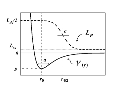
depends on three consecutive bp distances naturally as it represents the cooperativity of the stacking interaction lpcomment . The variation of depending on the bp distance is schematically illustrated in Fig. 1: When all of the three consecutive bps are outside the range , namely they are unbound one other, takes the single-stranded persistence length while as any one of the three are bound, is reduced to . The is comparable to the Morse potential width since the varies due to the bp unbinding.
Using this energy model, we simulate the dynamics and equilibrium distribution of the bp distance, via the Langevin equation,
| (5) |
where frictional coefficient per base is kg s-1 with 0.001 kgm for viscosity of water and nm for effective radius of nucleotide. is the Gaussian and white noise satisfying for each Cartesian component and . The values of potential depth and range, eV, nm are chosen so that the 300-bp DNA fragments have the melting temperature 350 K 9.J-Y KIM .
In our simulation, we employed 50 nm for the persistence length of dsDNA () and 4 nm for that of ssDNA () with the stretching constant pN/nm. For the parameters in , was chosen to nm where is the cutoff distance at which a bp is regarded as unbound in the simulation, and is 0.045 nm 9.J-Y KIM . We checked that small variation of the parameters values does not affect significantly the main result of our simulation.
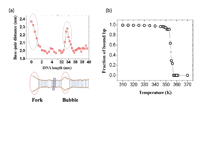
Figure 2 shows a snapshot profile of equilibrated bp distance along the contour of 300 bp long or about 100 nm, with free ends, and the fluctuating DNA thickness of about 2 nm therefrom constructed. The figure shows indeed the local denaturation exist in the forms of bubbles within the contour and fork at the ends. From the simulation we find the fraction of bound pairs undergoes a sharp transition from near unity to zero in agreement with experiment 9.3 at the melting temperature, which is about 350 K. It indicates that at the physiological temperature (310 K), the fraction of the bp that forms the local denaturation is less than 1%.
Shown in the Fig. 3 are the size distributions of the bubbles and forks for the various contour lengths of the DNA fragments. The distribution of the bubble size with bp open is remarkably independent of its position and the DNA length. For relatively large bubbles it follows the Poland-Sherega form 11.J-H Jeon
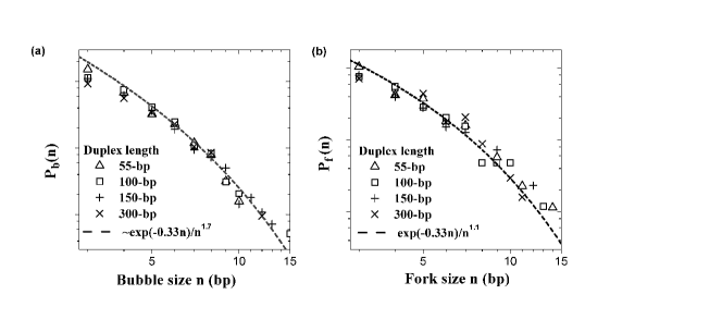
| (6) |
where is the statistical factor for a loop formation and is the average energy in to unbind a bp. These factors are independent of the length and given by those for a long length, , , at 310 K 9.J-Y KIM . The average size of the bubbles for various contour lengths is 1.7 bp at 310 K. This is in agreement with the Peyrard-Bishop-Dauxois model simulation 9.6 . The length-independence of their distribution and small average size implies that the thermal bubbles are transiently excited due to short-range elastic interaction in unconstrained DNA. This is to be contrasted with the dsDNA under mechanical constraints, where the average bubble size is found to be much larger depending on the (negative) twist as well as the contour length 9.7 . The distribution of bp open at a free end is given by
| (7) |
where , are also independent of the contour length within the errors. The average size of the forks is 1.9 bp. Since is 4 nm in these simulations, they are semiflexible forks with much larger than that of the flexible forks 9.75
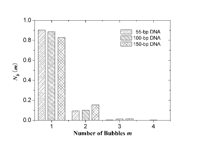
Last, we obtain the , the relative probability of finding bubbles simultaneously in a DNA of given length. The result obtained from simulation is presented in Fig. 4, showing that the single bubble occurrence becomes predominant as the DNA length gets shorter. This is mainly due to the large energy cost of bubble initiation; namely, once a bubble is formed, increasing its size is energetically more favorable than opening a new bubble elsewhere. Within the short DNAs of our interest, the number of bubbles, if they exist at all, can be regarded to be unity in a good approximation.
To focus on the effects of bubble (excluding the end fork effect) on the overall duplex persistence length, we consider an ensemble of fragments with given contour length randomly taken from inside a 300-bp long ( nm) dsDNA at a physiological temperature. The square of duplex end-to-end distance, , is defined as arithmetical average of the end-to-end distances of the two single strands. The thermal average of its square is taken over the ensemble of the duplex with a given . Suppose that the duplex to be a WLC with a uniform persistence length , we use the well-known relation 9.8
| (8) |
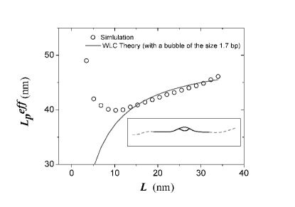
Comparing this with simulation data, we evaluate of our duplex for various values of the contour length . The result shown in Fig. 5 indicates that decreases from the value 50 nm to 40 nm as the decreases to 10 nm. It reflects the enhanced flexibility of the duplex, which we attribute to the presence of thermal bubbles. Along the DNA length shorter than 50 nm, if any, a single bubble is most likely to exist, as shown in , Fig. 4. If the single bubble survives as the contour length decreases, its mean size does not change as discussed earlier, consequently yielding larger flexibility.
To support this argument quantitatively, we note that the mean end-to-end distance is given by 9.8 , where is the unit tangent vector at the arclength from an end of the duplex. The tangent correlation function is given by if they are the positions within ds region and if and are the points within the bubble region. Integrating the correlation function along the contour with a bubble of the mean size, 1.7 bp, we evaluate the . Since a bubble occurs with equal probability along the contour we further average the mean square end-to-end distance over all possible position of the bubble, and relate it with the effective persistence length, which is shown by the curve in Fig. 5.
For the length larger than 10 nm, this analytical theory agrees with simulation result remarkably, suggesting that the persistence length reduction is indeed due to a single bubble. As the length decreases below that, this curve departs much from the simulation result for the length shorter than that. It is because even a single bubble is unlikely to occur within such a short length so that the persistence length sharply rises to the double strand value. For a long DNA fragment, on the other hand, only a bubble is found in most cases with its average size fixed independently of DNA length, which results in no significant perturbations on the persistence length.
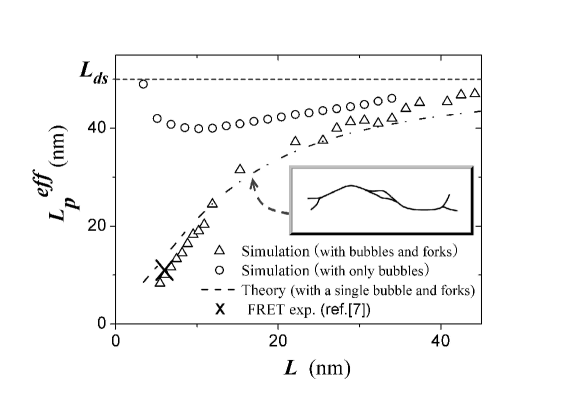
For the short duplex with free ends, we also have investigated the mean end-to-end distances of DNA fragments length shorter than 50 nm (147 bp) at 310 K. The persistence length calculated in a similar way from these data is shown by triangles in Fig. 6. It clearly shows that , in this range of the contour length, are shorter than nm but converges to the value as the contour length increases. The persistence length in this case is shorter than that of the duplex of the same contour length without the free ends discussed before (indicated by circles). This can be ascribed to the additional form of local denaturation, i.e., the forks at the free ends. To support this quantitatively, we analytically calculated the effective persistence length of the duplex with a single bubble of the size 1.7 bp and two forks of the size 1.9 bp at the ends by integrating the correlation function and following the procedure as before. The close agreement of the theoretical curve with the simulation evidences additional influence of the forks; the two forks combines with and increasingly dominates over a bubble, to enhance flexibility, as the duplex gets shorter.
In ref. 5.Yuan , the effective persistence length of dsDNAs of contour length bp (equivalently nm) was determined to be nm by a fluorescence resonance energy transfer (FRET) experiment, which is marked by a cross in Fig. 6. They considered the buffer conditions where the persistence lengths of non-interacting single stranded DNA have the range of the values 2.7-3 nm 13.J. B. Mills . In a duplex, however, the two unpaired single strands are subject to steric and electrostatic repulsions, which may enhance this ss persistence length effectively to about 4 nm. Indeed, our values of estimated from simulation with (=4 nm) are in a good agreement with this experimental value.
Simulating the Breathing DNA model that incorporates locally fluctuating persistence lengths depending upon the bp distances along the contour, we find that the distributions of bubbles and forks are nearly independent of the contour length, reducing the effective persistence length of the duplex for the short lengths. The forks dominate the bubbles in enhancing duplex flexibility and inducing unbinding transition.
Acknowledgements.
This work was supported by NCRC and BK 21. We thank L. A. Archer for a valuable communication.References
- (1) D. L. Nelson and M. M. Cox, Lehninger Principles of Biochemistry, 3rd ed. (W. H. Freeman, New York, 2000); M. Gueron, M. Kochoyan and J. L. Leroy, Nature 328, 89 (1987).
- (2) J. F. Marko and E. D. Siggia, Macromolecules 28, 8759 (1995).
- (3) T. E. Cloutier and J. Widom, Molecular cell 14, 355 (2004).
- (4) P. A. Wiggins et al., Nature Nanotechnology 1, 137 (2006).
- (5) C. Yuan, H. Chen, X. W. Lou, and L. A. Archer, Phys. Rev. Lett. 100, 018102 (2008).
- (6) G. Altan-Bonnet, A. Libchaber, and O. Krichevsky, Phys. Rev. Lett. 90, 138101 (2003).
- (7) J. Yan and J. F. Marko, Phys. Rev. Lett. 93, 108108 (2004); P. Ranjith, P. B. Sunil Kumar, and G. I. Menon, Phys. Rev. Lett. 94, 138102 (2005); H. Chen and J. Yan, Phys. Rev. E. 77, 041907 (2008).
- (8) J.-Y. Kim, J.-H. Jeon, and W. Sung, J. Chem. Phys. 128, 055101 (2008).
- (9) M. Peyrard and A. R. Bishop, Phys. Rev. Lett. 62, 2755 (1989).
- (10) N. Theodorakopoulos, T. Dauxois, and M. Peyrard, Phys. Rev. Lett. 85, 6 (2000).
- (11) J. B. Mills, E. Vacano, and P. J. Hagerman, J. Mol. Biol. 285, 245 (1999); M. C. Murphy et al., Biophys. J. 86, 2530 (2004).
- (12) A similar description was also introduced in the Peyrard-Bishop-Dauxois model Theo where the stacking energy is described by a harmonic energy with a bp-distance dependent spring constant, .
- (13) R. D. Blake and S. G. Delcourt, Nucleic Acids Res. 26, 3323 (1998).
- (14) D. Poland and H. A. Scheraga, J. Chem. Phys. 45, 1464 (1966).
- (15) S. Ares and G. Kalosakas, Nano Lett. 7, 307 (2007).
- (16) J.-H. Jeon and W. Sung, Biophys. J. 95, 3600 (2008).
- (17) M. Baiesi, E. Carlon, and A. L. Stella, Phys. Rev. E 66, 021804 (2002).
- (18) D. H. Boal, Mechanics of the cell, (Cambridge, UK, 2002).