Reentrant behavior of divalent counterion mediated DNA-DNA electrostatic interaction
Abstract
The problem of DNA-DNA interaction mediated by divalent counterions is studied using computer simulation. Although divalent counterions cannot condense free DNA molecules in solution, we show that if DNA configurational entropy is restricted, divalent counterions can cause DNA reentrant condensation similar to that caused by tri- or tetra-valent counterions. DNA-DNA interaction is strongly repulsive at small or large counterion concentration and is negligible or slightly attractive for a concentration in between. Implications of our results to experiments of DNA ejection from bacteriophages are discussed. The quantitative result serves to understand electrostatic effects in other experiments involving DNA and divalent counterions.
pacs:
87.19.xb, 87.14.gk, 87.16.A-The problem of DNA condensation has seen a strong revival of interest in recent years because of the need to develop effective ways of gene delivery for the growing field of genetic therapy. DNA viruses such as bacteriophages provide excellent study candidates for this purpose. One can package genomic DNA into viruses, then deliver and release the molecule into targeted individual cells. Recently there is a large biophysic literature dedicated to the problem of DNA condensation (packaging and ejection) inside bacteriophages *[Forareview; see]GelbartVirusReview2009.
Because DNA is a strongly charged molecule in aqueous solution, the process of ejection of DNA from bacteriophages can be strongly influenced by the screening condition of the solution. By varying the salinity of solution, one can vary the amount of DNA ejected. Interestingly, monovalent counterions such as Na+ have negligible effect on the DNA ejection process Evilevitch et al. (2003). In contrast, multivalent counterions such as Mg+2, CoHex+3, Spd+3, or Spm+4 exert strong and non-monotonic effects Evilevitch et al. (2008). There is an optimal counterion concentration, , where the least DNA genome is ejected from the phages. For counterion concentration, , higher or lower than , more DNA is ejected from phages. The case of divalent counterions is more marginal. The non-monotonicity is observed for MgSO4 salt but not for MgCl2 salt up to the concentration of 100mM.
The problem of DNA condensation by divalent counterions is a complex problem due to contributions from many physical factors. In the literature, most of the studies dealing with this problem have focused on the ion-specific effects. For example, the hydration effects have been proposed to explain the above dependence on the type of divalent salts Evilevitch et al. (2008). In this paper, we focus on role of non-specific electrostatic interactions between DNA and counterions. In a recent work Lee et al. , we suggested that some aspects of DNA ejection in the presence of divalent counterions can be accounted for from the electrostatic point of view. Specially the strong, non-monotonic influence of divalent counterions on DNA ejection mentioned above is expected to have the same physical origin as the phenomenon of reentrant DNA condensation in free solution Nguyen et al. (2000); Saminathan et al. (1999); *LivolantBJ1996. The fact that divalent counterions can have such strong influence on DNA ejection is not trivial. Unlike counterion of higher valences, Mg+2 counterions are known to not condense DNA Rau and Parsegian (1992), or to condense them only partially in free solution Hud and Downing (2001). However, DNA viruses provide a unique experimental setup. The constraint of the viral capsid strongly eliminates configurational entropic cost of packaging DNA. This allows divalent counterions to influence DNA condensation similar to that of tri- or tetra-valent counterions. In this paper, we use computer simulations to study the problem of DNA condensation in the presence of divalent counterions. We show that indeed, if one includes only the non-specific electrostatic contribution, divalent counterions can induce DNA reentrant condensation like those observed for higher counterion valences. We offer an explanation for the discrepancy between DNA condensation in free solution versus DNA condensation inside viruses. Our results show that, in addition to ion-specific effects, electrostatics exert a strong, non-negligible influence on qualitative and quantitative behaviors of this system. The results presented here can provide understanding of not only the electrostatics of DNA ejection problem, but also can serve as a starting point for investigating other systems involving DNA and divalent counterions where the physical pictures are still not very well understood.
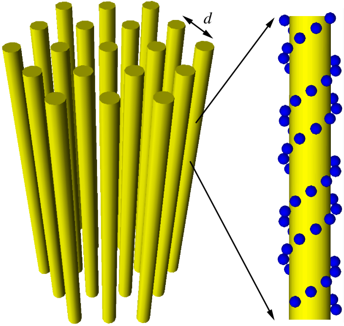
We model the DNA bundle in hexagonal packing as a number of DNA molecules arranged in parallel along the -axis. In the horizontal plane, the DNA molecules form a two dimensional hexagonal lattice with lattice constant (the DNADNA interaxial distance) (Fig. 1). An individual DNA molecule is modeled as an impenetrable cylinder with negative charges glued onto it. The charges are positioned in accordance with the locations of nucleotide groups along the double-helix structure of a BDNA. The hardcore cylinder has radius of 7Å. The negative charges are hard spheres of radius 2Å, charge , and lie at a distance of 9Å from the DNA axis. This gives an averaged DNA diameter of 1nm. The solvent water is treated as a dielectric medium with dielectric constant and temperature . The dielectric constant mismatch between water and DNA interior is neglected, and the cylinder only acts to prevent ion penetration. In our simulation, the positions of DNA molecules are fixed in space. This mimics the constraint on DNA configurational entropy inside viruses and other experiments of DNA condensation using divalent counterions.
In the experiment of DNA ejection from bacteriophages, there are both monovalent and divalent salts in solution. At very low concentration of divalent counterions, DNA is screened mostly by monovalent counterions. To account for this limit, we include both salts in our simulations. The mobile ions are modeled as hard spheres with unscreened Coulomb interaction (the primitive ion model). The radii of the coions and monovalent counterions are set to 2Å. (For simplicity, we assume the two salts have the same coions.) The divalent counterions radius is set to 2.5Å. The interaction between two ions, and , with radii, , and charges, , is given by
| (1) |
where is the distance between the ions.
The simulation is carried out using the periodic boundary condition. A periodic simulation cell with DNA molecules in the horizontal plane and 3 full helix periods in the direction is used. The dimensions of the box are , , and Å. The long-range electrostatic interactions between charges in neighboring cells are treated using the Ewald summation method. In Ref. Lyubartsev and Nordenskiöld (1995); *NordenskioldJCP86, it is shown that the macroscopic limit is reached when . Our simulation cell contains 12 DNA helices, hence it has enough DNA molecules to eliminate the finite size effect. We did test runs with 1, 4, 7, and 12 DNA molecules to verify that this is indeed the case. They are also used to check the correctness of our computer program by reproducing the results of DNA systems studied in Ref. Lyubartsev and Nordenskiöld (1995); *NordenskioldJCP86 in specific limits.
In a practical situation, the DNA bundle is in equilibrium with a water solution containing free mobile ions at a given concentration. Therefore we simulate the system using Grand Canonical Monte-Carlo (GCMC) simulation. The number of ions are not constant during the simulation. Instead their chemical potentials are fixed. The chemical potentials are chosen in advance by simulating a DNAfree salt solution and adjusting them so that the solution has the correct ion concentrations. In a simulation, the ions are inserted into or removed from the system in groups to maintain the charge neutrality Valleau and Cohen (1980). Following Ref. Valleau and Cohen (1980), instead of using individual chemical potentials, , , and , for each ion species, we use only the combined chemical potentials,
| (2) |
in the Metropolis acceptance criteria of a particle insertion/deletion move. In this paper, we simulate DNA bundles at varying concentrations . Both and are adjusted so that the monovalent salt bulk concentration, , in the DNAfree solution is always at 50mM (typical value of the DNA ejection experiment) and is at the desired value. Typical standard deviations in the final salt concentrations are about 10%.
To study DNADNA interactions, we use the Expanded Ensemble method Lyubartsev and Nordenskiöld (1995) to calculate the pressure of the DNA bundle. In this method, we calculate the difference of the system free energy at different volumes by sampling these volumes simultaneously in a simulation run. By calculating the free energy difference for two nearly equal volumes, and , we can calculate the total pressure of the system, (here is the set of chemical potentials of different ion species). The osmotic pressure of the DNA bundle is then obtained by subtracting the total pressure of the bulk DNAfree solution, , from the total pressure of the DNA system, .
In Fig. 2a, the osmotic pressure of the DNA bundle at different is plotted as a function of the interaxial DNA distance, . Because this osmotic pressure is directly related to the “effective” force between DNA molecules at that interaxial distance Lyubartsev and Nordenskiöld (1995); *NordenskioldJCP86, Fig. 2a also serves as a plot of DNADNA interaction. As one can see, when is greater than a value around 20mM, there is a shortrange attraction between two DNA molecules as they approach each other. This is the well-known phenomenon of like-charge attraction between macroions Naji et al. (2004); *GelbartPhysToday; Nguyen et al. (2000). It is the result of the electrostatic correlations between counterions condensed on the surface of each DNA molecule. The attraction appears when the distance between these surfaces is on the order of the lateral separation between counterions (about 14Å for divalent counterions). The maximal attraction occurs at the distance Å in good agreement with various theoretical and experimental results Rau and Parsegian (1992); Purohit et al. (2005). For smaller , the DNA-DNA interaction experiences a sharp increase due to the hardcore repulsion between the counterions. One also sees that the depth of attractive force between DNA molecules saturates at around atm as increases. This saturation is easily understood. At small , there are both monovalent and divalent counterions present in the bundle. As increases, divalent counterions replace monovalent ones in the bundle as the later ions are released into the bulk solution to increase the overall entropy of the solution. However, charge neutrality condition of the DNA macroscopic bundle and the hardcore repulsion between ions limit how many divalent counterions can be present inside the bundle. Once all monovalent counterions are released into solution (replaced by divalent counterions), further increase in does not significantly change the number of divalent counterions in the bundle. This leads to the observed saturation of DNADNA shortrange attraction with increasing .

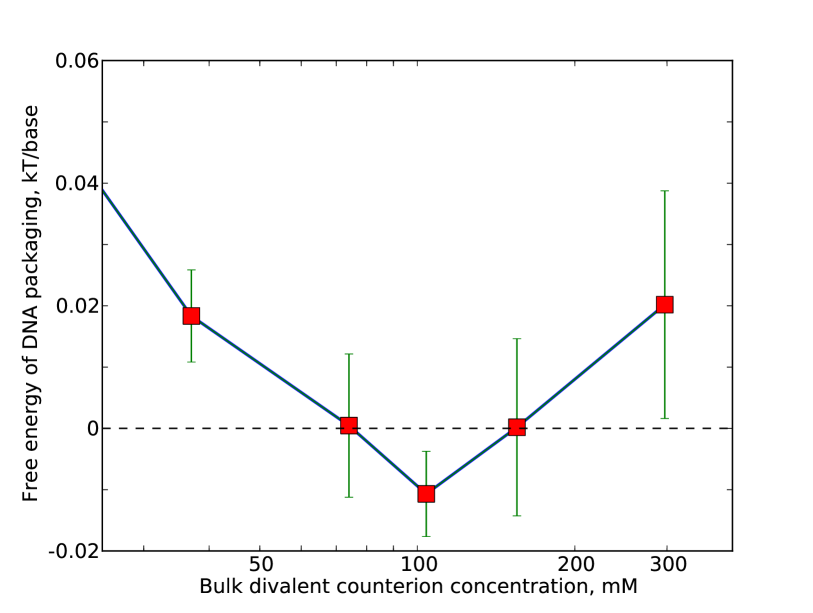
The strong influence of divalent counterions on DNA bundles can be seen by looking at the DNA-DNA “effective” interaction at larger . As evident from Fig. 2a for large , at small DNA-DNA interaction is repulsive. As increases, DNA-DNA interaction becomes less repulsive and reaches a minimum around 100mM. As increases further, DNA-DNA repulsion starts to increase again. This non-monotonic dependence of DNA-DNA “effective” interaction on the counterion concentration is even more clear if one calculates the free energy of packaging DNA into bundles. This free energy is the difference between the free energy of a DNA molecule in a bundle and that of an individual DNA molecule in the bulk solution. Per DNA nucleotide base, this free energy is given by:
| (3) |
here Å is the distance between DNA nucleotides along the axis of the DNA, and is the volume of our simulation box. The result for at the optimal bundle lattice constant Å is plotted in Fig. 2b as function of the NIn . Once again, there is an optimal concentration, , where the free energy cost of packaging DNA is lowest. It is even negative indicating the tendency of the divalent counterions to condense the DNA. At smaller or larger concentrations of the counterions, the free energy cost of DNA packaging is higher.
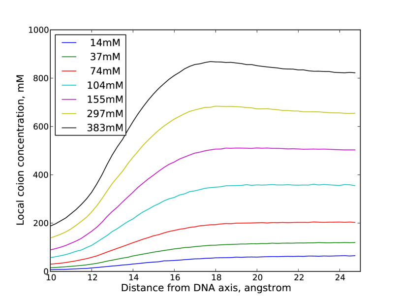
This reentrant behavior of DNA interaction can be understood. At large separations, the distribution of counterions in the bundle can be considered to be composed of two populations: condensed layers of counterions near the surfaces of the DNA molecules and diffuse layers of counterions further away. It is reasonable to expect the thickness of the condensed counterion layer to be on the order of the average lateral distance between counterions on the DNA surface (Å). So for Å, both counterion populations are present and one expects DNA-DNA interaction to be the standard screened Coulomb interaction between two charged cylinders with charge density . The qualitative dependence of on can be obtained by plotting the local coion concentration as a function of distance from DNA axis (Fig. 3). At low , decreases as decreases from suggesting is negative (undercharged DNA). At high , increases as decreases until the condensed counterion layers start to overlap at Å. This shows that is positive (overcharged DNA). In both cases, DNA repulsion is strong. For an intermediate value of , , DNA is almost neutral, and the repulsion is weakest. Furthermore, the like-charge attraction among DNA molecules mediated by the counterions Naji et al. (2004) is dominant in this concentration range, causing the electrostatic packaging free energy to become negative.
Figure 2b gives a value of /base for the shortrange attraction among DNA molecules at the optimal concentration. This is slightly less negative than previous theoretical fit of viral DNA ejection experiments Lee et al. . We believe this small difference is due our choice of the system’s physical parameters such as ion sizes Grønbech-Jensen et al. (1997); *NordenskioldPRL98; *DNA_reentrantPRE. The azimuthal orientation correlations of DNA Kornyshev et al. (2007) are another omission in our study. Relaxation along this degree of freedom can further lowering energy of the system. The non-electrostatic (such as van der Waals) interactions at small can also enhance DNA attraction. On the other hand, dielectric constant mismatch between water and DNA interior could push the condensed ions away from DNA interior and lower the attraction energy. However, these effects are minor at large DNADNA separations, thus do not change the qualitative reentrant condensation picture. More comprehensive studies that take these effects into account are the subjects of our future works. Nevertheless, the value range of obtained in this paper is significant. It explains why divalent counterions exert strong effect on DNA ejection from virus but are not able to condense DNA in free solution. This value corresponds to an attraction of a fraction of per one persistence length ( bases). This is too small to overcome thermal fluctuation of DNA (about one per persistence length), thus cannot condense them. Only inside the confinement of the viral capsid, where DNA configuration entropy is strongly suppressed, can divalent counterions cause strong influence. The non-monotonic behavior described above has the same physics as the phenomenon of reentrant DNA condensation by counterions Nguyen et al. (2000); Shklovskii (1999); *NguyenRMP2002 of high valences. In this paper, we demonstrate clearly that it can happen to divalent counterions if DNA configuration entropy is restricted. This correlates well with experimental data of DNA ejection from bacteriophages. We should mention here that DNA condensation by divalent counterions has also been observed in another environment where DNA configuration is constrained, namely the condensation of DNA in two dimensional systems Koltover et al. (2000). This fact once again strongly supports our argument.
In conclusion, in this paper, we use a computer simulation to study the electrostatics of DNA condensation in the presence of divalent counterions. The entropy of DNA configure fluctuation is suppressed in simulation. Such study can be applied directly to the experimental problem of DNA ejection from bacteriophages where DNA condensed in a strongly confined environment. Our results show that, even at the level of non-specific electrostatic interaction, divalent counterions can strongly influence DNA ejection. This potentially opens up an additional degree of freedom in controlling bacteriophages for the general purpose of gene therapy or viral diseases treatment. Beyond the scope of DNA ejection experiments, we believe the quantitative results of our paper can be used to understand many other experiments involving DNA and divalent counterions.
Acknowledgements.
We would like to thank Lyubartsev, Nordenskiöld, Shklovskii, Evilevitch, Fang, Gelbart, Phillips, Podgornick, Rau, and Parsegian for valuable discussions. TN acknowledges the support of junior faculty from the Georgia Institute of Technology. SL acknowledges financial support from Korean-American Scientists and Engineers Association (Georgia chapter). The authors are indebted to Dr. Lyubartsev for providing us with the source code of their simulation program. This code forms the basis of the simulation program used in this work. TN acknowledges the hospitality of the Aspen Center for Physics and the Fine Theoretical Physics Institute where part of the work is done.References
- Knobler and Gelbart (2009) C. M. Knobler and W. M. Gelbart, Annu. Rev. Phys. Chem. 60, 367 (2009).
- Evilevitch et al. (2003) A. Evilevitch, L. Lavelle, C. M. Knobler, E. Raspaud, and W. M. Gelbart, Proc. Nat. Acad. Sci. USA 100, 9292 (2003).
- Evilevitch et al. (2008) A. Evilevitch, L. T. Fang, A. M. Yoffe, M. Castelnovo, D. C. Rau, V. A. Parsegian, W. M. Gelbart, and C. M. Knobler, Biophys. J. 94, 1110 (2008).
- (4) S. Lee, C. V. Tran, and T. T. Nguyen, Biophys. J. (to be submitted) arXiv:cond-mat/0811.1296 .
- Nguyen et al. (2000) T. T. Nguyen, I. Rouzina, and B. I. Shklovskii, J. Chem. Phys. 112, 2562 (2000).
- Saminathan et al. (1999) M. Saminathan, T. Antony, A. Shirahata, L. H. Sigal, T. Thomas, and T. J. Thomas, Biochemistry 38, 3821–3830 (1999).
- Pelta et al. (1996) J. Pelta, D. Durand, J. Doucet, and F. Livolant, Biophys. J. 71, 48 (1996).
- Rau and Parsegian (1992) D. C. Rau and V. A. Parsegian, Biophys. J. 61, 246 (1992).
- Hud and Downing (2001) N. V. Hud and K. H. Downing, Proc. Nat. Acad. Sci. USA 98, 14925 (2001).
- Lyubartsev and Nordenskiöld (1995) A. P. Lyubartsev and L. Nordenskiöld, J. Phys. Chem. 99, 10373 (1995).
- Guldbrand et al. (1986) L. Guldbrand, L. G. Nilsson, and L. Nordenskiöld, J. Chem. Phys. 85, 6686 (1986).
- Valleau and Cohen (1980) J. P. Valleau and L. K. Cohen, J. Chem. Phys. 72, 5935 (1980).
- Naji et al. (2004) A. Naji, A. Arnold, C. Holm, and R. R. Netz, Eur. Phys. Lett. 67, 130 (2004).
- Gelbart et al. (2000) W. M. Gelbart, R. F. Bruinsma, P. A. Pincus, and A. V. Parsegian, Phys. Today 53, 38 (2000).
- Purohit et al. (2005) P. K. Purohit, M. M. Inamdar, P. D. Grayson, T. M. Squires, J. Kondev, and R. Phillips, Biophys. J. 88, 851 (2005).
- (16) Due to the limitation of computer simulations, the numerical integration is performed from Å to Å only. The omitted integration from Å to gives an almost constant shift to , and do not change the conclusion of this paper.
- Grønbech-Jensen et al. (1997) N. Grønbech-Jensen, R. J. Mashl, R. F. Bruinsma, and W. M. Gelbart, Phys. Rev. Lett. 78, 2477–2480 (1997).
- Lyubartsev et al. (1998) A. P. Lyubartsev, J. X. Tang, P. A. Janmey, and L. Nordenskiöld, Phys. Rev. Lett. 81, 5465 (1998).
- Le and Nguyen (2010) T. T. Le and T. T. Nguyen, Phys. Rev. E (to be submitted) (2010).
- Kornyshev et al. (2007) A. A. Kornyshev, D. J. Lee, S. Leikin, and A. Wynveen, Rev. Mod. Phys. 79, 943 (2007).
- Shklovskii (1999) B. I. Shklovskii, Phys. Rev. E 60, 5802 (1999).
- Grosberg et al. (2002) A. Y. Grosberg, T. T. Nguyen, and B. Shklovskii, Rev. Mod. Phys. 74, 329 (2002).
- Koltover et al. (2000) I. Koltover, K. Wagner, and C. R. Safinya, Proc. Nat. Acad. Sci. USA 97, 14046 (2000).