-stacking functionalization through micelles swelling: Application to the synthesis of single wall carbon nanotube/porphyrin complexes for energy transfer
Abstract
We report on a new, orginal and efficient method for "-stacking" functionalization of single wall carbon nanotubes. This method is applied to the synthesis of a high-yield light-harvesting system combining single wall carbon nanotubes and porphyrin molecules. We developed a micelle swelling technique that leads to controlled and stable complexes presenting an efficient energy transfer. We demonstrate the key role of the organic solvent in the functionalization mechanism. By swelling the micelles, the solvent helps the non water soluble porphyrins to reach the micelle core and allows a strong enhancement of the interaction between porphyrins and nanotubes. This technique opens new avenues for the functionalization of carbon nanostructures.
Molecular engineering of functionalized nano-materials is the object of intensive research in connection to a wide range of applications Nel2009 . Such complexes aim at combining the unique properties of nano-objects and the wide tunability of organic molecules properties. Especially, complexes presenting charge or energy transfer are intensively investigated for biology Taraska2009 ; Nel2009 ; Sacca2009 or optoelectronics Hardin2009 ; Ehli2009 ; Park2009 ; Ross2009 applications. For instance, fluorescence energy transfer (FRET) has been used to monitor structural rearrangements in proteins Taraska2009 or to enhance the excitation transfer in dye-sensitized solar cells Hardin2009 . In this context, due to their unique transport properties and especially their large carrier mobility, single-wall carbon nanotubes (SWNTs) are of particular interest for use in organic photovoltaïc devices Campidelli2008 ; Ehli2009 ; ehli06 ; alvaro06 ; hasobe06 ; Guldi2008 ; Prato2009 .
Excitation transfer between "-stacked" porphyrin molecules and carbon nanotubes was demonstrated very recently magadur2008 ; casey2008 . The main advantage of "-stacking" functionalization is to hardly perturb the electronic structure of nanotubes (and therefore their optical and transport properties). In contrast, covalent functionalization is known to alter the mobility of carriers and to quench the nanotube luminescence herranz06 . In the other hand, "-stacking" functionalization performances are quite poor regarding the yield of functionalization, the reproducibility and the stability of the suspensions. These difficulties are due to the weakness of the interactions involved in "-stacking" functionalization and become problematic for scaling up a controlled process.
In this paper, we report on the synthesis of controlled and stable SWNT/porphyrin complexes in micelles showing an efficient energy transfer. The samples are obtained from aqueous suspensions of individual nanotubes in sodium cholate micelles. Transmission electron microscopy combined with electron energy loss spectroscopy demonstrate the presence of porphyrin molecules in the area of nanotubes. We investigate the functionalization process by means of optical absorption spectroscopy (OAS) of suspensions of various concentrations and solvent/water volume ratios. We point out the crucial role of the organic solvent that acts as a vector for the insertion of the porphyrin molecules into the micelle core. Finally, by means of photoluminescence excitation (PLE) experiments, we demonstrate an efficient energy transfer from the excited porphyrin to the nanotube.
Results
I Structural characterization
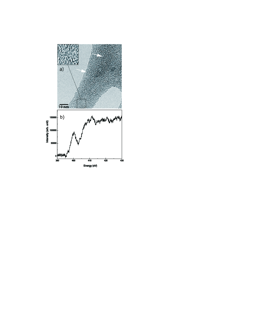
In a first approach, the composition of the SWNT/TPP suspensions is investigated by TEM.
Figure 1 a) shows a typical area where we identify the presence of individual nanotubes embedded in surfactant molecules. Nanotubes are difficult to distinguish since their contrast is shrouded by the amorphous contrast of the surfactant molecules. However as pointed by the arrows in figure 1 a), one can distinguish the presence of straight lines with spacing close to 1 nm which is consistent with the nanotube contrast springer2006 .
Additional EELS measurements have been recorded on the whole zone of figure 1 a). The corresponding spectrum is displayed on figure 1 b). An absorption edge close to 400 eV typical of the N-K edge is observed indicating the presence of nitrogen. This element is never observed in pristine nanotube. Furthermore, the fine structure of the absorption edge displays two features identified as a peak at 397 eV and a peak at 400 eV. These features are characteristic of nitrogen bonded to carbon atoms in configuration, which is the case in the TPP molecule. Since no other component of the suspension is expected to contain nitrogen, this band is interpreted as the signature of porphyrin molecules: porphyrin molecules are present in the very close area of the nanotubes.
II Optical properties
II.1 Optical absorption
Figure 2 displays the OAS of the SWNT/TPP suspension together with the OAS of a suspension of pure SWNTs and a suspension of pure TPP, both in SC micelles. The TPP suspension shows the so-called Soret band at 420 nm and the four weaker Q bands in the 500-700 nm region.

The OAS of SWNTs consists in a group of lines in the 1000 nm region corresponding to the so-called (lowest) transitions in semi-conducting nanotubes. Each line in this group stems from a given chiral family. Additional lines near 600 nm and at smaller wavelengths come from transitions superimposed to the onset of transitions in metallic nanotubes. The background in OAS mainly stems from light scattering and is substracted when analysing the line amplitudes.
The OAS of the SWNT/TPP suspension shows a band at 438 nm with a shoulder at 420 nm. These lines are attributed to the Soret absorption band of TPP encased in micelles (at 420 nm, in agreement with the OAS of pure TPP in micelles) and of TPP stacked on nanotubes (438 nm). This 18 nm red-shift of the Soret band when porphyrin molecules are "-stacked" on nanotubes is consistent with previous observations magadur2008 ; cambre2008 and is related to a conformation change of TPP molecules. The presence of these two bands shows that the suspension contains both pure TPP molecules encased in micelles and TPP stacked on nanotubes in micelles. The absorption features around 1000 nm are consistent with absorption lines in semi-conducting nanotubes. We note however a 7 nm red-shift compared to the reference SWNT suspension. This bathochromic shift is due to the presence of TPP close to the nanotubes and to the resulting interaction.
II.2 Control of the functionalization
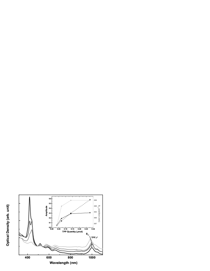
In contrast with previous reports on SWNT/TPP stacking, the key of our work is to provide a reliable method to produce samples of controlled and reproducible quality. The control of the process is investigated by studying the functionalization yield as a function of amount of TPP. Figure 3 shows the OAS of suspensions made from the same reference SWNT suspension mixed with a fixed volume of DCM (2.5 mL of SWNT suspension with 0.85 mL of DCM: volume ratio 34%, see below) containing a quantity of TPP ranging from 0.04 mol up to 0.28 mol. The four curves are arbitrary shifted (background correction) in order to match at 490 nm and to facilitate the comparison of the peak amplitudes. For low TPP concentrations (light grey and grey curves), the band at 438 nm (stacked TPP) is higher than the one at 420 nm (free TPP). The two bands have the same intensity for a TPP quantity of 0.14 mol (dark grey curve). For a larger amount of TPP, the band at 420 nm becomes predominant. The amplitude of each band as a function of TPP concentration are plotted in the inset of figure 3. The free TPP band (420 nm) grows regularly with the initial TPP concentration whereas the amplitude of the stacked TPP band reaches a plateau above 0.14 mol.
These results are interpreted as follows. At low TPP concentration, TPP molecules are preferentially "-stacked" on SWNTs rather than encased alone in micelles, therefore the amplitude of the 438 nm band is higher than the one at 420 nm. For an amount of TPP larger than 0.14 mol, no more porphyrin can stack onto nanotubes and TPP molecules mostly aggregate in micelles due to the excess of SC: the amplitude of the 438 nm band saturates while the one at 420 nm grows regularly with the TPP quantity (inset of figure 3). The position of the band of SWNTs is another way to track the stacking of TPP onto SWNTs. For a small amount of TPP, the band (near 1000 nm) is identical to the one in the reference SWNT suspension: most of the nanotubes are not functionalized. This band progressively red-shifts with increasing TPP concentration. Above the saturation threshold ( 0.14 mol), the position of reaches a plateau: most of the nanotubes are functionalized (grey curve in the inset of figure 3).
Importantly, the absorption amplitude of the band is almost identical in both the SWNT/TPP and the reference SWNT suspensions. This point shows that very few nanotubes are lost in the process. The increase in TPP quantity only affects the fonctionalization degree of nanotubes. This is due to the stability of micelle suspensions of nanotubes. The use of such a stable starting material is the key for the reproducibility and control of our method, in contrast to previous attempts where soluble TPP molecules where used for both solubilization and functionalization of SWNTs magadur2008 .
II.3 Functionalization mechanism
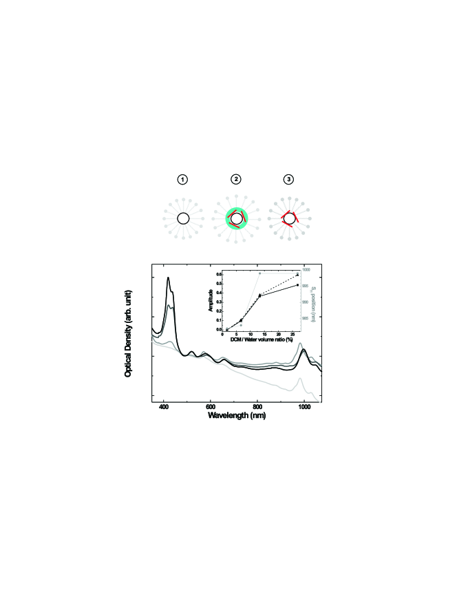
In view of further developments of this technique, a good description of the functionalization mechanism is needed. The SWNT/TPP complex is hydrophobic and is thus expected to stay in the organic core of the micelles in water suspensions. However, the formation mechanism of such a complex from pre-existing SWNT micelles and non water soluble TPP is less straightforward since TPP molecules have to diffuse through water to reach the core of the micelles. We show that an organic solvent can act as a vector and help TPP molecules to penetrate into the micelles. The control parameter is the DCM/water volume ratio which drives the exchange area between the two immiscible liquids during the sonication process. The OAS of SWNT/TPP suspensions for various volumes of DCM but with constant quantity of TPP (0.14 mol) is displayed in figure 4 note . For a DCM/water volume ratio of 2 % no functionalization is observed (light grey curve in figure 4). For a volume ratio of 7%, both the 420 nm and 438 nm Soret bands are observable but with a weak amplitude (grey curve). For increasing DCM/water volume ratio up to 27% (dark grey and black curves), both bands rise meaning that both the stacking of porphyrin onto nanotubes and the formation of TPP aggregates in micelles become efficient. Bearing in mind that the quantity of TPP is kept constant, this reveals the central role of DCM in the functionalization mechanism.
Wang et al. have shown that water-immiscible organic solvents swell the micelle hydrophobic core surrounding the SWNT wang09 . The authors have mixed a micelle suspension of nanotubes with immiscible organic solvents. The presence of the organic solvent inside the micelle is investigated by means of optical spectroscopy. Typical solvatochromic shifts are observed in the PL spectra and interpreted as a signature of nanotubes coated by the solvent. The organic solvent evaporates over time and after several hours the initial optical spectra are recovered showing the reversibility of the mechanism. The solvent/water ratio in Ref.wang09 is on the order of 50%, close to the one used in our studies (34%) for optimal synthesis conditions. Therefore we can confidently extend their conclusions to our work. When mixing the SWNT suspension with the TPP solution, DCM swells the sodium cholate micelle and therefore acts as a vector for the interaction between the porphyrin molecules and the nanotubes. This mechanism is schematically depicted in the upper panel of figure 4 . The starting material is a nanotube encased in a micelle (step 1). After addition of the DCM solution of TPP, the DCM swells the hydrophobic core of the micelle bringing porphyrin molecules in contact with the nanotube (step 2). As described in reference wang09 , DCM rapidly evaporates : TPP molecules stack onto the nanotubes (step 3) and the new complex remains in the micelle core.
Optical absorption spectra recorded just after and several hours after the synthesis do not show any noticeable change. We can therefore conclude that the samples investigated in this study correspond to step 3 of figure 4.
This micelle core swelling by organic solvents as a way to enhance the interaction between ""-conjugated molecules and nanotubes is very promising and opens new avenues for nanotube functionalization.
III Stability
Spectroscopic measurements of SWNT/TPP suspensions were recorded at different time delays (up to several months) after the synthesis and no noticeable evolution could be detected. An important point is that the photoluminescence (PL) signal is stable for months (see figure S1 in Supplementary Information), which is the major improvement of our method compared with previous reports casey2008 ; magadur2008 . Furthermore, the PL intensity of the SWNT/TPP suspension corresponding to the emission of nanotubes is on the same order of magnitude as the one of the reference SWNT suspension. It is one order of magnitude higher than for suspensions obtained with the soluble TPP stacking method magadur2008 . In contrast with this earlier work, this observation confirms that our micelle-based synthesis ensures that most of the nanotubes remain in the final product.
IV Energy transfer
In this section we probe the interaction between the porphyrin and the nanotube by means of photoluminescence experiments. The PL spectra of the SWNT reference suspension and of the SWNT/TPP suspension are almost identical, except that all bands are red-shifted by nearly 20 nm as already mentionned for absorption spectra.
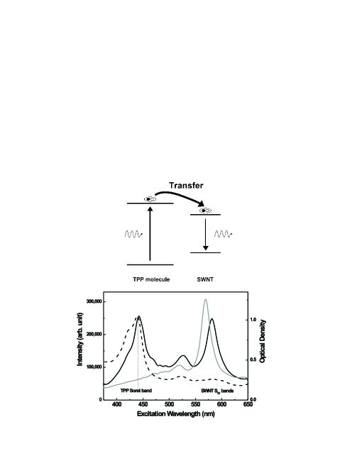
The PLE spectrum of the reference SWNT suspension detected at 984 nm (figure 5, grey curve) consists in a broad line near 560 nm. This couple of PL/PLE wavelengths or equivalently and energies allows us to identify a specific chiral family namely the (6,5) family bachilo02 .
The PLE spectrum of SWNT/TPP complexes (black curve) strongly contrasts with the one of the reference SWNT suspension. The PLE spectrum shows a strong additional line at 441 nm. This line corresponds to the absorption of stacked TPP (dashed curve). This PLE peak is the signature of the energy transfer from the photo-sensitized porphyrin to the nanotube casey2008 ; magadur2008 : when photons are absorbed by a stacked porphyrin, they give rise to photons emitted by the nanotube (as depicted in the upper panel of figure 5)). The line at 580 nm is clearly the line of the nanotubes. Interestingly, this line is red-shifted of about 10 nm as compared to the reference SWNT suspension. This result is similar to the shift observed for the lines. Therefore we conclude that this effect arises from a dielectric screening of excitons and not from a strain-induced band-shift (as observed for some surfactants Kiowski2007 ; Ohno2007 ) that would result in opposite shifts for and lines berger2009 .
Remarkably, the amplitudes of PLE lines corresponding to porphyrin Soret band and to S22 nanotubes transitions are of the same order of magnitude. This means that the number of photons emitted by the SWNTs is the same when the SWNTs are excited directly ( absorption) or through TPP absorption. We conclude that the energy transfer from the photo-sensitized porphyrin molecules to the nanotubes is very efficient. Nevertheless, further measurements are required in order to quantify precisely the transfer quantum yield.
In conclusion, we have developed a new method for "-stacking" functionalization of carbon nanotubes by organic molecules. An organic solvent is used to swell the micelle surrounding the nanotube and to bring the organic molecules onto the nanotube in the core of the micelle. This new method allows the production of controlled, reproducible and stable suspensions of SWNT/TPP complexes. These complexes show an efficient energy transfer from the photo-sensitized porphyrin molecules to the nanotubes confirming the potentialities of this system for light-harvesting applications. This physico-chemical functionalization method combines the advantages of "-stacking" functionalization together with a remarkable stability of the obtained complexes. This is a significant step towards scaling up a controlled process and designing functional nano-devices. Finally, this method is neither specific to nanotubes nor to porphyrins and can therefore be generalized to a wide range of nano-objects and dye molecules.
Method
Preparation of SWNT/TPP suspensions: The nanotubes used in this study are synthesized by the CoMoCAT process resasco1 and produced by SouthWest Nanotechnologies. The mean diameter of these tubes is about 0.8 nm. The nanotube suspensions are prepared by adding raw nanotubes at 0.15 mg.mL-1 in a pH 8 Normadose buffer (10-2 M, Prolabo) plus 2 wt% of sodium cholate (Sigma-Aldrich). The mixture is sonicated for 1h30 with an ultrasonic tip and ultracentrifuged at 120,000 g for 1h00. Then, the supernatant is drawn out. It consists in a suspension of isolated nanotubes oconnel02 .
Nanotubes functionalization is achieved by mixing the nanotubes suspension with a solution of porphyrin in dichloromethane (DCM). The porphyrin molecules used in this study are tetraphenylporphyrin (TPP) purified twice by column chromatography. After adding the TPP solution to the nanotubes suspension, the mixture is sonicated for 2 hours with an ultrasonic tip. The sample is placed in a thermostat at 12oC during sonication. The phase corresponding to DCM in excess is removed and the sample is centrifuged at 3,000 g for 10 minutes. The supernatant is drawn out and a suspension of SWNT/pophyrin complexes is obtained.
Spectroscopic measurements: Optical absorption spectra
are recorded with a spectrophotometer (lambda 900 Perkin-Elmer).
A laser diode emitting at 532 nm is used as excitation source for
photoluminescence experiments. The signal is dispersed in a
spectrograph (Spectrapro 2300i, Roper Scientific) and detected by
an IR CCD (OMA V, PI Acton). A UV-VIS Xe lamp and a monochromator
(Spectrapro 2150i, Roper Scientific) are used as tunable light
source for the photoluminescence excitation experiments.
TEM measurements: Transmission electron microscopy (TEM)
images and electron energy loss spectroscopy (EELS) are recorded
using a Libra200 transmission electron microscope at an
accelerating voltage of 200 kV. A three times diluted suspension
is used for preparing deposits on TEM grids. Then, the grid is
rinsed with water to remove the excess of sodium cholate (SC).
References
- (1) Nel, A.E. et al. Understanding biophysicochemical interactions at the nanobio interface. Nature Materials 8, 543 (2009).
- (2) Taraska, J.W., Puljung, M.C., Olivier, N.B., Flynn, G.E. & Zagotta, W.N. Mapping the structure and conformational movements of proteins with transition metal ion FRET. Nature Methods 6, 532 (2009).
- (3) Saccá, B., Meyer, R. & Niemeyer, C.M. Temperature-dependent FRET spectroscopy for the high-throughput analysis of self-assembled DNA nanostructures in real time. Nature Protocols 4, 271 (2009).
- (4) Hardin, B.E. et al. Increased light harvesting in dye-sensitized solar cells with energy relay dyes. Nature Photonics 3, 406 (2009).
- (5) Ehli, C., et al. Manipulating single-wall carbon nanotubes by chemical doping and charge transfer with perylene dyes. Nature Chemistry 1, 243 (2009).
- (6) Park, S.H. et al. Bulk heterojunction solar cells with internal quantum efficiency approaching 100%. Nature Photonics 3, 297 (2009).
- (7) Ross, R.B. et al. Endohedral fullerenes for organic photovoltaic devices. Nature Materials 8, 208 (2009).
- (8) Campidelli, S. et al. Facile decoration of functionalized single-wall carbon nanotubes with phthalocyanines "via click chemistry". J. Am. Chem. Soc. 130, 11503 (2008).
- (9) Ehli, C. et al. Interactions in single wall carbon nanotubes/pyrene/porphyrin nanohybrids. J. Am. Chem. Soc. 128, 11222 (2006).
- (10) Alvaro, M. et al. Synthesis, photochemistry, and electrochemistry of single-wall carbon nanotubes with pendent pyridyl groups and of their metal complexes with zinc porphyrin. comparison with pyridyl-bearing fullerenes. J. Am. Chem. Soc. 128, 6626 (2006).
- (11) Hasobe, T., Fukuzumi, S. & Kamat, P.V. Organized assemblies of single wall carbon nanotubes and porphyrin for photochemical solar cells: Charge injection from excited porphyrin into single-walled carbon nanotubes. J. Phys. Chem. B 110, 25477 (2006).
- (12) Aminur Rahman, G.M.et al. Improving photocurrent generation: Supramolecularly and covalently functionalized single-wall carbon nanotubes-polymer/porphyrin donor-acceptor nanohybrids. Chem. Eur. J 14, 8837 (2008).
- (13) Giordani, S. et al. Multifunctional hybrid materials composed of [60]fullerene-based functionalized-single-walled carbon nanotubes. Carbon 47, 578 (2009).
- (14) Magadur, G. et al. Excitation transfer in functionalized carbon nanotubes. ChemPhysChem 9, 1250 (2008).
- (15) Casey, J.P., Bachilo, S.M. & Weisman, R.B. Efficient photosensitized energy transfer and near-ir fluorescence from porphyrin-swnt complexes. Journal of Material Chemistry 18, 1510–1516 (2008).
- (16) Herranz, M.A., Martin, N., Campidelli, S., Prato, M., Brehm, G. & Guldi, D.M. Control over electron transfer in tetrathiafulvalene-modified single-walled carbon nanotubes. Angew. Chem. Int. Ed. 45, 4478 (2006).
- (17) Lambin, P., Loiseau, A., Monthioux, M., Thibault, J. Understanding Carbon Nanotubes (Springer, 2006).
- (18) Cambré, S.et al. Characterisation of nanohybrids of porphyrins with metallic and semiconducting carbon nanotubes by EPR and optical spectroscopy. ChemPhysChem 9, 1930 (2008).
- (19) As shown in the previous section, this amount of TPP is sufficient for a full functionalization of all the SWNTs.
- (20) Wang, R.K., Chen, WC., Campos, D.K. & Ziegler, K.J. Swelling the micelle core surrounding single-walled carbon nanotubes with water-immiscible organic solvents. J. Am. Chem. Soc. 130, 16330 (2009).
- (21) Bachilo, S.M., Strano, M.S., Kittrell, C., Hauge, R.H., Smalley, R.E. & Weisman, R.B. Structure-assigned optical spectra of single-walled carbon nanotubes. Science 298, 2361 (2002).
- (22) Kiowski, 0. et al. Photoluminescence microscopy of carbon nanotubes grown by chemical vapor deposition: Influence of external dielectric screening on optical transition energies. Phys. Rev. B 75, 075421 (2007).
- (23) Ohno, Y., Iwasaki, S., Murakami, Y., Kishimoto, S., Maruyama, S. & Mizutani, T. Excitonic transition energies in single-walled carbon nanotubes: Dependence on environmental dielectric constant. Phys. Status Solidi B 244, 4002 (2007).
- (24) Berger, S. et al. Optical properties of carbon nanotubes in a composite material: The role of dielectric screening and thermal expansion. J. App. Phys. 105, 094323 (2009).
- (25) Lolli, G., Zhang, L., Balzano, L., Sakulchaicharoen, N., Tan, Y. & Resasco, D.E. Tailoring (n,m) structure of single-walled carbon nanotubes by modifying reaction conditions and the nature of the support of como catalysts. J. Phys. Chem. B 110, 2108 (2006).
- (26) O’Connel, M.J. et al. Band gap fluorescence from individual single-walled carbon nanotubes. Science 297, 593 (2002).
Acknowledgement
The authors are grateful to D.E. Resasco for providing the CoMoCAT nanotubes produced by SouthWest Nanotechnologies. LPQM, PPSM and LPA are "Unités mixtes" de recherche associées au CNRS (UMR8537; UMR8531; UMR8551). This work was supported by the GDR-E "nanotube" (GDRE2756), grant "C’Nano IdF EPONAD" from "Région Ile de France" and ANR grant "CEDONA".