Epitaxial Growth of NdFeAsO Thin Films by Molecular Beam Epitaxy
Abstract
Epitaxial films of NdFeAsO were grown on GaAs substrates by molecular beam epitaxy (MBE). All elements including oxygen were supplied from solid sources using Knudsen cells. The x-ray diffraction pattern of the film prepared with the optimum growth condition showed no indication of impurity phases. Only (00) peaks were observed, indicating that NdFeAsO was grown with the -axis perpendicular to the substrate. The window of optimum growth condition was very narrow, but the NdFeAsO phase was grown with a very good reproducibility. Despite the absence of any appreciable secondary phase, the resistivity showed an increase with decreasing temperature.
The discovery of high temperature superconductivity in F-doped LaFeAsO Y. Kamihara et al. (2008) has ignited a great deal of excitement and an explosive flow of studies on iron-pnictides and related materials. The worldwide efforts led to the increase in the critical temperature by substituting other lanthanide elements for La, resulting in a thus far record high of 56 K X. H. Chen et al. (2008); Z.-A. Ren et al. (2008a); H. Kito et al. (2008); Z.-A. Ren et al. (2008b); C. Wang et al. (2008), and to the finding of other superconductors with related structures M. Rotter et al. (2008); J. H. Tapp et al. (2008); F.-C. Hsu et al. (2008). High-quality epitaxial films are indispensable both for exploring the intrinsic properties of these materials and for electronic device applications. Efforts of preparing thin films of the new superconductors started soon after their discovery H. Hiramatsu et al. (2008); E. Backen et al. (2008). Thin films with similar to the bulk values have already been reported for Co-doped Fe2As2 (=Sr, Ba) H. Hiramatsu et al. (2008); T. Katase et al. (2009) and iron-chalcogens Y. Han et al. (2009); M. J. Wang et al. (2009); Y. F. Nie et al. (2009); E. Bellingeri et al. (2009); P. Mele et al. (2009). However, the thin film preparation of the FeAsO (=lanthanide) family, which exhibits the highest to date among the iron-pnictide superconductors, seems to be more difficult. Hiramatsu et al. were the first to successfully prepare epitaxial films of LaFeAsO H. Hiramatsu et al. (2008), although no superconductivity was observed. Superconducting F-doped LaFeAsO thin films were reported from another group, but only after the films were post-annealed at significantly high temperatures E. Backen et al. (2008); S. Haindl et al. (2009). Obviously, there is a large room for improving the film quality of FeAsO.
All the above mentioned thin films were prepared by pulsed-laser deposition (PLD) on oxide crystalline substrates. Among the various thin film preparation techniques, molecular beam epitaxy (MBE) has been proven as a reliable growth method for the fabrication of high quality films of many different materials. The flux from each deposition source can be independently and precisely controlled, which is a large advantage for fine tuning the growth condition. Therefore, we employed the MBE method in this study, and report on the first successful growth of an epitaxial film of the FeAsO family by MBE.
NdFeAsO was chosen as the material to be grown because of its high potential in terms of . GaAs(001) were used as substrates. The lattice matching between GaAs and NdFeAsO is very good, because the lattice constant of NdFeAsO (0.3963 nm H. Kito et al. (2008)) multiplied by is close to the lattice constant of GaAs (0.5653 nm). First, an about 300 nm thick GaAs buffer layer was grown at 610∘C on the substrate. NdFeAsO was then grown at 670∘C by supplying all elements from solid sources charged in Knudsen cells; Fe, As, NdF3, and Fe2O3. Here, Fe2O3 was used as an oxygen source. A certain amount of flux was observed when the cell temperature of Fe2O3 was varied between 500 and 800∘C, although the vapor pressure of Fe is very low in this temperature range. In fact, the Fe content of the film did not change appreciably when only the temperature of the Fe2O3 cell was altered. Therefore, we think that Fe2O3 merely supplies oxygen without increasing the amount of Fe flux for the deposition conditions adopted in this study. This is reasonable because Fe2O3 is expected to reduce to Fe3O4 for the present experimental conditions from a thermodynamic consideration based on the Ellingham diagram Gaskell (2003).
The Nd and Fe contents of the prepared films were examined by electron probe micro-analysis (EPMA) using NdFeAs(O,F) powders as a reference, which were prepared by the method reported before . Takenaka et al. (2009). To determine the thickness, part of the film was removed by etching with hydrochloric acid (HCl), and the height of the step was measured using an atomic force microscope (AFM). The film thickness thus determined was about 30 nm for a film grown for 2 hours. Except for this film, all other films reported below were grown for 1 hour. The relation between the vapor pressure and the cell temperature was determined using an ion gauge beam flux monitor. Resistivity was measured by a four-probe method.
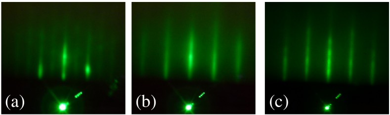
Figure 1 shows reflection high-energy electron diffraction (RHEED) patterns taken along the GaAs[110] azimuth during the film growth. A streaky pattern with a clear (24) structure was observed after the buffer layer was grown (Fig. 1(a)). The streaks elongated when the growth of NdFeAsO started (Fig. 1(b)), and were well maintained until the end of the growth (Fig. 1(c)). These results suggest that the film was grown with a flat surface, which is very important for device applications. The RHEED patterns in the [10] direction were very similar to that along [110] azimuth, implying that the NdFeAsO film was grown epitaxially on the GaAs buffer layer.
Since NdFeAsO consists of four elements, there were many parameters that had to be controlled to successfully grow an epitaxial film. We first changed the flux of Nd and Fe and adjusted their compositional ratio based on the EPMA data. After that, the vapor pressures of As and oxygen were changed. Figure 2 shows x-ray diffraction (XRD) profiles of films that were grown by changing only the oxygen flux. The vapor pressures of Fe, As and NdF3 were 1.910-6 Pa, 1.510-3 Pa, and 2.710-6 Pa, respectively. Only few peaks that can be assigned to FeAs were observed beside the substrate peaks for the film grown with the largest oxygen flux (Fig. 2(a)). Peaks that can be indexed as (00) reflections of NdFeAsO emerged when the oxygen flux was reduced, but NdAs was observed as the secondary phase when the oxygen flux was too small (Fig. 2(c)). Only within a small window, a single-phased NdFeAsO film was obtained as shown in Fig. 2(b).

A similar experiment was conducted for the vapor pressure dependence of As as well. Figures 3(a) and (b) show the XRD profiles of films that were grown with an As vapor pressure larger and smaller than that of the best film (Fig. 2(b)), respectively. All the other conditions were the same as that of the film of Fig. 2(b). When As was oversupplied, NdAs was observed as an impurity phase (Fig. 3(a)). On the other hand, almost no crystalline phase was observed for the film grown with the lowest As vapor pressure (Fig. 3(b)), similarly to the film for which oxygen was oversupplied (Fig. 2(a)).

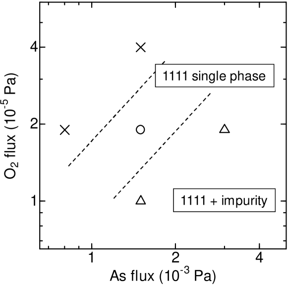
Figure 4 summarizes the results of the XRD observations. Interestingly, increasing oxygen flux had an effect similar to reducing As flux. This is probably because arsenic oxides, which have high vapor pressures, are formed when the oxygen flux is large. The growth window for a single-phased NdFeAsO film is very narrow, as is obvious from the diagram. Nevertheless, NdFeAsO was always the dominant phase when the growth condition was close to the optimum one. This includes films that were grown with different vapor pressures of Fe and/or NdF3 (and therefore which cannot be plotted on Fig. 4). In other words, NdFeAsO was grown with a very good reproducibility, in contrast to the report on PLD preparation of LaFeAsO films H. Hiramatsu et al. (2008).
The -axis lattice constant calculated from the peaks labeled as 002, 004, and 005 of the optimized film shown in Fig. 2(b) was 0.8570.002 nm, which is close to the value reported for bulk samples H. Kito et al. (2008). On the other hand, the film thickness was estimated to be 15 nm as mentioned above. This implies that the growth rate is very low, but is consistent to the fact that growth of only small single crystals has been reported for FeAsO so far J. Karpinski et al. (2009).

Figure 5 shows the temperature dependence of resistivity of the single-phased film of Fig. 2(b). While a metallic temperature dependence with a drop at about 150 K due to a structural transition is reported for bulk samples Y. Kamihara et al. (2008), the resistivity of our film increased with temperature decreasing. Such semiconductor-like behaviors were observed for all the so-far prepared films for which NdFeAsO was the dominant phase (e.g. Fig. 2(c) and Fig. 3(a)). It is also similar to the temperature dependence of the as-grown films prepared by PLD H. Hiramatsu et al. (2008). The reason of the semiconductor-like behavior despite the absence of any obvious secondary phase is not clear but several possibilities may be pointed out. For instance, the film might be structurally deteriorated because the thickness is only 15 nm, the composition might be slightly deviated from the ideal one and the carrier content is not appropriate, or Ga might be diffused from the GaAs substrate because the growth temperature was rather high. A further optimization of the growth condition is highly desired.
In conclusion, we have grown NdFeAsO films epitaxially on GaAs substrates. This is the first report of a successful growth of FeAsO films by MBE. All elements including oxygen were supplied from solid sources. For the growth of the NdFeAsO phase, it was important to precisely adjust the flux of the constituting elements, particularly the balance between oxygen and As. Even a small deviation from the optimum condition resulted in a formation of impurities, or in the worse case, absence of the NdFeAsO phase. Despite the narrow window of optimum growth condition, a very good reproducibility was obtained. The RHEED observation suggests that the growth proceeded with maintaining a flat surface, which is favorable for device applications.
This work was supported by Transformative Research Project on Iron Pnictides (TRIP), JST.
References
- Y. Kamihara et al. (2008) Y. Kamihara et al., J. Am. Chem. Soc. 130, 3296 (2008).
- X. H. Chen et al. (2008) X. H. Chen et al., Nature 453, 761 (2008).
- Z.-A. Ren et al. (2008a) Z.-A. Ren et al., Europhys. Lett. 83, 17002 (2008a).
- H. Kito et al. (2008) H. Kito et al., J. Phys. Soc. Jpn. 77, 063707 (2008).
- Z.-A. Ren et al. (2008b) Z.-A. Ren et al., Chin. Phys. Lett. 25, 2215 (2008b).
- C. Wang et al. (2008) C. Wang et al., Europhys. Lett. 83, 67006 (2008).
- M. Rotter et al. (2008) M. Rotter et al., Phys. Rev. Lett. 101, 107006 (2008).
- J. H. Tapp et al. (2008) J. H. Tapp et al., Phys. Rev. B 78, 060505 (2008).
- F.-C. Hsu et al. (2008) F.-C. Hsu et al., Proc. Natl. Acad. Sci. USA. 105, 14262 (2008).
- H. Hiramatsu et al. (2008) H. Hiramatsu et al., Appl. Phys. Lett. 93, 162504 (2008).
- E. Backen et al. (2008) E. Backen et al., Supercond. Sci. Technol. 21, 122001 (2008).
- H. Hiramatsu et al. (2008) H. Hiramatsu et al., Appl. Phys. Express 1, 101702 (2008).
- T. Katase et al. (2009) T. Katase et al., arXiv:0907.0666.
- Y. Han et al. (2009) Y. Han et al., J. Phys.: Condens. Matter 21, 235702 (2009).
- M. J. Wang et al. (2009) M. J. Wang et al., arXiv.org:0904.1858.
- Y. F. Nie et al. (2009) Y. F. Nie et al., Appl. Phys. Lett. 94, 242505 (2009).
- E. Bellingeri et al. (2009) E. Bellingeri et al., arXiv:0906.3221.
- P. Mele et al. (2009) P. Mele et al., Appl. Phys. Express 2, 073002 (2009).
- S. Haindl et al. (2009) S. Haindl et al., arXiv:0907.2271.
- Gaskell (2003) D. R. Gaskell, Introduction to the thermodynamics of materials, 4th ed. (Taylor & Francis, New York, 2003).
- . Takenaka et al. (2009) K. Takenaka et al., J. Phys. Soc. Jpn. 78, 073701 (2009).
- J. Karpinski et al. (2009) J. Karpinski et al., Physica C 469, 370 (2009).