Patch coalescence as a mechanism for eukaryotic directional sensing
Abstract
Eukaryotic cells possess a sensible chemical compass allowing them to orient toward sources of soluble chemicals. The extracellular chemical signal triggers separation of the cell membrane into two domains populated by different phospholipid molecules and oriented along the signal anisotropy. We propose a theory of this polarization process, which is articulated into subsequent stages of germ nucleation, patch coarsening and merging into a single domain. We find that the polarization time, , depends on the anisotropy degree through the power law , and that in a cell of radius there should exist a threshold value for the smallest detectable anisotropy.
pacs:
64.60.My, 64.60.Qb, 87.16.Xa, 87.17.Jj, 82.39.Rt, 82.40.NpThe cells of multicellular organisms are endowed with a chemical compass of amazing sensitivity, formed as a result of billion years of evolution. Concentration differences of the order of a few percent in the extracellular soluble attractant chemicals from side to side are sufficient to induce a chemical polarization of the membrane leading to cell migration towards the signal source Song et al. (2006). This way, a sensible amplifier of slight gradients in the distribution of chemicals in the surrounding environment is realized. Its relevance is easily understood if one recognizes that no multicellular organism could exist without the constituent cells being able of sensing directional signals. Directional sensing is actually essential both in embryo development, where tissue formation is realized through coordinated migration of specific cells guided by chemical signals, and in the adult organism, where chemical signals guide white blood cells to the sites of inflammation and platelets to sites of wound repair. The main steps of the process are as follows (see the reviews Ridley et al. (2003) and Lauffenburger and Horwitz (1996)). As a response to the attractant signal, the cell membrane is polarized, afterwards inducing differentiated polymerization of the cell cytoskeleton in its proximity. The resulting unbalance, triggered by a well characterized cascade of chemical reactions, leads to the formation of a growing head and a retracting tail, in such a way that the cell starts to drift towards the source of the signal. The initial part of this process is constituted by the early chemical polarization of the cell membrane. In this letter we propose a simple phenomenological scheme providing a universal description of this fundamental phenomenon.
Membrane polarization can be recognized as a self-organization process governed by a network of diffusion-controlled chemical reactions. It is known that reaction-diffusion networks may become bistable in the presence of chemical feedback loops Aurell and Sneppen (2002); Angeli et al. (2004). In spatially extended systems bistability may lead to the formation of competing phases and to a phenomenology typical of first order phase transitions, such as metastability, nucleation and coarsening Cross and Hohenberg (1993); Bray (1995). The polarized membrane state observed during directional sensing can therefore be interpreted as the coexistence of domains of two different phases.
Let us briefly describe the chemical reactions which are responsible for directional sensing. The chemical factors clustering in complementary membrane domains are the phospholipids PIP2 and PIP3. Two enzymes, PI3K and PTEN, respectively transform PIP2 into PIP3 and viceversa. The phospholipids are permanently bound to the inner face of the cell membrane, while PI3K and PTEN diffuse in the cell volume and are active only when they are adsorbed by the membrane. PI3K adsorption takes place through binding to receptors activated by the extracellular attractant signal. This way, the external attractant field is coupled to the inner dynamic of the cell. PTEN adsorption takes place through binding to the PTEN product, PIP2. This process introduces a positive feedback loop in the system dynamics Iijima and Devreotes (2002); Gamba et al. (2005). When the cell is not stimulated by an attractant signal the cell membrane is uniformly populated by PTEN and PIP2 molecules. When a uniform receptor stimulation of a suitable amplitude is switched on, PI3K molecules bind to the membrane and shift its chemical balance toward a PIP3-rich phase, while PTEN desorbs. PIP3-rich germs are then nucleated in the PIP2-rich sea and PIP3-rich regions start to coexist with PIP2-rich ones Postma et al. (2004).
Two different regimes of cell polarization may be distinguished. Anisotropy driven polarization induced by the presence of an attractant gradient is realized in a time of the order of a few minutes, and results in the formation of a PIP3-rich domain on the membrane side closer to the attractant source and of a PIP2-rich domain in the complementary region Iijima and Devreotes (2002); Gamba et al. (2005). On the other hand, cells exposed to uniform distributions of an attractant polarize in random directions, in times of the order of an hour (see e.g. Shields and Haston (1985)). The existence of two clearly separated polarization regimes is confirmed by the recent observation of a sensitivity threshold of the order of a few percent difference in the attractant molecule concentration from side to side Song et al. (2006). Direct observation of the polarization process (Iijima and Devreotes, 2002, Fig. 7a) implies the bound for PTEN diffusion time in the cell volume, which is therefore much less than the polarization time for both regimes. In this process, the amplitude of the cell stimulation is of crucial importance. At very low stimulation levels PTEN is not desorbed in a significant amount and no directional sensing takes place. At very high stimulation levels a homogeneous PIP3-rich phase is realized and directional sensing again does not take place. There exists therefore an optimal attractant concentration, such that below it the minority phase is PIP3-rich and above it is PIP2-rich.
Numerical simulations of the directional sensing network performed with the use of realistic physical and kinetic parameters have shown that under appropriate conditions the biochemical network is indeed bistable, and that it undergoes spontaneous separation in chemically different phases, rich in PIP2 and PIP3, respectively Gamba et al. (2005); de Candia et al. (2007). A 5% anisotropic component in the cell stimulation accelerates cell polarization and correspondingly decreases the characteristic time needed for complete phase separation by more than one order of magnitude: fast, anisotropy driven polarization is realized in times of the order of a minute, while slow, stochastic polarization is realized in times of the order of one hour, in accordance with experimentally observed times. In the numerical experiments, when PIP2 is the minority phase, the evolution leading to phase separation consists of an early nucleation regime, resulting in the formation of isolated PIP2-rich patches and a late coarsening process, where large patches of the PIP2-rich phase grow at the expense of the evaporation of smaller ones, similarly to what happens in the case of first order phase transitions in a liquid-gas system or in the precipitation of a supersaturated solution Lifshitz and Pitaevskii (1981). Finally, the patches condense into a single large cluster, leading to a stationary state characterized by the coexistence of a PIP2- and a PIP3-rich domain. However, the dynamics of the directional sensing network differs from that of otherwise similar processes, such as the precipitation of a supersaturated solution, under one important respect. When precipitation nuclei in a supersaturated solution dissolve, matter is transferred to larger nuclei through diffusion in the surrounding medium. In contrast, in the directional sensing network enzyme-substrate patches evaporate through desorption of the PTEN enzyme from the membrane, which is then transferred to other patches through diffusion in the cell volume. Therefore the transformation of PIP3 into PIP2 molecules cannot be described at the membrane level as a local, diffusion-like process as is the case with the adsorption-desorption process from precipitation nuclei; in particular, there is no local conservation of the number of PIP2 molecules.
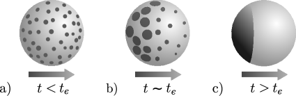
The above summarized scenario can be put on a firm analytical ground resorting to the kinetic theory of first order phase transitions Lifshitz and Slyozov (1961); Lifshitz and Pitaevskii (1981); Bray (1995). In this theory, after germ nucleation larger patches of the stable phase grow at the expense of smaller patches which shrink, leading to scaling laws and universal probability distribution of patch sizes. We shall now show how the ideas of the Lifshitz-Slyozov theory Lifshitz and Slyozov (1961) may be adapted to our problem to deduce simple scaling laws for the membrane polarization time and explain most of the observed phenomenology. We discuss here the case when PIP2 is the minority phase (the other case being symmetric). In this case PIP2-rich patches are formed inside the PIP3-rich sea (see Fig. 1a). We restrict our consideration to approximately circular patches of the PIP2-rich phase, which are expected to dominate over different geometries due to the presence of a linear tension between the two phases. The free energy of a PIP2-rich patch of radius can be written on phenomenological grounds as , where is the linear tension of the interface with the surrounding PIP3-rich phase and represents the degree of metastability Lifshitz and Pitaevskii (1981), which is a function of the concentration of PTEN molecules in the cell volume and of the concentration of extracellular attractant.
According to the kinetic theory of first order phase transitions, the equation of growth of a patch is dissipative. In the absence of a local conservation law the equation for a circular patch can be written as , where is a damping coefficient Bray (1995). Since energy dissipation occurs mainly along the perimeter of the interface between the two phases, may be written as , where is a constant, and we get
| (1) |
where the noise term represents thermal fluctuations. The fluctuations are responsible for the formation of an initial population of patches with varying radii (Lifshitz and Pitaevskii, 1981, §99). Patches with smaller than the critical radius are mainly dissolved while most patches with survive and grow because of the gain in free energy. At initial time, is of the order of the thickness of the interface between the two phases Lifshitz and Pitaevskii (1981); Bray (1995). As long as the area occupied by patches of the PIP2-rich phase grows, the degree of metastability decreases, some of the patches that were initially growing become undercritical and shrink, large patches start “feeding” on smaller ones, and the total number of patches diminishes (Lifshitz and Pitaevskii, 1981, §100). In the final stage of this process a single domain of the PIP2-rich phase is formed coexisting with the PIP3-rich phase, see Fig. 1c. However, the details of the process leading to this final stage depend on the external conditions, and, particularly, on the degree of anisotropy of the attractant signal.
The population of patches can be described in terms of the size distribution function such that gives the number of patches with sizes in the interval . An important simplification comes from the fact that for patches with the noise term in (1) becomes negligible Lifshitz and Pitaevskii (1981). This means that the stochastic nature of the problem enters mainly in the formation of the initial distribution of patch sizes , while for the time evolution of is dictated by the deterministic part of (1), from which the kinetic equation
| (2) |
follows Lifshitz and Pitaevskii (1981); Bray (1995). With the chosen normalization represents the total number of patches, a quantity which is monotonically decreasing in time according to the previously described “coarsening” dynamics Lifshitz and Slyozov (1961). Eq. (2) is valid as long as is much larger than Lifshitz and Pitaevskii (1981).
To obtain a closed system of equations we need an additional equation for the time evolution of the metastability degree Lifshitz and Pitaevskii (1981). In the case of isotropic stimulation does not depend on the position on the membrane and is instead only a function of time. Since diffusion of PTEN molecules in the cell volume is faster than phospholipid diffusion on the membrane we can regard the concentration of PTEN molecules in the volume as uniform Gamba et al. (2005). Moreover, fast PTEN diffusion also implies that instantaneously adjusts to the changes in the size distribution function. While the total number of patches diminishes as an effect of the coarsening dynamics, the total area occupied by the patches, as well as the total number of PIP2 molecules found in the patches, monotonically increases towards their respective equilibrium values. The metastability degree is equal to zero in equilibrium, and tends to zero in accordance with
| (3) |
as the total patch area tends to its limit value . Eq. (3) reflects the fact that in the asymptotic region is proportional to the excess concentration of PTEN molecules in the volume with respect to the equilibrium value, and therefore to the difference between the area occupied by the PIP2-rich phase at equilibrium and at current time. The law (3) is valid for , where is the characteristic time needed for the formation of a germ of an alternative phase that can be estimated as Lifshitz and Pitaevskii (1981); Bray (1995).
Asymptotically, (2,3) lead to the self-similar solution
| (4) |
where if and if . Similarly to what happens in Lifshitz-Slyozov theory Lifshitz and Slyozov (1961) the total number of patches decreases in time due to the evaporation of small patches: , and from (4) one gets . The evolution of the size distribution function governed by (4) stops at times of order , where is defined as the instant when the average patch size reaches the cell size . From the scaling law we get . Eventually, at , a single PIP2-rich patch survives. Its orientation is determined by the random unbalance in the initial germ distribution. Notice that in this derivation, following the lines of Lifshitz and Slyozov (1961); Lifshitz and Pitaevskii (1981); Bray (1995), isotropy was essential to assume that was uniform along the whole membrane surface.
Let us now consider the case of an inhomogeneous activation pattern. The inhomogeneity of the concentration distribution modifies the degree of metastability, which becomes a function of the position on the membrane surface. Since the distribution of PTEN molecules in the cell volume is homogeneous, it influences only the isotropic part of the metastability degree , which is a function of time, as previously. In contrast, the anisotropic part of the metastability degree, , related to the external attractant inhomogeneity, does not depend on time. If the cell membrane has a nearly spherical form and a radius much smaller than the characteristic scale of the extracellular attractant distribution, then . Here is the initial metastability degree, is a dimensionless factor measuring the initial anisotropy degree, and is the azimuthal angle on the cell surface. This way we obtain the equation
| (5) |
generalizing (1). As long as , the first stage of patch growth proceeds approximately as in the isotropic case and decreases as . However, at a time of order , where is defined by the equation , the perturbation becomes comparable to and the process of polarization becomes anisotropic, so that patches in different regions get different average sizes, see Fig. 1b. From the scaling law (4) for one gets . For , the leading term in (5) becomes the perturbation , implying that in the region closer to the source of the stimulation () the PIP2-rich phase evaporates in a time which is easily estimated as being again of order , leading to the formation of a single PIP2-rich patch in the region further from the source of the stimulation () and realizing complete polarization, as shown in Fig. 1c.
The above scheme is valid as soon as the initial nucleation time is significantly smaller than , an assumption which is compatible with the results of numerical experiments Gamba et al. (2005). On the other hand, the second stage of patch evolution occurs only if . Otherwise, the presence of a gradient of attractant becomes irrelevant and only the stage of isotropic patch growth actually occurs. This condition implies that a smallest detectable gradient exists, such that directional sensing is impossible below it. The threshold value for is found by letting . Since the product is a time-independent constant, we can simply compare its value at initial and final time when , obtaining , which gives us the expression for the threshold anisotropy.
It is interesting to estimate , and, consequently, , in terms of observable parameters. Comparing the characteristic patch surface and perimeter energy as a function of the phospholipid diffusion coefficient , surface phospholipid concentration , surface concentration of activated receptors , and the characteristic catalytic time , one gets . Using parameter values from Ref. Gamba et al. (2005) one gets and . The value for is compatible with the observations (the data from Ref. Song et al. (2006) imply for dictyostelium).
One may wonder whether a cell may become polarized by the anisotropy produced by a spontaneous fluctuation in the extracellular distribution of attractant molecules or fluctuations in receptor-ligand binding Lauffenburger and Horwitz (1996). Since eukaryotic cells typically carry - receptors for attractant factors, one expects spontaneous fluctuations in the fraction of activated receptors to be of the order of , a value which is comparable to observed anisotropy thresholds. However, to actually produce directed polarization the fluctuation should sustain itself for several minutes, i.e. for a time comparable to the characteristic polarization time. Such an event has very low probability of being observed since the correlation time of the fluctuations determined by attractant diffusion at the cell scale and the characteristic times of receptor-ligand kinetics are much less than the polarization time. Indeed, the diffusion time is at the typical cell size , and the characteristic times of receptor-ligand kinetics are also (see online supporting information to Ref. Song et al. (2006)). Therefore, the direction of cell polarization in the case of a homogeneous distribution of attractant can only be determined by the inhomogeneity in the initial distribution of the positions of PIP2-rich germs produced by thermal fluctuations.
In conclusion, we have constructed a universal phenomenological description of the mechanism of directional sensing in the eukaryotes based on the process of patch coarsening. This description implies the existence of two clearly separated polarization regimes depending on the presence or absence of an anisotropic component in the activation pattern produced by the extracellular attractant factor, and the existence of a sensitivity threshold for the anisotropic component. Both results are in reasonable agreement with experimental observations. Moreover, we predict that directed polarization time should scale as the inverse square of the relative signal anisotropy, a law that should be verifiable by direct observation. Our picture suggests that directed and stochastic polarization share a common mechanism, and that stochastic polarization should be the result of noise in subcellular and not in extracellular dynamics. Importantly, our picture does not depend on the details of the reactions involved, but only on the general structure of the directional sensing network and on its bistability. This means that the picture is robust not only with respect to variations of the kinetic and physical parameters, but also with respect to the identity of the chemical species involved. Indeed, PI3K and PTEN could be substituted by, or synergize with, molecules endowed with similar enzymatic activity. An interesting speculation is that the bound may explain why spatial directional sensing was developed only in the large eukaryotic cells and not in smaller prokaryotes, whose directional sensing mechanisms rely instead on the measurement of temporal variations in concentration gradients Alon et al. (1999). Our bound derives from the intrinsic properties of polarization dynamics and is independent of the size criterion formulated in Ref. Berg and Purcell (1977). The experimental observation of selforganized phospholipid patches Postma et al. (2004) following uniform attractant stimulation provides an initial confirmation of the validity of our scheme. To check the predictions of our theory, similar observations should be performed for the longer times characteristic of random and directed polarization, both under uniform attractant activation and in the presence of accurately controlled concentration gradients. Experimental modulation of PTEN levels could be used to modify the overall size of patches and eventually switch off the patch formation mechanism.
Acknowledgements.
A.G. likes to thank Guido Serini and Stefano Di Talia for many helpful discussions. I.K. and V.L. acknowledge partial support of RFBR grant 06-02-17408-a.References
- (1)
- Song et al. (2006) L. Song et al., Eur. J. Cell Biol. 85, 981 (2006).
- Ridley et al. (2003) A. Ridley et al., Science 302, 1704 (2003).
- Lauffenburger and Horwitz (1996) D. Lauffenburger and A. Horwitz, Cell 84, 359 (1996).
- Aurell and Sneppen (2002) E. Aurell and K. Sneppen, Phys. Rev. Lett. 88, 048101 (2002).
- Angeli et al. (2004) D. Angeli, J. Ferrell, and E. Sontag, Proc. Nat. Acad. Sci. U.S.A. 101, 1822 (2004).
- Cross and Hohenberg (1993) M. Cross and P. Hohenberg, Rev. Mod. Phys. 65, 851 (1993).
- Bray (1995) A. Bray, Adv. Phys. 43, 357 (1995).
- Iijima and Devreotes (2002) M. Iijima and P. Devreotes, Cell 109, 599 (2002).
- Gamba et al. (2005) A. Gamba et al., P.N.A.S. 102, 16927 (2005).
- Postma et al. (2004) M. Postma et al., J. Cell Science 117, 2925-35 (2004).
- Shields and Haston (1985) J. Shields and W. Haston, J. Cell Sci. 74, 75 (1985).
- de Candia et al. (2007) A. de Candia et al., Sci. STKE 378, pl1 (2007).
- Lifshitz and Pitaevskii (1981) E. Lifshitz and L. Pitaevskii, Physical Kinetics (Butterworth-Heinemann, 1981).
- Lifshitz and Slyozov (1961) I. Lifshitz and V. Slyozov, ZhETF 35, 479 (1958); J. Phys. Chem. Solids 19, 35 (1961).
- Alon et al. (1999) U. Alon et al., Nature 397, 168 (1999).
- Berg and Purcell (1977) H. Berg and E. Purcell, Biophys. J. 20, 193 (1977).