Sharp optical phonon softening close to optimal doping in La2-xBaxCuO4+δ
Abstract
We report a direct observation of a sharp Kohn-like anomaly in the doubly degenerate copper-oxygen bond-stretching phonon mode occurring at in La2-xBaxCuO4+δ with , thanks to the high resolution of inelastic x-ray scattering. This anomaly is clearly seen when the inelastic signal is analysed using a single mode but is also consistent with a two mode hypothesis possibly due to a splitting of the degenerate modes due to symmetry breaking stripes. Our observation shows that the effect persists at the stripe propagation vector in a superconducting system close to optimal doping.
pacs:
74.25.Kc, 74.72.Dn, 63.20.Dj, 63.20.Kr, 78.70.CkI Introduction
Copper-oxygen bond stretching modes in high-temperature superconducting cuprate (HTCS) have long been known to show anomalies related to dopingPintschovius and Reichardt (1998). The origin of the anomalies is still an open issue. Along with electron-phonon coupling effects, a mechanism involving the formation of an inhomogeneous charge state, also known as stripesZaanen and Gunnarsson (1989); Machida (1989); Kato et al. (1990); Kivelson et al. (1998, 2003) has been discussed McQueeney et al. (1999); Pintschovius and Braden (1999). At the outset let us clarify the anomalies seen in this phonon mode. Earlier worksPintschovius and Reichardt (1998) focused on the gradual softening of this mode with doping, a phenomenon seen quite universally in HTCS and now relatively well understood, even theoretically Giustino et al. (2008) on the basis of electron-phonon coupling and screening mechanisms which come into play on doping. This softening can be well described by a cosine-like behaviour of the mode with its minimum at the zone boundary. Even in the earliest works, however, there were indications that the dispersion might be more complicated, with deviations from the cosine-like shape McQueeney et al. (1999). The possibility that these deviations are caused by stripes has been invoked and adopted in a recent paper Reznik et al. (2006) in which previous data on HTCS, are compared to newer ones, in particular for with x=1/8. In this system, for x1/8, an anomalous suppression of the superconductivity reported in Ref. Moodenbaugh et al., 1988 was later shown to be associated with charge and spin stripe orderFujita et al. (2004); Abbamonte et al. (2005); Kim et al. (2008). Signatures of stripes in superconducting samples remain elusive, though it was proposed that stripes are difficult to detect in these because they are no longer staticVojta et al. (2006); Hinkov et al. (2007). Using phonons to probe charge fluctuations can poten- tially lead to a reliable signature of static and dynamic stripes in HTCS. The authors of Ref. Reznik et al., 2006 in describing their high resolution inelastic neutron scattering data of the copper-oxygen bond stretching mode, show how one of its two normally degenerate components follows the expected cosine-like dispersion while the other deviates presenting a much sharper dip. They interpret this behaviour as a Kohn-like anomaly due to stripes which lift the degeneracy. 111The “classical” Kohn-anomaly is due to electron phonon coupling and in its strongest form can result in a charge density wave. The wave vector related to this effect is given by a nesting vector of the Fermi surfaceGrüner (1994). In the case of a stripe related anomaly, the difference is that stripes are supposed to originate from strong on-site coulomb repulsionKivelson et al. (1998) and are not linked to the Fermi surface topology. This scenario is very intriguing, nevertheless, the dip is not directly visible in the data of Ref. Reznik et al., 2006, and the authors deduce its existence from a broad shoulder on the low energy side of the Cu-O bond stretching phonon mode. Thus, as we show in a recent paper Graf et al. (2007), it is desirable to have a direct measurement of the dip since without it, other reasons could be invoked to explain the observed signal. To clarify this point we have carried out a high -resolution measurement in using Inelastic X-ray Scattering (IXS), with a comparable energy resolution, but with a higher resolution in reciprocal space compared to previous inelastic neutron scattering experiments.
This allowed us not only to confirm the presence of a dip around in the dispersion of the copper-oxygen bond stretching mode, but also to extend this observation to superconducting doping value (x = 0.14). This doping level leads to a stripe-propagation-vector Kivelson et al. (2003), in striking agreement with our findings, and supports a picture based on the interaction with stripes McQueeney et al. (1999); Reznik et al. (2006).
II Experiment
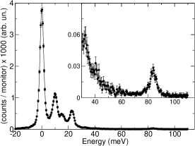
We studied a single crystal of grown from the melt in an image furnace by the traveling solvent zone method, under a pressure of 3 bar oxygen. The measured = 18.1 K with = 7 K defined as the temperature range from to of the maximum Meissner signal, is consistent with micro-probe results indicating a content of Axe et al. (1989). The experiment was carried out at the IXS beamline II (ID28) at the European Synchrotron Radiation Facility in Grenoble. The sample was mounted in reflection geometry, on the cold finger of a closed-loop helium cryostat, in order to kept it at constant temperature during the experiment. The low temperature was chosen so as to optimize the signal of the high energy modes over the tails of low energy ones. The sample crystal axe was perpendicular to the scattering plane. We consider the tetragonal unit cell with = = = 90∘ and the axes and along the Cu-O bond. The sample was aligned along (H,0,0), and we refined the parameter =3.792 0.001 Å, according to the scan on the (4, 0, 0) reflection, and adopted the value of = 13.235 Å. The rocking curve at the (4,0,0) reflection had a FWHM , indicating a very low mosaic spread in the small volume probed. The IXS multi-analyzer spectrometerMasciovecchio et al. (1996); Krisch and Sette (2007) configurations chosen were of type , with simultaneous measurements from 7 analyzers. We collected data from the first 5 analyzers closer to the beam direction, which are at fixed angular spacing of ( 0.07), with the third one in longitudinal condition = 0 while for the others 0.045. This setup corresponds to the 3rd extended Brillouin Zone, where the zone center is at (4,0,0) and the extended zone boundary at (3,0,0). The standard or folded zone boundary is at the point M=(3.5, 0, 0). The scattering vector resolution was set using a slit opening in front of the analyzers of h v = 20 60 mm corresponding to a solid angle of = 0.19∘ 0.57∘. The resolution in the reciprocal space for 0.6968 Å wave-length was (, 0, 0) with 0.009. The high energy resolution is obtained using the back-scattering silicon monochromator aligned along the (1 1 1) directionVerbeni et al. (1996, 2008). In the present experiment we choose to work with the Si (9 9 9) reflection order, with a wave-length of 0.6968 Å(17794 eV) and an energy resolution E = 3.0 0.2 meV. An example of a raw IXS spectrum is given in Fig. 1. The energy scans were fitted using a sum of pseudo-Voigt for elastic and resolution-limited inelastic contributions, with parameters fixed to match the instrumental function, while a Lorentzian line-shape was used to fit modes with an intrinsic width larger than the instrumental resolution. In order to assign the measured phonon modes, we performed a lattice dynamical calculation using a computer code Mirone (2001) based on a shell modelChaplot et al. (1995). More details on analysis and simulation are described elsewhere Graf et al. (2007).
III Results and discussion
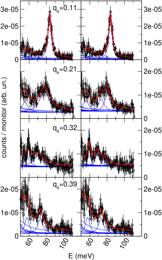
The high energy portion of some typical measurements is shown in Fig. 2. In this spectral window, at least two modes could be identified: a mode at about 60 meV, corresponding to the 5th longitudinal optic mode (in order from the lowest energy), and a second dispersing from about 90 meV down to about 70 meV, the highest (6th) energy longitudinal Cu-O bond-stretching (or half breathing) optic mode. The fast downward dispersion is accompanied by a rapid broadening from 5 meV to about 15 meV FWHM. This mode is doubly degenerate, and could eventually split if the local symmetry is lowered by some anisotropy. Such splitting has been suggested Reznik et al. (2006) for a q 0.2 - 0.3, corresponding to the propagation vector of a charge modulation (“stripe”). Other possible origins of the double modes have been previously reportedMcQueeney et al. (1999); Tranquada et al. (2002) always in connection with microscopic phase-separation. The doubling of the modes can contribute to the observed broadening if the energy resolution is not enough to distinguish the two modes as in our case. In most of the measured spectra it is then not possible to have independent fitted parameters for frequency, intensity and width of each mode. We have used the error estimation of the fitted widths as a goodness of fit criterion and only in the case of qx = 0.32 (see Fig. 2), the two mode fit appears to be better than the single mode fit for our data.
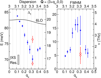
In Fig. 3 we show the results of our fitting for the frequencies (left panel) and width (right panel) for the single mode (blue circles). A minimum at qx = 0.32 0.04 is clearly observed. In the same figure, we also show the results for the fit in a double mode hypothesis (red squares), for qx = 0.32.
The difficulty of the analysis (the impossibility of reliably fitting two closely placed peaks) is inherent to the experimental strategy chosen: in order to follow in detail the dispersion we chose a higher -resolution with comparable energy resolution to Ref. Reznik et al., 2006. This also results in a significantly different line-shape. In Ref. Reznik et al., 2006, using inelastic neutron scattering, the highest mode appears more intense because it is weakly dispersing, and thus benefits from a large integration, while the low energy one appears broader because the supposed Kohn-like dip in the dispersion is only as large as the experimental -resolution. Assuming that the two-mode hypothesis is valid let us examine the consequences for our data. A -resolution narrower than the dip width results in a sharper low energy mode. We have already seen that the intensity of the high energy mode decreases due to the better -resolution. This results in two modes of similar intensity and width, more difficult to separate with this energy resolution. Returning to the results shown in Fig. 3, in a one mode hypothesis, we obtain a maximum of the width around q 0.3, coinciding with the dispersion minimum. Indeed, even a one peak fit gives a well pronounced anomalous dip that is inconsistent with a cosine-like dispersion. The dip is observed even with a one peak fit because the dispersion of the concerned mode and its intensity are strong enough and dominate even after averaging with the less dispersing higher energy mode. The maximum in the width is, in fact, a measure of the energy distance between the modes which is associated with the dip, as already pointed out (see Fig. 4 in Ref. Reznik et al., 2006). Note that we extended the observation of this anomaly to a doping range which is well beyond the singular 1/8th level and crucially in a superconducting range where the stripes should be dynamic. Consistent with this higher doping level we observe that the dip is more spread out over the BZ, already starting at q 0.1 with a FWHM of meV and that the softening is reduced even in the two mode hypothesis. These observations are thus consistent with a persisting anomaly at the stripe propagation wave-vector in a more metallic system, where both the doubling and the shift of the low energy mode appear to be reduced. Previous results obtained by IXS Fukuda et al. (2005) on for different doping level are interpreted in the standard cosine-like dispersion picture, but corresponds to a set-up with only 6 meV resolution, about twice the present one. A review of this anomaly in other HTCS is reported in Ref. Reznik et al., 2006. Finally, we note here that a possible link has recently been suggested in single layer Bi2Sr1.6La0.4Cu2O6+δ Graf et al. (2008) between the maximum of the Cu-O bond stretching softening and width, and a Fermi surface nesting vector at 0.22, with a clear kink in the electron dispersion at the energy of the phonon mode. The vector is close to the stripe propagation vector in double layer Bi2-xSr2CaCu2O8+δ reported by STM in Ref. Hoffman et al., 2002 and very recently in single layer Bi2-yPbySr2-zLazCu2O6+x Wise et al. (2008). This correlation suggests that stripes are closer to the classical charge-density-wave, which in fact fully develops in others systems as bismuthates, with similar effects on the Bi-O bond stretching modes Braden et al. (1996). In order to confirm this hypothesis, however, further investigations, similar to the one of Ref. Graf et al., 2008, on others systems and dopings are required.
IV Conclusions
We directly observe a sharp dip in the dispersion of the Cu-O bond-stretching or half-breathing mode at the wave-vector q 0.3 associated with the stripe propagation-vector, as previously suggested in Ref. Reznik et al., 2006. Our data are consistent with the two mode hypothesis, which would describe this softening as associated with a doubling of the mode, possibly due to a splitting of the doubly degenerate Cu-O bond-stretching mode Reznik et al. (2006). Our observation shows that the effect persists in a superconducting system close to optimal doping.
Acknowledgements.
We acknowledge D. Gambetti for technical help. This work was supported by ESRF through experiment HS-3460 The IXS measurements and data analysis were partially supported by the Director, Office of Science, Office of Basic Energy Sciences, Materials Sciences and Engineering Division, of the U.S. Department of Energy under Contract No. DE-AC02-05CH11231. We acknowledge the support of the National Science Foundation through Grant No. DMR-0349361 and DMR-0405682, as well as of the University of California, Berkeley, through France Berkeley Fund Grant.References
- Pintschovius and Reichardt (1998) L. Pintschovius and W. Reichardt, in Physics and Chemistry of Materials with Low-Dimensional Structures, edited by A. Furrer (Kluwer Academic Publishers, Dordrecht, 1998), vol. 20, p. 165.
- Zaanen and Gunnarsson (1989) J. Zaanen and O. Gunnarsson, Phys. Rev. B 40, 7391 (1989).
- Machida (1989) K. Machida, Physica C158, 192 (1989).
- Kato et al. (1990) M. Kato, K. Machida, H. Nakanishi, and M. Fujita, J. Phys. Soc. Jpn. 59, 1047 (1990).
- Kivelson et al. (1998) S. A. Kivelson, E. Fradkin, and V. J. Emery, Nature 393, 550 (1998).
- Kivelson et al. (2003) S. A. Kivelson, I. P. Bindloss, E. Fradkin, V. Oganesyan, J. M. Tranquada, A. Kapitulnik, and C. Howald, Rev. Mod. Phys. 75, 1201 (2003).
- McQueeney et al. (1999) R. J. McQueeney, Y. Petrov, T. Egami, M. Yethiraj, G. Shirane, and Y. Endoh, Phys. Rev. Lett. 82, 628 (1999).
- Pintschovius and Braden (1999) L. Pintschovius and M. Braden, Phys. Rev. B 60, R15039 (1999).
- Giustino et al. (2008) F. Giustino, M. L. Cohen, and S. G. Louie, Nature 452, 975 (2008).
- Reznik et al. (2006) D. Reznik, L. Pintschovius, M. Ito, S. Iikubo, M. Sato, H. Goka, M. Fujita, K. Yamada, G. Gu, and J. Tranquada, Nature 440, 1170 (2006).
- Moodenbaugh et al. (1988) A. R. Moodenbaugh, Y. Xu, M. Suenaga, T. J. Folkerts, and R. N. Shelton, Phys. Rev. B 38, 4596 (1988).
- Fujita et al. (2004) M. Fujita, H. Goka, K. Yamada, J. M. Tranquada, and L. P. Regnault, Phys. Rev. B 70, 104517 (2004).
- Abbamonte et al. (2005) P. Abbamonte, A. Rusydi, S. Smadici, G. Gu, G. Sawatzky, and D. Feng, Nature Physics 1, 155 (2005).
- Kim et al. (2008) Y.-J. Kim, G. D. Gu, T. Gog, and D. Casa, Physical Review B (Condensed Matter and Materials Physics) 77, 064520 (2008).
- Vojta et al. (2006) M. Vojta, T. Vojta, and R. K. Kaul, Physical Review Letters 97, 097001 (2006).
- Hinkov et al. (2007) V. Hinkov, P. Bourges, S. Pailhès, Y. Sidis, A. Ivanov, C. D. Frost, T. G. Perring, C. T. Lin, D. P. Chen1, and B. Keimer, Nature Physics 3, 780 (2007).
- Graf et al. (2007) J. Graf, M. d’Astuto, P. Giura, A. Shukla, N. L. Saini, A. Bossak, M. Krisch, S.-W. Cheong, T. Sasagawa, and A. Lanzara, Phys. Rev. B 76, 172507 (2007).
- Axe et al. (1989) J. D. Axe, A. H. Moudden, D. Hohlwein, D. E. Cox, K. M. Mohanty, A. R. Moodenbaugh, and Y. Xu, Phys. Rev. Lett. 62, 2751 (1989).
- Masciovecchio et al. (1996) C. Masciovecchio, U. Bergmann, M. Krisch, G. Ruocco, F. Sette, and R. Verbeni, Nucl. Instrum. Meth. B 111, 181 (1996).
- Krisch and Sette (2007) M. Krisch and F. Sette, in Light Scattering in Solids IX, edited by M. Cardona and R. Merlin (Springer-Verlag Berlin, Heidelberger, 2007), vol. 108 of Topics in Applied Physics, p. 317.
- Verbeni et al. (1996) R. Verbeni, F. Sette, M. Krisch, U. Bergmann, B. Gorges, C. Halcoussis, K. Martel, C. Masciovecchio, J. Ribois, G. Ruocco, et al., J. Synchrotron Radiation 3, 62 (1996).
- Verbeni et al. (2008) R. Verbeni, M. d’Astuto, M. Krisch, M. Lorenzen, A. Mermet, G. Monaco, H. Requardt, and F. Sette, Rev. Sci. Instrum. 79, 083902 (2008).
- Mirone (2001) A. Mirone, OpenPhonon code source (2001), available on http://www.esrf.fr/computing/scientific/.
- Chaplot et al. (1995) S. L. Chaplot, W. Reichardt, L. Pintschovius, and N. Pyka, Phys. Rev. B 52, 7230 (1995).
- Tranquada et al. (2002) J. M. Tranquada, K. Nakajima, M. Braden, L. Pintschovius, and R. J. McQueeney, Phys. Rev. Lett. 88, 075505 (2002).
- Fukuda et al. (2005) T. Fukuda, J. Mizuki, K. Ikeuchi, K. Yamada, A. Q. R. Baron, and S. Tsutsui, Physical Review B (Condensed Matter and Materials Physics) 71, 060501(R) (2005).
- Graf et al. (2008) J. Graf, M. d’Astuto, C. Jozwiak, D. R. Garcia, N. L. Saini, M. Krisch, K. Ikeuchi, A. Q. R. Baron, H. Eisaki, and A. Lanzara, Physical Review Letters 100, 227002 (2008).
- Hoffman et al. (2002) J. E. Hoffman, E. W. Hudson, K. M. Lang, V. Madhavan, H. Eisaki, S. Uchida, and J. C. Davis, Science 295, 466 (2002).
- Wise et al. (2008) W. D. Wise, M. C. Boyer, T. K. K. Chatterjee, T. Takeuchi, H. Ikuta, Y. Wang, and E. W. Hudson, Nature Physics 4, 696 (2008).
- Braden et al. (1996) M. Braden, W. Reichardt, A. S. Ivanov, and A. Y. Rumiantsev, Europhys. Lett. 34, 531 (1996).
- Grüner (1994) G. Grüner, Density Waves in Solids (Addison-Weasley, Reading, MA, 1994).