Weak Ferromagnetism in Fe1-xCoxSb2
Abstract
Weak ferromagnetism in Fe1-xCoxSb2 is studied by magnetization and Mössbauer measurements. A small spontaneous magnetic moment of the order of appears along the -axis for . Based on the structural analysis, we argue against extrinsic sources of weak ferromagnetism. We discuss our results in the framework of the nearly magnetic electronic structure of the parent compound FeSb2.
pacs:
75.30.-m, 76.80.+y, 71.28.+dI Introduction
FeSi and FeSb2 are semiconductors that show crossover from a nonmagnetic semiconducting ground state with a narrow gap to a thermally induced paramagnetic metal with enhanced susceptibility. Hulliger ,Petrovic1 The magnetic properties of FeSi have instigated considerable theoretical interest, starting with the narrow-band model of Jaccarino. Jaccarino Further models include a nearly ferromagnetic semiconductor model of Takahashi and MoriyaTakahashi in which the state was sustained by thermally induced spin fluctuations found in neutron scattering experiments. Shirane ,Tajima Moreover, the nearly ferromagnetic semiconductor picture was supported by LDA+U band structure calculations by Mattheiss and HamannMattheiss and Anisimov et al. Anisimov1 At the same time, Aeppli and FiskFiskAeppli pointed out that the magnetic properties of FeSi are analogous to the physics of Kondo insulators, albeit with a reduced on-site Coulomb repulsion . The basis of their argument was a model, ruled out by Jaccarino in his original work, of the narrow gap and high density of states. Experiments of Mandrus et al.Mandrus and Park et al.Park confirmed the validity of the model of Aeppli and Fisk.
A search for new model systems, where the applicability of the Kondo insulator framework to 3d transition metals can be investigated, led to the synthesis of large single crystals of FeSb2. Furthermore, a crossover was discovered similar to the one in FeSi, for the magnetic and electrical transport properties. Petrovic1 ,Petrovic2 Subsequent alloying studies have shown heavy fermion metallic state induced in FeSb2-xSnx, just as in FeSi1-xAlx. Danes ,DiTusa In both materials the optical conductivity revealed unconventional charge gap formation. That is, a complete recovery of spectral weight in FeSi and FeSb2 occurs over an energy range of few eV, suggesting contributions of larger energy scales. Schlesinger ; Leo This is in sharp contrast to metal-insulator transitions in band insulators where thermal excitations of charge carriers through the gap redistribute just above the gap.
One of the key predictions of the LDA+U approach was the close proximity of FeSi to a ferromagnetic state. Anisimov2 In analogy to FeSi, recent ab-initio calculations predicted the nearly ferromagnetic nature of the FeSb2 ground state. Lukoyanov In FeSi the ferromagnetic state has been induced by lattice expansion in FeSi1-xGex Sunmog or by carrier insertion in Fe1-xCoxSi. Dave In contrast, FeSb2 has not yet been tuned to a ferromagnetic state by any external parameters. In this work, we demonstrate the presence of the weak ferromagnetism (WFM) in Fe1-xCoxSb2 (). The origins of the WFM are discussed. Extensive structural analysis shows no evidence of extrinsic impurity induced WFM. We argue that instead the WFM is a consequence of the nearly ferromagnetic electronic structure of the parent compound FeSb2.
II Experiment
The Fe1-xCoxSb2 single crystals were grown from excess Sb flux. Petrovic1 Powder X-ray diffraction (XRD) patterns of the ground samples were taken with Cu Kα radiation ( Å) using a Rigaku Miniflex X-ray diffractometer. The lattice parameters were obtained using Rietica software. Hunter High resolution XRD patterns were taken at the beamline X7A of the National Synchrotron Light Source at the Brookhaven National Laboratory using monochromatic synchrotron X-ray and gas-proportional position-sensitive detector. Rietveld refinements were performed using GSAS.GSAS A JEOL JSM-6500 SEM microprobe with resolution of 1.5 nm was used for verifying the Co concentrations and investigating the microstructure. Single crystals were oriented using a Laue Camera. Magnetization measurements were performed in a Quantum Design MPMS XL 5 instrument. The iron-57 Mössbauer spectra, at temperatures ranging from 2.8 to 295 K, were measured on a constant acceleration spectrometer that utilized a rhodium matrix cobalt-57 source. The instrument was calibrated at 295 K with -iron powder. The isomer shifts reported herein are relative to -iron at 295 K. The thickness of the absorber was 23 and 72 mg/cm2 for FeSb2 and Fe0.75Co0.25Sb2, respectively. The sample temperature in the Janis SV-300 cryostat was controlled with a LakeShore 330 temperature controller and a silicon diode mounted on the copper sample holder. The accuracy of the sample temperature is better than %.
The powder X-ray patterns show that the Fe1-xCoxSb2 () samples crystallize in the Pnnm structure without any additional crystalline peaks introduced by Co alloying. The effect of Co substitution on the Fe site is to expand the unit cell volume as compared to FeSb2. This expansion is anisotropic and results from a contraction in the basal a-b plane and an expansion along the c-axis upon substitution of Fe by Co. Rongwei
III Magnetic properties
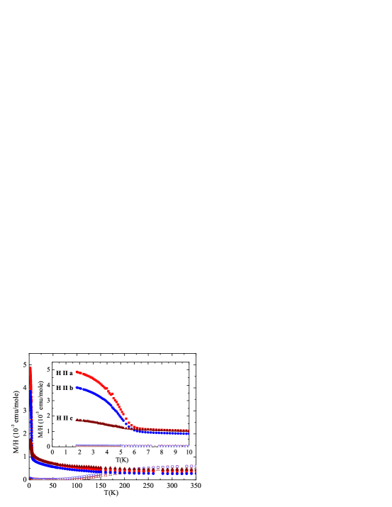
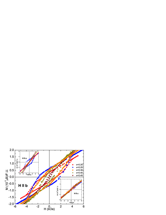
At low temperature, the parent compound FeSb2 is a narrow gap semiconductor with a rather small and temperature independent magnetic susceptibility. Petrovic1 Similar to FeSi, above 100 K there is a temperature induced paramagnetic susceptibility and an enhanced electronic conduction. The magnetic susceptibility can be described by both a thermally induced Pauli susceptibility and a low to high spin transition. Petrovic1 ,Petrovic2 ,Grandjean In the temperature () range from 1.7 to 150 K the Fe0.75Co0.25Sb2 magnetic susceptibility is larger than that of FeSb2. For above 6 K, it shows little anisotropy with the magnetic field applied along the different crystallographic axes. As shown in Fig. 1, the temperature dependence of the susceptibility indicates Pauli paramagnetism at high temperature. A clear ferromagnetic transition at K for a field of 1 kOe applied along any of the three crystallographic axes is illustrated in the inset to Fig. 1. These observations are in agreement with ferromagnetic long range order of the small magnetic moments below K.Rongwei The ferromagnetic nature of the transition is supported by the hysteresis loop measured at K and displayed in Fig. 2. For field strength varying between -6 and 6 kOe applied along the - axis, hysteresis loops are observed for . The width of the hysteresis loop grows initially with increasing from , peaks at , and becomes progressively smaller upon further Co substitution. Hysteresis loops are absent for field applied along the - axis and are observed only for for field applied along the - axis. By extrapolating the magnetization of Fe0.75Co0.25Sb2 to H=0, a lower estimate of the saturation magnetization along the b-axis of MLL = /F.U.) or ( /Fe), where F.U. refers to the FeSb2 formula unit, is obtained.

| Fe0.75Co0.25Sb2 | FeSb2 | |||||
| T(K) | ,mm/s | EQ,mm/s | ,mm/s | ,mm/s | EQ,mm/s | ,mm/s |
| 296b | - | - | - | 0.450(6) | 1.286(6) | - |
| 295 | 0.433(2) | 1.343(2) | 0.264(5) | 0.449(1) | 1.275(2) | 0.262(3) |
| 240 | 0.483(2) | 1.394(2) | 0.265(2) | - | - | - |
| 190 | 0.509(2) | 1.422(2) | 0.267(2) | - | - | - |
| 140 | 0.538(2) | 1.449(2) | 0.273(2) | - | - | - |
| 90 | 0.455(2) | 1.364(2) | 0.284(2) | - | - | - |
| 50 | 0.569(2) | 1.474(3) | 0.290(4) | - | - | - |
| 6.4b | - | - | - | 0.572(6) | 1.575(6) | - |
| 4.2 | 0.560(2) | 1.483(2) | 0.291(2) | 0.572(1) | 1.573(3) | 0.270(4) |
| 2.8 | 0.558(2) | 1.483(3) | 0.338(4) | - | - | - |
The Mössbauer spectra of FeSb2 single crystals exhibit a doublet at T=295 and 4.2 K. No impurity, and in particular no impurity with a large hyperfine field, is observed in the Mössbauer spectra. Furthermore, the Mössbauer spectral parameters for FeSb2 obtained herein are in excellent agreement with the previously reported parameters (see Table I). Raphael For Fe0.75Co0.25Sb2, the Mössbauer spectra, shown in Fig. 3, exhibit a doublet for temperatures ranging from K to 2.8 K. Again no impurity contribution is observed. Its spectral parameters, obtained at K and 4 K, are close to those observed in the FeSb2. The isomer shift observed in Fe0.75Co0.25Sb2 is ca. 0.01 mm/s smaller than in FeSb2. This indicates a somewhat larger s-electron density at the 57Fe nucleus.

The variation of the quadrupole splitting from 295 to 4.2 K is larger in FeSb2 than in Fe0.75Co0.25Sb2. This strong temperature dependence of the quadrupole splitting in FeSb2 is consistent with a scenario of electron delocalization appearing with increasing temperature, with a gap E of 380 K. Grandjean ,Goodenough As illustrated in Fig. 4, a fit of the Fe0.75Co0.25Sb2 quadrupole splitting as a function of temperature with the delocalization model described in Ref. 24 yields a somewhat larger gap energy K than that observed in FeSb2. The difference between the hyperfine parameters in FeSb2 and Fe0.75Co0.25Sb2 indicates that there is indeed a modification of the FeSb2 structure, and that no phase segregation is present. The Mossbauer spectra show that the investigated phase is (Fe,Co)Sb2 and not FeSb2+CoSb2 since the hyperfine parameters are significantly different. Furthermore no iron-bearing impurity is observed in Fe0.75Co0.25Sb2.
Apparently, the T = 2.8 K spectrum of Fe0.75Co0.25Sb2 is a doublet, which is somewhat surprising. Our interpretation is that either the iron experiences no magnetic hyperfine field or that the hyperfine field is below the detection limit. If the small broadening of ca. 0.047(6) mm/s of the 2.8 K spectrum, when compared to the 4.2 K spectrum, was associated to a magnetic hyperfine field, it would correspond to a 1.5+/-0.2 kOe hyperfine field. With a linewidth constrained to 0.29 mm/s, a fit of this spectrum, with both a quadrupole interaction and a hyperfine field yields a field of 2.8+/-1.2 kOe. Taking the usual proportionality of ca. 150 kOe/, these values can be used to estimate an upper limit of about MUL = 0.01 . for the magnetic moment on Fe.
IV Intrinsic vs. Extrinsic Magnetism
Given the small value of the saturated moment, it is possible that WFM originates from extrinsic sources, such as artifacts of the measurement process or the presence of a small amount of ferromagnetic impurity, e.g. elemental Fe. The former can be excluded based on the lack of sample dependence, both in magnetization and in heat capacity data. Rongwei Below we discuss the possibility of undetected second phases as extrinsic sources of the WFM.
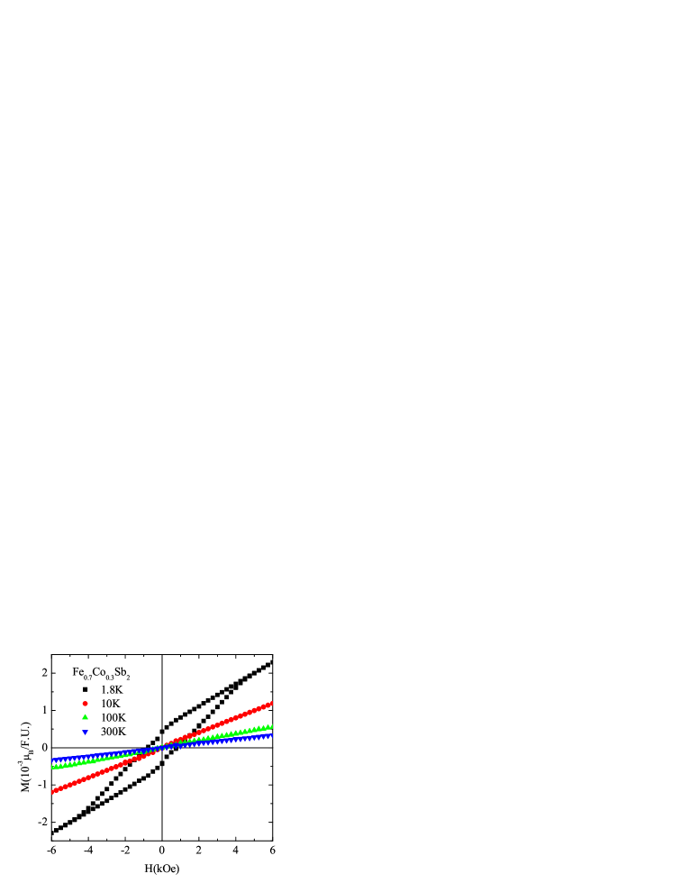
No hysteresis loops are observed for temperatures above for (example shown in Fig. 5). No known Fe-Sb, Co-Sb, Fe-Co, Fe-O, or Co-O phases show a ferromagnetic transition in this temperature range. FeCo alloys have large hyperfine fields (200-400 kOe) that would have been detected by Mössbauer measurement. We can calculate the X-ray patterns expected in the presence of bulk crystalline Fe impurities by superimposing the strongest peak of 0.3% elemental Fe to the measured patterns. No overlap between the calculated and measured Fe0.75Co0.25Sb2 X-ray patterns was observed (Fig. 6). Any other unknown Fe-O, Fe-Co-O, Co-O, Fe-Co, Fe-Sb-Co, etc. phase with the same atomic ratio in the mixture would have been detected and refined by synchrotron powder X-ray diffraction because its contribution to the scattering mixture would be higher than that of Fe. Though observed in magnetic hysteresis loops could be caused by Fe impurities of the order of the synchrotron powder X - ray diffraction detection limit, absence of hysteresis loops above 6 K strongly argues against such scenario.
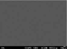
Another possibility is that magnetism in Fe1-xCoxSb2 is caused by magnetic nanoparticles. Mössbauer measurement shows no evidence of iron bearing nanoparticles (either FeCo or Fe-oxide). Such nanoparticles would have a paramagnetic spectrum with different isomer shift and quadrupole splitting at room temperature, which would be detected with a 0.3% limit. Below the nanoparticle blocking temperature the field would be large, typically 500 kOe for typical oxides. Solid evidence against nanoparticles or bulk extrinsic phases comes from energy dispersive scanning electron microscope (SEM) measurements. Among the samples grown from several different batches for , the uncertainty in Co concentration is . SEM data taken with resolution down to 1.5 nm exclude the presence of either bulk secondary phases or embedded nanoparticles. This is because high resolution SEM images of several randomly chosen polished crystals and crystalline surfaces show no trace of nanosize inclusions, clusters or inhomogeneities (example shown in Fig. 7). The images were taken in the “composition” mode with a solid state detector consisting of paired PN junctions. This type of detector is very sensitive to back-scattered electrons which in turn are sensitive to local variations in atomic number. If nano-crystallites of Fe or other elements were present, they would have been visible as bright dots in the high magnification image.
Taken in conjunction, our results argue against extrinsic sources of WFM in Fe1-xCoxSb2. Recent muon spin relaxation measurements indicate that the WFM state is spread throughout the full sample volume for Fe0.7Co0.3Sb2, further supporting our results.Uemura
V Discussion and Conclusions
Examples of intrinsic WFM states in narrow band materials are abundant in nature. DeLong Besides numerous oxide compounds, many intermetallic systems also exhibit intrinsic weak ferromagnetism, such as YbRhSb, YbRhSb MnS, MnS and Yb0.8YInCu4. YbInCu4 Magnetism in FeSb2 in analogy to FeSi has been predicted by LDA+U calculations. Lukoyanov Besides the use of an external magnetic field, one interesting possibility would be to induce the ferromagnetic state by lattice expansion and band narrowing, as in FeSi1-xGex. Sunmog , Anisimov3 Unfortunately, isoelectronic lattice expansion is limited to rather small values of in FeSb2-xBix. Our preliminary data show that the ferromagnetic state is not reached for . As in Fe1-xCoxSi, ferromagnetic state is induced with Co substitution in FeSb2. In both alloy systems critical temperature TC exhibits a characteristic peak as a function of Co concentration. Rongwei ,Grigoriev Whereas metallicity simultaneously appears with ferromagnetism in Fe1-xCoxSi at , Dave in Fe1-xCoxSb2 alloys transport and spin gap vanish at and respectively. Rongwei
What could be the mechanism of the WFM in Fe1-xCoxSb2 alloys? Knowing that there is an inversion symmetry at the Fe site in the Pnnm space group of FeSb2, we can exclude the presence of the Dzayloshinskii - Moriya (DM) type of interactions. This is in contrast to the doped FeSi where the DM interaction is believed to be responsible for the WFM. DM ,Anisimov3 A canted antiferromagnetism can be excluded based on the observed field dependence of the transition temperature. That is, the ferromagnetic tail at low temperature is insensitive to variation of the applied field. However, it is possible to ascribe the low magnetic moment in Co doped FeSb2 to the partial ordering of Co2+ ions. This scenario is in agreement with detailed analysis of the magnetic and thermodynamic properties of Fe1-xCoxSb2. Rongwei
Besides the obvious lattice expansion, the effect of the Co insertion is to introduce extra carriers in the system. The carriers cause a closing of the gap by . Rongwei Thus, the WFM appearance could be a consequence of carrier-induced metallicity. This claim is further supported by discarding another well known scenario for the WFM induction. More precisely, one can imagine that the WFM is induced by an “inverted metal-insulator” scenario. Anisimov2 ,Anisimov3 In this scenario, magnetic order exists only in the metallic phase. Furthermore, the metallicity is a direct consequence of transition to the ferromagnetic state where a bulk moment of 1 develops out of small gap semiconductor with small susceptibility. Anisimov2 ,Anisimov3 However, in Fe1-xCoxSb2 for a small ordered moment is induced. Therefore, the presence of the small moment excludes the “inverted metal-insulator” scenario and leaves as the only possibility that the WFM arises as a consequence of carrier-induced metallicity.
In conclusion, detailed structural and magnetic measurements argue against extrinsic sources of WFM in Co - substituted FeSb2. The ordered moment below the WFM transition for Fe0.75Co0.25Sb2 is . As opposed to FeSi where the metallic state is caused by band narrowing of nearly ferromagnetic parent electronic structure, weak ferromagnetism in Fe1-xCoxSb2 could be a consequence of carrier induced metallic state. In order to fully understand the magnetic structure, magnitude of moments, and mechanism of magnetic ordering, further neutron scattering and/or nuclear magnetic resonance measurements are envisaged.
We thank Yasutomo Uemura, Paul Canfield, T. M. Rice and Maxim Dzero for useful communications and Dr. L. Rebbouh for assistance in the Mössbauer spectral measurements. This work was carried out at the Brookhaven National Laboratory which is operated for the U.S. Department of Energy by Brookhaven Science Associates (DE-Ac02-98CH10886), and at Department of Physics, Université de Liège, Belgium. This work was also supported in part by the National Science Foundation DMR-0547938 (V. F. M.).
∗Present address: Institut für Festkörperforschung, Forschungzentrum Jülich GmbH, D-52425 Jülich, Germany
References
- (1) F. Hulliger, Nature (London), 198, 1081 (1963).
- (2) C. Petrovic, J. W. Kim, S. L. Bud’ko, A. I. Goldman, P. C. Canfield, W. Choe and G. J. Miller, Phys. Rev. B 67, 155205 (2003).
- (3) V. Jaccarino, G. K. Wertheim, J. H. Werinick, L. R. Walker and S. Arajs, Phys. Rev. 160, 476 (1967).
- (4) Y. Takahashi and T. Moriya, J. Phys. Soc. Jpn. 46, 1451 (1979).
- (5) G. Shirane, J. E. Fischer, Y. Endoh and K. Tajima, Phys. Rev. Lett. 59, 351 (1987).
- (6) K. Tajima, Y. Endoh, J. E. Fischer and G. Shirane, Phys. Rev. B 38, 6954 (1988).
- (7) L. F. Mattheis and D. R. Hamann, Phys. Rev. B 47, 13114 (1993 ).
- (8) V. I. Anisimov, J. Zaanen and O. K. Anderson, Phys. Rev. B 44, 943 (1991).
- (9) G. Aeppli and Z. Fisk, Comments Cond. Mat. Phys. 16, 155 (1992).
- (10) D. Mandrus, J. L. Sarrao, A. Migliori, J. D. Thompson, and Z. Fisk, Phys. Rev. B 51, 4763 (1995).
- (11) C.-H. Park, Z.-X. Shen, A. G. Loeser, D. S. Dessau, D. G. Mandrus, A. Migliori, J. Sarrao, and Z. Risk, Phys. Rev. B 52, 16981 (1995).
- (12) C. Petrovic, Y. Lee, T. Vogt, N. Dj. Lazarov, S. L. Bud’ko and P. C. Canfield, Phys. Rev. B 72, 045103 (2005).
- (13) A. Bentien, G. K. H. Madsen, S. Johnsen, and B. B. Iversen., Phys. Rev. B 74, 205105 (2006).
- (14) J. F. DiTusa, K. Friemelt, E. Bucher, G. Aeppli and A. P. Ramirez, Phys. Rev. B 58, 10288 (1998).
- (15) Z. Schlesinger, Z. Fisk, H-T. Zhang, M. B. Maple, J. F. DiTusa and G. Aeppli, Phys. Rev. Lett. 71, 1748 (1993).
- (16) A. Perucchi, L. Degiorgi, Rongwei Hu, C. Petrovic and V. F. Mitrovic, European Physical Journal B 54, 175 (2006).
- (17) V. I. Anisimov, S. Yu. Ezhov, I. S. Elfimov, I. V. Solovyev and T. M. Rice, Phys. Rev. Lett. 76, 1735 (1996).
- (18) A. V. Lukoyanov, V. V. Mazurenko, V. I. Anisimov, M. Sigrist and T. M. Rice, European Physical Journal B 53, 205 (2006).
- (19) S. Yeo, S. Nakatsuji, A. D. Bianchi, P. Schlotmann, Z. Fisk, L. Balicas, P. A. Stampe and R. J. Kennedy, Phys. Rev. Lett. 91, 046401 (2003).
- (20) N. Manayala, Y. Sidis, J. F. DiTusa, G. Aeppli, D. P. Young and Z. Fisk, Nature 404, 581 (2000).
- (21) Hunter B., ”Rietica - A visual Rietveld program”, International Union of Crystallography Commission on Powder Diffraction Newsletter No. 20, (Summer) http://www.rietica.org (1998).
- (22) A. C. Larson and R. B. VonDreele, (Report LAUR 86-748, Los Alamos National Laboratory, New Mexico, 1986). B. H. Toby, J. Appl. Crystallogr. 34, 210 (2001).
- (23) Rongwei Hu, V. F. Mitrovic, and C. Petrovic, Phys. Rev. B 74, 195130 (2006).
- (24) S. V. Grigoriev, S. V. Maleyev, V. A. Dyadkin, D. Menzel, J. Schoenes, and H. Eckerlebe, Phys Rev. B 76, 092407 (2007).
- (25) A. Gérard and F. Grandjean, J. Phys. Chem. Solids 36, 1365 (1975).
- (26) J. Steger and E. Kostiner, J. Sol. St. Chem. 5, 131 (1972).
- (27) J. B. Goodenough, J. Sol. State Chem. 5, 144 (1972).
- (28) T. Uemura, private communication
- (29) L. E. De Long, J. G. Huber and K. S. Bedell, J. Magn. Magn. Mater 99, 171 (1991).
- (30) Y. Muro, Y. Haizaki, M. S. Kim, K. Umeo, H. Tou, M. Sera, and T. Takabatake, Phys Rev. B 69, 020401 (2004).
- (31) S. S. Aplesnin, L. I. Ryabinkina, G. M. Abramova, O. B. Romanova, A. M. Vorotynov, D. A. Velikanov, N. I. Kiselev, and A. D. Balaev, Phys. Rev B 71, 125204 (2005).
- (32) A. Mitsuda, T. Goto, K. Yoshimura, W. Zhang, N. Sato, K. Kosuge and H. Wada, J. Phys. Chem. Solids, 63, 1211 (2002).
- (33) B. Lebech, J. Bernhard and T. Freltoft, J. Phys. Cond. Matter 1, 6105 (1989).
- (34) V. I. Anisimov, R. Hlubina, M. A. Korotin, V. Mazurenko, T. M. Rice, A. O. Shorikov and M. Sigrist, Phys. Rev. Lett. 89, 257203 (2002).