EIT and diffusion of atomic coherence
Abstract
We study experimentally the effect of diffusion of Rb atoms on Electromagnetically Induced Transparency (EIT) in a buffer gas vapor cell. In particular, we find that diffusion of atomic coherence in-and-out of the laser beam plays a crucial role in determining the EIT resonance lineshape and the stored light lifetime.
In this paper we address an important issue for electromagnetically induced transparency (EIT) harris'97pt ; scullybook and stored light fleischhauer02pra ; lukin03rmp in alkali vapor cells (i.e., warm atoms with buffer gas): the practical role of the diffusion of atomic coherence in-and-out of the laser fields that prepare and probe the atoms. EIT occurs in a three-level -system in which two coherent electromagnetic fields (the probe and control fields) are in two-photon Raman resonance with two atomic ground-state sublevels, as shown in Fig. 1(a). The atoms are optically pumped into a “dark state”, a coherent superposition of the two ground-state sublevels which is decoupled from the optical fields. Typically, a buffer gas is included to restrict the motion of the EIT atoms and thus lengthen the atomic interaction time with the laser fields arimondo'96pra ; wynands'97 ; helm'01 ; and also to pressure broaden the electronic excited state so as to allow co-propagating probe and control fields that interact with most of the atoms despite Doppler broadening javan'02 ; lee'03 .
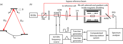
As shown in Fig. 1, we employed an external cavity diode laser (ECDL) tuned to the line of 87Rb. The total available laser power was about mW.
In this regime, atomic diffusion is important, yet rather subtle to model effectively. To date EIT models have treated diffusion phenomenologically, by assuming a simple homogeneous decay of the ground state coherence characterized by the timescale of the lowest order diffusion mode across the laser beam happer72 ; arimondo'96pra ; helm'01 . Some recent experiments have seen indications of the inadequacy of this simple approach zibrov'01ol ; zibrovmatsko02pra ; novikova05josab . Here we report experiments that show that diffusion of atomic coherence in-and-out of the laser beam (i.e., from the region illuminated by the optical fields to the surrounding, unilluminated region, and then back into the laser beam) plays a crucial role in determining the EIT resonance lineshape and the lifetime of stored light. As described below, we observed a narrow central peak in the EIT resonance (much narrower than ) due to the contribution of atoms diffusing in-and-out of the laser beam. We then eliminated this narrow peak by applying transverse gradients in the longitudinal magnetic field to decohere atoms that diffuse well out of the laser beam. Finally, we measured the influence of atomic coherence diffusion on light pulses that have been stored in the atomic ensemble using a dynamic EIT technique.
I Experimental setup
We used a polarizing beam splitter (PBS) to create a bypass reference beam. We modulated the phase of the main laser field at GHz (near the 87Rb ground-state hyperfine splitting) using an electro-optical modulator (EOM), so that approximately of the total laser power was transferred to each first order sideband. All three optical fields then passed through an acousto-optical modulator (AOM), shifting the frequencies of all fields by MHz. We tuned the laser such that the main carrier frequency field was resonant with the transition of 87Rb; in this case the sideband was resonant with the transition. We neglected any influence of the far-detuned sideband.
Before entering the Rb vapor cell the laser beam was weakly focused to a mm diameter spot and circularly polarized using a quarter-wave plate . We mounted the cylindrical glass cell, containing isotopically enriched 87Rb and either Torr or Torr Ne buffer gas ( and respectively vanier74pra ; franz76 ), inside a three-layer magnetic shield, which reduced stray magnetic fields to less than over the interaction region. A solenoid inside the magnetic shields provided a weak bias magnetic field ( mG). We controlled the temperature of the cell to K using a blown-air oven. For the EIT lineshape measurements we kept the temperature low enough (C for the Torr Ne cell, and C for the Torr Ne cell) to ensure an optically thin Rb vapor; for stored light experiments we operated with the cell at C to provide significant pulse delay and storage.
After traversing the cell, the laser beam was combined with the bypass reference beam on a non-polarizing beam-splitter (NPBS) and sent to a fast photodetector (FPD). Since the frequency of the reference field was MHz lower than the control field, we detected the sideband by measuring the amplitude of the beat note at GHz using a microwave spectrum analyzer.
II Gradient coils
Pulsed magnetic field gradients are a well established tool in NMR for imaging (MRI) and for measuring diffusion and coherent flow in liquids and gases.
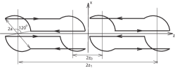
In our experiment we used a standard gradient coil design (see Fig. 2) callaghan_book ; jin_book to produce a controllable linear gradient of the longitudinal magnetic field in the transverse direction: up to , with excellent linearity at the center of the coils.
Note that away from the coil center a transverse magnetic field is also created such that . In traditional high-field NMR applications, the effect of the transverse magnetic field is negligible. In our experiment the weak bias magnetic field limits the gradient strength we can use without distorting the EIT resonance: , where is the length of the cell.
III Steady-state EIT in buffer gas cells
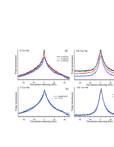
Fig. 3 shows a typical example of the measured EIT lineshape (i.e. the probe field transmission as a function of two-photon detuning) for the Torr Ne cell and no applied magnetic fields. Note the sharp, narrow peak on-resonance. Also note that the FWHM is significantly narrower that kHz happer72 ; arimondo'96pra , i.e., the linewidth set naively by single-pass diffusion across the laser beam.
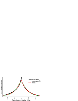
The shape of this EIT resonance is clearly not the Lorentzian function predicted by simple three-level EIT theory lee'03 :
| (1) |
where and are the probe and control field Rabi frequencies, is proportional to the probe field transmission intensity, is the two-photon detuning, and and are respectively the excited-state and ground-state coherence relaxation rates (see Fig. 1a).
Recent extensions of three-level EIT theory have considered the effect of atomic motion in a laser beam with a Gaussian intensity profile in the transverse direction. In the regime of high buffer gas pressure and moderately high laser intensity, the motion of alkali atoms across the laser beam is assumed to be slower than the excited and ground-state relaxation rates, and the atomic coherence is assumed to adjust instantaneously to the light intensity at each point of the laser beam. Thus atoms in the center of the laser beam, where the laser intensity is maximum, have greater power broadening than atoms in the wings. For a Gaussian laser intensity profile, the probe field transmission lineshape is then found to be taichenachev'02iqec :
| (2) |
which we refer to as the “TY-fit”. In the regime of low buffer gas pressure and very low light intensity, there is negligible power broadening and the EIT lineshape is calculated to be pfleghaar93 :
| (3) |
where is the average alkali atom transit time through the laser beam, and is the average thermal radial velocity of the alkali atoms. Interestingly, the lineshapes described by Eqs. (2) and (3) are very similar for typical conditions for Rb vapor EIT, despite being obtained in very different limits. In Fig. 3 we plot the best Lorentzian fit (Eq. (1)) and TY-fit (Eq. (2)), which is more relevant to our experimental conditions. The Lorentzian fit provides a poor approximation to our measurements; the TY-fit is superior to the Lorentzian fit, but it fails to reproduce the sharp structure on resonance.
The observed EIT lineshape may be understood if we assume that the diffusion of Rb atoms in-and-out of the laser beam creates a broad range of interaction times rather than a single average diffusion time . That is, we must account not only for atoms that diffuse once through the laser beam and then decohere (never return), but also those that diffuse out of the laser beam and return, and thus interact with the laser fields multiple times. For these returning atoms the interaction with the optical fields resembles a Ramsey separated-oscillatory-field experiment,with significant coherent evolution “in-the-dark”, leading to much narrower EIT resonances.
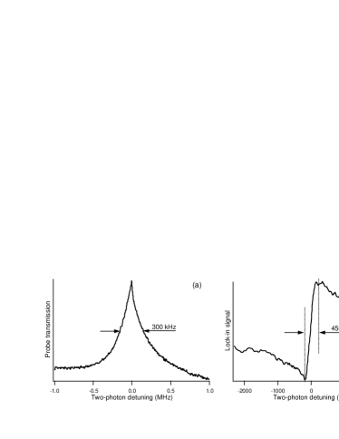
To better characterize the sharp central peak of the EIT resonance, we employed phase-sensitive detection. We used audio-frequency-modulation of the GHz synthesizer ( Hz), and detected changes in the laser transmission using a slow photodetector and lock-in amplifier. Fig. 4 shows EIT resonance measurements made both with a spectrum analyzer (covering the entire resonance) and phase-sensitive detection (restricted to the narrow center of the resonance). For both the Torr and Torr Ne cell, we measured the spectral width of the narrow central EIT peak to be Hz, and to be largely insensitive to laser power, which is consistent with the central peak reflecting coherent atomic evolution “in-the-dark”.
To test the hypothesis that atoms diffusing in and out of the laser beam contribute significantly to the EIT lineshape (particularly the sharp central peak), we measured the EIT resonance in the presence of a linear transverse gradient in the longitudinal magnetic field , using the gradient coils described above. For these measurements we also applied a uniform longitudinal field ( mG) which splits the Rb ground-state Zeeman sublevels, and makes negligible the effects of the transverse component of the magnetic field created by the gradient coils (for mG/cm). We studied EIT formed on the ground-state Zeeman sublevels (i.e., coherence between the and levels). The associated transition frequency between those levels has a gyromagnetic ratio MHz/G. Therefore, induces an inhomogeneous broadening to the EIT coherence; and for suitable magnitudes of this gradient, the broadening across the laser beam waist ( mm) can be small compared to the narrow central peak, while the larger magnetic field deviation outside the laser beam causes most atoms to decohere if they diffuse out of and then back in the laser field.
Fig. 5 shows measured EIT resonances at several gradient strengths. For stronger gradients, the central peak becomes less sharp, and the TY-fit of the EIT lineshape is improved, which is consistent with diffusion-induced Ramsey-narrowing being destroyed by gradient-field-induced decoherence of atoms that diffuse out of the laser beam.
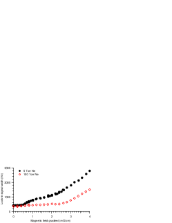
Fig. 6 illustrates the greater dependence on applied gradient of the spectral width of the central EIT peak for the Torr Ne cell, in comparison to the Torr Ne cell. These measurements are also consistent with diffusion-induced Ramsey-narrowing, which would be more thoroughly inhibited by the applied magnetic field gradient for the more rapid Rb diffusion in the Torr Ne cell. Modelling and quantitative analysis of these diffusion effects will be reported in a future publication.
IV Stored light in a buffer gas cell
Under EIT conditions, the strength of the control field determines the coherent coupling between the probe field and the ground-state atomic coherence and thus the group velocity of probe pulse propagation through the medium boydPiO2002 . Appropriate variation of the control field allows storage of the probe pulse in the form of an atomic ensemble coherence fleischhauer02pra ; lukin03rmp . In realistic atomic systems, the maximum storage time is determined by the atomic coherence lifetime, including the effect of coherence diffusion.
To avoid large additional absorption and pulse reshaping, the bandwidth of the probe pulse should be less than the the sharp peak observed in the static probe field transmission SPIEproc05 . We employed a Gaussian waveform for the probe pulse with a full width of ms, which experienced a group delay s for a control field power of W. This delay is not great enough to trap the entire probe pulse inside the atomic vapor cell ( cm). We typically stored about half the Gaussian pulse, as shown in Fig. 7(a). Note that we used a larger control field intensity at the retrieving stage (W) to increase the EIT width of the atomic medium and thereby minimize losses during release and propagation of the retrieved pulse.

The measured amplitude of the probe pulse for various storage intervals is shown in Fig. 7(b). The decay time of the retrieved pulse area is s (see Fig. 7(c)), corresponding to a decoherence rate Hz, in agreement with the width of the sharp central peak of the EIT resonances at low light intensity, and much narrower than the rate kHz associated with diffusion of Rb atoms out of the laser beam.
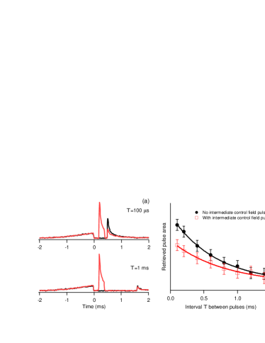
The large spatial distribution of atomic coherence stored outside the laser beam allows multiple retrieved pulses to be observed by turning the control field on and off several times during the retrieval process. As a demonstration, we stored a signal pulse in the atomic ensemble; turned the control field off for s; then on for another s to retrieve a first pulse; next turned off the control field again for a variable time T; and finally turned the control field on and retrieved a second pulse. Fig. 8 shows the results of these measurements and a comparison to experiments with no intermediate control field pulse. For small T, there is a large difference between the retrieved pulse areas with and without an intermediate control field application, since the second application of the control field mostly interacts with the same atoms from which coherence has already been retrieved by the first pulse. For larger values of T, however, atomic coherence diffuses back into the laser beam, and the difference between the pulses retrieved with and without an intermediate control field pulse decreases.
V Conclusions
We have presented an initial experimental study of the effect of atomic coherence diffusion in-and-out of the laser beam on EIT and light storage in a Rb vapor cell with Ne buffer gas. We found that the EIT resonance lineshape is not well described by models that account for atomic diffusion with a simple homogeneous decay rate of the ground state. We also observed a sharp, narrow peak of the EIT resonance that can be understood qualitatively due to the coherent atomic evolution “in-the-dark” as atoms diffuse in-and-out of the laser beam. We found that this diffusion-induced Ramsey-narrowing could be reduced by application of a suitable magnetic field gradient, , which decoheres most atoms that diffuse out of the laser beam. We also measured light storage times consistent with the inverse of the linewidth of the narrow central peak of the EIT resonance, and observed “replenishment” of stored light signals due to atomic coherence diffusion from the region outside of the laser beam.
The authors are grateful to F. Cané, M. Klein, C. Smallwood, M. D. Lukin, M. D. Eisaman, M. Bajcsy, L. Childress, A. André, M. Crescimanno, A. Weis, and A. S. Zibrov for useful discussions, and to Christine Y.-T. Wang for construction of the gradient coils. This work was supported by DARPA, ONR and the Smithsonian Institution.
References
- (1) S. E. Harris, Phys. Today 50 (7), 36 (1997).
- (2) M. O. Scully, and M. S. Zubairy, Quantum Optics (Cambridge University Press, Cambridge, UK, 1997).
- (3) M. Fleischhauer, and M. D. Lukin, Phys. Rev. A 65, 022314 (2002).
- (4) M. D. Lukin, Rev. Mod. Phys. 75, 457 (2003).
- (5) E. Arimondo, Phys. Rev. A 54, 2216 (1996).
- (6) S. Brandt, A. Nagel, R. Wynands, and D. Meschede, Phys. Rev. A 56, R1063 (1997); R. Wynands, and A. Nagel, Appl. Phys. B 68, 1 (1998).
- (7) M. Erhard, and H. Helm, Phys. Rev. A 63, 043813 (2001).
- (8) A. Javan, O. Kocharovskaya, H. Lee, and M. O. Scully, Phys. Rev. A 66, 013805 (2002).
- (9) H. Lee, Y. Rostovtsev, C. J. Bednar, and A. Javan, Appl. Phys. B 76, 33 (2003).
- (10) W. Happer, Rev. Mod. Phys. 44, 169 (1972).
- (11) A. S. Zibrov, I. Novikova, and A. B. Matsko, Opt. Lett. 26, 1311 (2001).
- (12) A. S. Zibrov, and A. B. Matsko, Phys. Rev. A 65, 013814 (2002).
- (13) I. Novikova, A. B. Matsko, and G. R. Welch, J. Opt. Soc. Am. B 22, 44 (2005).
- (14) J. Vanier, J.-F. Simard, and J.-S. Boulanger, Phys. Rev. A 9, 1031 (1974).
- (15) F. A. Franz, and C. Volk, Phys. Rev. A 14, 1711 (1976).
- (16) P. T. Callaghan, Principles of Nuclear Magnetic Resonance Microscopy (Clarendon, Oxford, 1991).
- (17) J. Jing, Electromagnetic analysis and design in magnetic resonance imaging (CRC Press, Boca Raton, 1998).
- (18) A. V. Taichenachev, A. M. Tumaikin, V. I. Yudin, M. Stahler, R. Wynands, J. Kitching, and L. Hollberg, Phys. Rev. A 69, 024501 (2004).
- (19) E. Pfleghaar, J. Wurster, S. Kanorsky, and A. Weis, Opt. Commun. 99, 303 (1993).
- (20) R. W. Boyd, and D. J. Gauthier, in Progress in Optics, edited by E. Wolf (Elsevier, Amsterdam, 2002), Vol. 43, pp. 497- 530.
- (21) D. F. Phillips, A. Fleischhauer, A. Mair, R. L. Walsworth, and M. D. Lukin, Phys. Rev. Lett. 86, 783 (2001).
- (22) I. Novikova, M. Klein, D. F. Phillips, and R. L. Walsworth, Proc. SPIE International Symposium Integrated Optoelectronic Devices, 5735, 87 (2005).