ACT-8/00
CTP-TAMU-18/00
quant-ph/0007088n
June 2000
Quantum Brain?
Andreas Mershin,1***mershin@rainbow.physics.tamu.edu
Dimitri V. Nanopoulos1,2,3†††dimitri@soda.physics.tamu.edu
and Efthimios M.C. Skoulakis4‡‡‡eskoulakis@bio.tamu.edu
1 Center for Theoretical Physics,
Dept. of Physics, Texas A&M University,
College Station, TX 77843-4242, USA
2 Astro Particle Physics Group,
Houston Advanced Research Center (HARC),
The Mitchell Campus,
Woodlands, TX 77381, USA
3 Academy of Athens, Chair of Theoretical Physics,
Division of Natural Sciences,
28 Panepistimiou Avenue, Athens 10679, Greece
4 Center for Advanced Invertebrate Molecular Science,
Dept. of Biology, Texas A&M University,
College Station, TX 77843-2475, USA
In order to create a novel model of memory and brain function, we focus our approach on the sub-molecular (electron), molecular (tubulin) and macromolecular (microtubule) components of the neural cytoskeleton. Due to their size and geometry, these systems may be approached using the principles of quantum physics. We identify quantum-physics derived mechanisms conceivably underlying the integrated yet differentiated aspects of memory encoding/recall as well as the molecular basis of the engram. We treat the tubulin molecule as the fundamental computation unit (qubit) in a quantum-computational network that consists of microtubules (MTs), networks of MTs and ultimately entire neurons and neural networks.
We derive experimentally testable predictions of our quantum brain hypothesis and perform experiments on these.
1 INTRODUCTION
1.1 Overview of the Field
During the last decade or so, it has become increasingly popular among researchers to look for manifestations of quantum physics in neurobiological processes associated with brain function. Recent works in this field by Penrose[1, 2], Hameroff[3], Mavromatos and Nanopoulos[4, 5] and others[6] as well as earlier research (as early as 1968) on coherent excitations by Frölich[7, 8, 9], have been seminal to this new approach to brain function research. The arguments for the necessity of this unconventional approach have been greatly elaborated upon in the literature by its advocates and yet the very existence of ”quantum brain” effects is still challenged by physicists and biologists alike. To date, at least to our knowledge, experiments targeted at investigating the existence of neurobiological quantum phenomena have not been performed. Most of the research has been of theoretical and computational nature[10, 11, 12]and as a result, there has been no clear answer. The nature of the subject under investigation is interdisciplinary and consequently the target audience has widely varying scientific backgrounds and expectations. The effort described here has included research by experts in both fields and aspires to provide a bridge for experimental and theoretical scientists from both disciplines.
Understanding memory will bring us one step closer to finding out how the external world is coded in the microscopic structure of the brain and eventually, we will be able to appreciate how unique experiences make unique individuals even though the basic genetic, molecular and physical processes are shared by all.
1.2 Problems
There are certain aspects of brain function that appear to have no obvious explanation based on traditional neuroscience. There exist many biological models of memory function but all call for some sort of ”Differentiated Yet Integrated”[13] (DYI) function. Anatomical and neurobiological evidence clearly shows that specific memories are not precisely localized in the brain. Although certain structures such as the hippocampus[13, 14, 15] have traditionally been implicated in memory formation more than others, it is clear that individual components (for instance correlated visual and auditory memories) are stored at macroscopically separated regions of the neural network. This is the ”differentiated” part of memory. During recall, large numbers of neurons fire in tandem to produce an ”integrated” picture. By extension, we expect that during the initial recording of a memory, a process which results in the engram, there must also have been correlations between distant neurons. This lies at the root of the binding problem where a single stimulus activates neurons located far apart from each other ”simultaneously” or at least faster than chemical neurotransmission allows. To date, what all proposed biological memory models lack in common, is a plausible mechanism for establishing these fast correlations between distant neurons and explaining the speed at which information is processed. This is a feeling shared at least by some biologists who have started looking for non-neurotransmitter based communication pathways, such as electrical[16] and phase couplings[17]. It is our purpose here to suggest another, quantum physics-derived mechanism for the DYI operation of memory. The property of non-locality exhibited by certain quantum systems may produce a solution to the binding problem as well as to the speed problem.
Learning and memory are manifested as modifications of behavior produced by experience of environmental stimuli and they reflect the function of the brain. Although it is generally accepted that changes in the biochemical properties of neurons (especially their synapses) mediate changes in brain function and memory encoding, we have yet to have a satisfactory understanding of how molecular events effect or influence these changes. This is the molecular engram problem. A prediction of our quantum approach gives the neural cytoskeleton and its associated proteins a major role during engram formation and thus proposes experimentally testable molecular mechanisms of memory formation.
Classical approaches to digitally simulating biological neural networks (each neuron roughly playing the role of a switch whose connections/synapses to other neurons are ”weighted” according to past experiences) have so far proved insufficient to adequately explain how the biological efficiency of recall occurs as well as the observed complexity, capacity and versatility of a biological brain. On the other hand, new developments in theoretical quantum computation, learning, storage and retrieval algorithms, have shown that by using quantum bits or qubits, one resolves the capacity problem of classical computers as well as speeds up these processes[18]. By modeling the brain as a quantum computer we envision to resolve the problems of recall, complexity, capacity and versatility.
Assuming our suggestion that quantum phenomena underlie biological function is correct, it is yet unclear at exactly which level the transition to classical, purely biological processes takes place. With virtually no experimental data in this field, it is impossible to precisely define the model but certain testable predictions can nevertheless be derived.
Our quantum mechanical model of brain function differs significantly from the classical approach to conventional neural networks but it is not in competition with the well established neurobiology of chemical and electrical neurotransmission, synaptic function etc. The main difference is that in our model, a single neuron is upgraded from a relatively simple (yet adjustable) switch to a device capable of information processing. In addition, within the context of our model, (at least some) neurons are capable of launching fast connections to establish correlations with distant neurons using the principles of quantum entanglement and/or photon interactions (both discussed later).
1.3 Why Use Quantum Mechanics?
The connection between quantum physical events and biological function has been studied for quite some time, for instance Frölich’s[7, 8, 9]work on protein conformational changes linked to quantum level interactions/events such as dipole oscillation and electron mobility in a protein’s hydrophobic pocket. As discussed in great detail in references [8] and [19], electron density localization inside a hydrophobic pocket dictates protein conformation. This should come as no surprise as the van der Waals forces arising from a change in the electron localization will push/pull against the charged parts of the molecule. As such, this process seems of limited quantum-physical interest since analytic solutions to Schrödinger equation for such many-body systems are extremely difficult to obtain. Motivation for work on quantum mechanics and protein conformational changes comes from a defining property of quantum systems discussed later, namely their ability to be in a superposition of states i.e. being in two (or more) states at once.
In particular, the tubulin protein, the structural block of microtubules (MTs), has the ability to switch (”flip”) from one conformation to another as a result of a shift in the electron density localization from one resonance orbital to another (figure1). The tubulin system has only two possible basis states labeled and according to whether the electrons inside the tubulin hydrophobic pocket are localized closer to the or monomers. These two states are distinguished from each other by a flip in the electric dipole moment vector of the tubulin molecule[6] by .
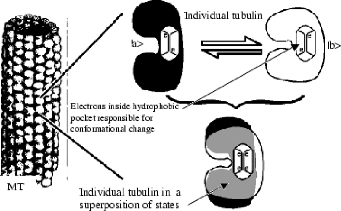
The tubulin system described above could easily serve as a textbook example of how a biological qubit should look like! The two tubulin conformations make for a simple binary qubit with the ability of entanglement with similar neighboring qubits/dimers in the protofilaments giving us a quantum cluster! The timescale for the spontaneous conformational changes in the tubulin dimers is of order
Once in an entangled state, a ”measurement” or interaction with the environment will collapse the state into one of its basis states leaving each tubulin in either the or conformations. Yet, the correlations can be communicated instantaneously among the tubulin qubits as described in Section 4, spanning entire MTs or conceivably whole neurons or neural networks.
1.4 Coincidences?
Alzheimer’s Disease
Damage to neural MTs resulting from hyperphosphorylation of tau () which is a microtubule associated protein (MAP), results in memory loss in Alzheimer’s Disease (AD) patients[20] suggesting a connection between MTs and memory. Neurofibrillary Tangles (NFTs) are bundles of twisted MTs that are no longer held apart by their MAPs. Post-mortem histological examination of AD patients shows a clear and direct correlation between NFTs and duration and severity of the disease[21].
Anesthesia
It is a rather remarkable fact that general anesthesia can be induced by a large number of completely different substances of no chemical similarity whatsoever, from ether to chloroform to xenon. Purely biophysical studies on the mechanisms of anesthesia[22, 23] have shown unequivocally that the long debated action of anesthetics is not on the lipid membrane proteins but on the dynamic conformational functions of proteins (such as ion channel operation, receptor activation and cytoskeletal function). An extension of these findings[24] has produced computer simulations strongly suggesting that anesthetic molecules bind to the hydrophobic pocket of the tubulin dimer. This is directly relevant to our suggestion regarding the role of the tubulin conformational changes as follows: binding of an anesthetic molecule to the hydrophobic pocket of the tubulin dimer may have the effect of preventing changing the electron orbitals (i.e. the tubulin’s ability to flip) thus shutting the whole system down. Therefore, in our model, it is just the electric dipole properties of these anesthetic substances that need to be similar (which is the case) and not necessarily their chemical properties. Furthermore, if the general anesthetic concentrations are not too high, complete reversibility of anesthetic effects is possible, indicating that the temporary van der Waals blockage of the crucial tubulin electron(s) has ended and conformational changes are free to occur again.
Geometry of Microtubules
There has been speculation for quite some time that MTs are involved in information processing: it has been shown that the particular geometrical arrangement (packing) of the tubulin protofilaments obeys an error-correcting mathematical code known as the code[25] (K-code). Error correcting codes are also used in classical computers to protect against errors while in quantum computers special error correcting algorithms are used to protect against errors by preserving quantum coherence among qubits. Furthermore, it has been recently suggested that the geometric curvature of MTs may also play a role in information processing[26].
1.5 Our Motivation
On the one hand, protein conformational changes are directly related to quantum level phenomena and on the other, those same protein functions are directly related to system-wide phenomena such as anesthesia and (potentially) memory. Therefore, it seems reasonable for us to look for the effects of quantum processes on neuronal (and) brain- wide function. Lastly, recent theoretical and experimental advances in the field of quantum computation call for molecular switches/qubits, the parameters of which fit nicely with the proposed role of tubulin dimers. The anticipated quantum clusters also sound very much like the MT protofilaments.
It seems credible that we have uncovered the elementary components of a quantum computation network inside the biological brain.
1.6 Our Research Approach
Our target system has been the microtubule. We claim that the long and characteristically ordered MTs that comprise the bulk of proteins in the axons of neurons are the microsites of computation.
During the last few years, physicists have been investigating MTs as physical systems applying the principles of Electromagnetic, Quantum and even String Theory[4, 5, 10, 11]. In the model under discussion here, the MTs’ periodic, paracrystalline structure, augmented by the K-code, makes them able to support a superposition of coherent quantum states among their component tubulin dimers. This quantum superposition may collapse spontaneously[4, 5]or dynamically through interactions with the environment such as neurotransmitter binding and action potential firing. As a result of quantum mechanical entanglement interactions, the MT network in the neuron’s axon acts in an ”orchestrated” or ”coherent” way possibly setting up fast communication pathways among neurons that do not depend directly on chemical or electrical synaptic signal transmission. When the quantum entangled state collapses, the result can be synchronous synaptic release of neurotransmitter molecules, and/or feedback information about each neurons’ environment. The combined effect of such events may be translated into orchestrated action and changes in large parts of a neural network.
Entanglement-based communication would allow MTs to work in tandem and it is conceivable that coherence might span macroscopic distances for long times in the brain within the context of a particular environment. Although there is a suggestive theoretical background[4, 5, 10, 11] to justify such assumptions, more experimental data is needed before we can say with certainty that quantum coherence is preserved for appreciable times and for more than the spatial extent of a few tubulins.
1.7 Phenomenology of the Quantum Brain
As the main areas where we expect to see direct manifestation of quantum phenomena are memory encoding, storage and retrieval, these are the points our research concentrates upon. If MTs are indeed quantum computing devices, then memory encoding would have to be affected by their dynamics. We envision that the role of the MAPs, especially MAP-2, is to ”tune” the MT network, allowing individual MT states to entangle and collapse in specific ways. We expect a redistribution of MAPs to be one of the results of memory encoding. We have named this the ”guitar string model” (GSM) of memory encoding as MAPs can be thought of as the fingers on guitar strings (MTs). By changing the binding sites, which in our model represent distinct memory encoding events, we change memory encoding (the engram). This is in analogy to different finger configurations on guitar strings producing different chords while the strings and fingers remain the same. This model predicts a redistribution of MAP-2 concentration in neurons as a result of learning. This has never been conclusively shown and is the goal of our experiments described in Section 4. The model also predicts MAP-2 production and breakdown as a result of learning and there is some preliminary evidence from other groups that this is indeed the case[27, 28].
1.8 The Quantum Brain Hypothesis
What follows is a qualitative description of the proposals of our model in their entirety. These are justified later in the text but are included here for completeness.
-
•
We propose that the tubulin dimers comprising MTs act as molecular binary switches (qubits) and the two conformations of the tubulin dimer are the equivalent of a 0 and 1 in a binary quantum computer.
-
•
We propose that information can (at least temporarily) be stored as patterns of 0’s and 1’s corresponding to the conformational states of the tubulin dimers. We propose that protofilaments and whole microtubules act as memory clusters analogous to RAM (Random Access Memory) in digital computers.
-
•
We propose that the cytoskeleton in general and the axonal and dendritic microtubules in particular, are the microsites of information manipulation via electromagnetic and quantum mechanical interactions between tubulin dimers, protofilaments, MTs and MAPs. We further propose that at least part of an intermediate or permanent memory trace (the engram) is achieved by means of a redistribution of microtubule associated proteins along the cytoskeleton. The pattern of MAP binding is the engram.
-
•
Following engram formation, neurons that have been simultaneously restructured are expected to share similar patterns of MAP binding. During recall, neurotransmitter activation of key neuron(s) by a stimulus results in instantaneous co-activation of most or all other relevant neurons containing a similar cytoskeletal geometry (MAP distribution) via quantum coherence phenomena. Thus, large numbers of neurons relevant to a particular memory trace can be activated synchronously after which ordinary neurotransmitter-based communication sets in.
To summarize, these are the problems we will be addressing and the quantum- physics derived paths to their solution that we propose:
| Problem | Relevant quantum property | Research approach |
| Binding problem including | Quantum coherence, non- | Theoretical investigation |
| the DYI aspect of memory | locality and entanglement. | yields testable predictions. |
| Section 3 | Sections 3&4 | |
| Recall | Quantum coherence, non- | Testable predictions |
| locality and entanglement. | derived. | |
| Section 4 | Sections 3&4 | |
| Capacity, versatility, speed | Quantum entanglement. | Investigation of quantum- |
| Quantum computer-like | computing algorithms | |
| operation of biological | shown to maintain | |
| brain. | coherence, increase capacity | |
| Section 4 | and speed up recall. | |
| Section 4 | ||
| Molecular basis of the | Quantum effects in neural | Experiments |
| engram | cytoskeletal function. | involving associative |
| Tuning of MT network by | learning in fruit-flies are | |
| MAPs | underway. | |
| Sections 1–4 | Section 5 |
1.9 Where does our Model Fit in with Classical Neuroscience?
Existing biological memory models can be complemented by taking advantage of the processes suggested in our quantum brain hypothesis. For instance in her experiments using rats, Nancy Woolf[28, 29, 30], observed degradation of MAP-2 in the rat brain following an associative learning task. This can be interpreted as follows: MAP-2 degradation is the first logical step required for the redistribution of this protein along axons and dendrites. Such a redistribution will alter the local geometry of the cytoskeleton and this is important for our proposed quantum coherence mechanisms. Regrettably, Wolf’s analysis was complicated by the multitude and complexity of neuronal connections within the mammalian brain, the lack of defined genetic background of the animals and the lack of mutants to investigate the mechanism and interaction with biochemical pathways known to be operand in learning and memory.
Traditionally, memory is thought to be manifested as Long Term Potentiation (LTP). LTP is the process by which a synapse is potentiated meaning that the probability of Action Potential (AP) initiation by the follower neuron is increased for given excitation of the sending neuron. Our proposed additional role for MAPs in memory encoding is not in discord with the LTP hypothesis. It is conceivable that the altered dendritic and axonal MT geometry affects synaptic weight and efficacy of signal transduction and thus, effects LTP and memory plasticity as an emergent property, necessary for consolidation of memory.
1.10 Structure of this paper
In Section 2, we give a brief account of fundamental relevant concepts in neurobiology. In Section 3, we offer a simplified, qualitative explanation of the formation of coherent quantum modes in MTs. In Section 4, we present an elementary introduction to some pertinent concepts of quantum computing and quantum mechanics while maintaining the focus on the biological connection. In Section 5, we present our experimental design and some preliminary results. Finally, in Section 6 we summarize our findings and address discussions of this work by others.
2 FUNDAMENTALS OF NEUROBIOLOGY
2.1 Cells of the Nervous System
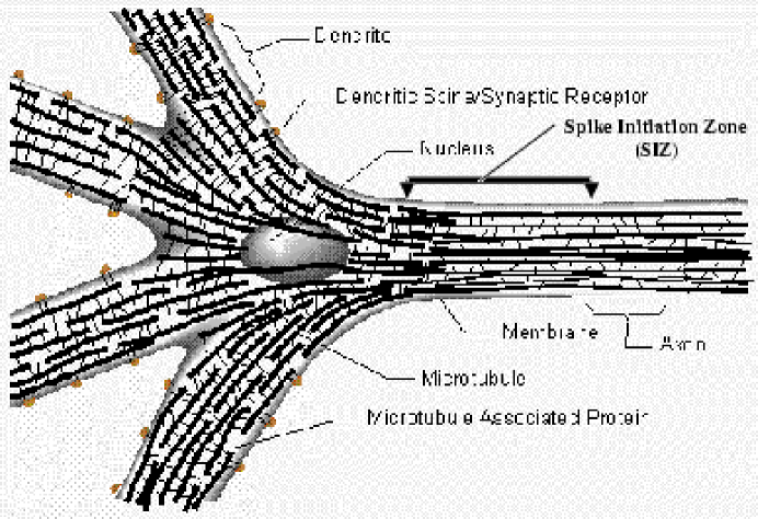
Neurons are polarized cells which are highly specialized to receive, process, transfer and store information. They are subdivided into three major parts. The soma contains the nucleus while the dendrites are relatively short, multiply branched extensions. The single axon is a long extension that branches at the neuron’s distal end. This differential architecture reflects functional differences between the two types of projections. Information is generally received by the dendrites and transmitted through the soma to the axon where it is relayed to the dendrites of neighboring neurons. Multiple neurons may be stimulated by one axon and one neuron may receive multiple axonal stimulations in each of its dendrites (see figure 2). This complexity is directly correlated to the vast capacity of the brain as a whole to receive, process and store information. It seems that there is a signal integration zone at the beginning of the axon known as the ”spike initiation zone” (SIZ) indicated by the arrows in figure 2. The SIZ is where action potentials are initiated.
2.2 Neuronal Signaling
Neurons relay information to each other via specialized structures at the dendritic and axonal termini known as synapses. The number of synapses and efficacy of information flow through them is a function of the frequency and ”importance” of information exchanged between these neurons. This neuronal property is plastic in the sense that the number of synapses and their efficacy changes as a result of prior and frequent use and this reflects memory at the cellular level. These structural changes are mediated by biochemical signaling pathways that relay the information flow to various areas of the cytoplasm and the nucleus, which in turn responds by altering gene activity to mediate the aforementioned events that underlie neuronal plasticity. The evidence supporting this classical neurobiological model is overwhelming. However, despite the vast number of possible connections within the brain, it is still difficult to explain the amount and speed at which information is processed and stored within this tissue based purely on amount and strength of synapses available. The hypotheses and proposals presented here are not in competition with the well-established neurobiological properties of cells. We hope that most if not all observed neurobiological phenomena can be explained as emergent properties of our model. Our proposals extend traditional findings by focusing on the generally neglected role of the neuronal (microtubular) cytoskeleton and its accessory proteins during information storage and retrieval.
2.3 Cytoskeleton and Microtubules
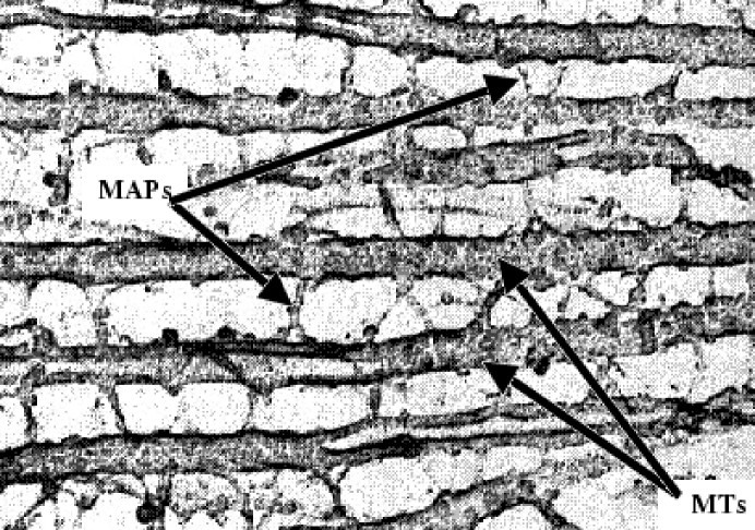
Neurons as well as all other eukaryotic cells are internally organized and held together by a scaffold made up of a network of protein polymers called the cytoskeleton. The cytoskeleton consists of microtubules, actin filaments, intermediate filaments and microtubule associated proteins (MAPs), which among other functions, link parallel arrays of MTs into networks (figure 3).
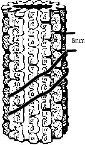
The MT’s cylindrical walls (outer diameter 25nm, inner diameter 15nm) are comprised of 13 longitudinal protofilaments. These protofilaments are constructed from a series of subunit proteins known as tubulins (figure 4). Each tubulin subunit is a polar dimer of length of about 8nm and it consists of two slightly different classes of a 4nm, 55kD (kilo-Dalton) monomer known as and -tubulin. The tubulin dimer subunits within MTs are arranged in a hexagonal twisted lattice, and helical pathways that repeat every 3, 5 and 8 rows (figure 4).
Microtubules are major components of the cell’s cytoskeleton and are involved in a variety of functions such as mitosis, axonal protein transport, signal transduction and –we claim– quantum computation. These processes are dependent on the distinctive structure of the MT.
We will concentrate our analysis to axonal MTs of neurons. The axonal MT is typically long (hundreds of nm) (figures 2 and 3) and also it is characteristically stable (compared to other cytoskeletal MTs which exhibit great dynamic instability). We have compelling theoretical indications that MTs are ferroelectric[31] and experiments are currently underway to confirm our predictions.
Furthermore, it has been suggested[5] that the ordered arrangement of water molecules provides isolation from thermal oscillations, and other potential decohering mechanisms, thus creating an environment that can support quantum entangled states of the component tubulin molecules. This is discussed in more detail in Sections 3 and 4.
2.4 Microtubule Associated Proteins
There are many types of MAPs each with different roles in cell function[32]. We are particularly interested in MAP-2 and MAP-tau, as in our model, MAP-2 phosphorylation (breakdown) and de-phosphorylation seems to play a major role in memory encoding and this has been suggested for some time[33]. MAP-2 consists of a pair of high molecular mass (280kD) proteins (isoforms a and b) and a low mass (70kD) polypeptide (isoform c). There is experimental evidence that MAP-2c may be dephosphorylated following contextual memory training in rodents[33]. Phosphorylation of MAP-2 decreases its co-assembly (binding) to MTs[28, 29, 30, 32] thus enabling cytoskeletal restructuring and favoring (dendrite) plasticity.
3 FORMATION OF COHERENT STATES IN MICROTUBULES
3.1 Ordered Water and Superradiance
There is evidence that the hollow interior of MTs may be capable of supporting a very special state of ”ordered” water molecules both inside and outside of the MT[5, 31]. We also notice that it has been recently confirmed experimentally that at the exterior of the MT cylinders, there do exist thin layers of charged ions, of thickness of order 7-8 Å, in which the electrostatic interaction energy is larger than the thermal energy due to the interaction with the environment[34] meaning that electrostatic interaction effects are dominant. In view of such results, we have previously conjectured[5] that similar layers might also exist in the interior of the MT cylinders, which provide us with the necessary thermal isolation to sustain quantum coherent states over time scales comparable to the dynamical timescales of neural cells, namely of order . This would make the MT interior act as a waveguide to photons of special frequencies and would also thermally isolate the MT interior from the environment, so that it may act in a laser-like way, a property called superradiance[35, 36, 37]. Due to the strong suppression of such couplings in the disordered states of regular, liquid water, this is not ordinarily observed. It is however quite plausible that such behavior characterizes the ordered water molecules that exist in the interior of MTs. The presence of unpaired electrons in the tubulin molecule is crucial to such a phenomenon. If true, then this coupling of the tubulins’ electric dipole to the quantum radiation will be responsible for the appearance of collective quantum coherent modes[31]. Such modes are termed ‘dipole quanta’. This mechanism has been applied to microtubules[37], with the conclusion that such coherent modes cause superradiance, i.e. create a special quantum-mechanical ordering of the water molecules with characteristic collapse times much shorter than those of thermal interaction and thus make the interior of MTs transparent to photons of certain frequencies. This has been conjectured as early as 1978 and MTs have been theorized to play the role of ‘dielectric waveguides’ for photons[38].
Such a coupling implies a ‘laser-like’ behavior. The interaction of the dipole- quanta coherent modes with the protein dimers results in an entanglement which we claim is responsible for the emergence of soliton***Solitons are special pulse-like waveforms that have well-established yet unusual physical properties including non-dispersion over large distances of propagation. Solitons are extremely resilient to noise and propagate unaltered even after interaction with other solitons. quantum coherent states, extending over large scales, e.g. the MT or even the entire MT network. An explicit mathematical construction of such solitonic states has been made in the quantum field-theoretic model for MT dynamics of Mavromatos and Nanopoulos[10, 11] in 1997, which was based on classical ferroelectric models for the displacement field discussed in other works[6]. The quantum-mechanical picture described here should be viewed as a simplification of the field-theoretic formalism, it is however sufficient for qualitative estimates of emergent properties including expected decoherence times.
3.2 How is this relevant to information processing?
To summarize, at least theoretically, there exists a credible mechanism for the formation of quantum coherent modes in the water in and around the MT, inspired by earlier suggestions of ‘laser-like’ behavior of the water[35, 36, 37] arising from the interaction of the electric dipole moments of the water molecules with photons of specific frequencies (i.e. selected modes of the quantized elelctromagnetic radiation). This is important as it provides the needed isolation from environmental noise to preserve the delicate quantum coherence between the qubits/tubulin dimers.
3.3 Possible Role for the Photons
This is a variation of the mechanism suggested earlier to establish fast (speed of light) connections between distant neurons and integrate information processing inside the MT network of a single neuron or a network of neurons. In this scenario, entanglement of neural cells over macroscopic distances is not required. Conceivably, photons emitted by MTs can be absorbed by distant neurons that are ’tuned’ to receive at specially modulated frequencies. Again, the tuning/modulation can happen via the binding of MAPs which brings us back to the GSM model of memory encoding. It is exciting to note that in most of the various suggested quantum computer designs, photons are used to communicate information from qubits to detectors.
3.4 Why no data?
Whether this is the case in all cell MTs, or only in certain areas, such as the characteristically long and stable neural MTs, is something that cannot be determined presently. Questions like these can only be answered once detailed information at the atomic scale, becomes available on the structure of tubulin dimers, on the precise magnitude of their electric dipole moments, and on the detailed structure of the water interior to MTs. As a first step in this direction we mention the atomic resolution map of tubulin, which became available only recently by means of electron crystallogaphy[39].
4 BASICS OF QUANTUM COMPUTATION
4.1 Why Quantum Computation?
In our quantum brain hypothesis the brain is modeled as a vast network of interconnecting neurons which have the potential of isolated and parallel quantum computation. As a result, in order to understand this hypothesis it is necessary to grasp the basics of quantum computation.
The possibility of creating quantum computers is being thoroughly investigated by numerous research groups around the world both theoretically[40, 41] and experimentally[42] (for a review see Preskill[43]). Quantum computation envisions quantum computers utilizing qubits rather than conventional bits. Some of the advantages of quantum computers of the future are better speed, gigantic memory capacity and immense computing power. The most intriguing aspect of quantum computers is their ability to perform tasks that are simply impossible using classical computers. The two most celebrated such abilities are efficient factorization of large numbers[40] and search through unordered data lists in times faster than allowable classically[41]. Behind both these feats lies an integrated and ”delocalized” way of handling data that makes the machine capable of retrieving stored information in much the same way the human brain does during pattern recognition. Furthermore, the existence of a quantum-error correcting code is needed to protect the delicate coherent qubits from decoherence. This has been the major problem of quantum computers until the works of Shor and Steane have independently shown that such a code can be implemented[40, 44]. We conjecture that the K-code apparent in the packing of the tubulin dimers and protofilaments is partially responsible for keeping coherence among the tubulin dimers. By simulating the brain as a quantum computer it seems we are capable of obtaining a more accurate picture than if we simulate the brain as a classical, digital computer.
Although based on the well-established physical principles of quantum mechanics, quantum computers are yet to be experimentally realized. This has not deterred theoretical work in the field and even the writing of ”quantum software” in the form of mathematical algorithms that take advantage of quantum computers yet to be developed[18, 40, 41]. Note that recently there has been progress towards creating the necessary apparatus that will ultimately provide us with quantum logic gates[42]
4.2 Quantum Mechanics and Quantum Computing
What’s so different about quantum computers?
The main difference between classical, digital computation (on which the traditional approach to simulating the brain using neural networks is based) and quantum computation is the latter’s usage of quantum bits rather than ordinary bits. In our approach, we treat the tubulin dimer as a quantum two-state system which represents one qubit i.e. the building block of a quantum cluster. Tubulin protofilaments that make up MTs play the role of quantum clusters and whole MTs can be thought of as blocks of clusters.
What’s so different about Quantum Mechanics?
For three quarters of a century, quantum physics has been universally accepted as the most accurate account of the phenomena of the microcosm and arguably the most accurate and precise scientific theory ever. Atoms, nuclei and elementary particles can be correctly described only by using the mathematical framework of quantum physics called quantum mechanics. The name is an analogy to classical Newtonian mechanics, which can be derived as an approximation to quantum mechanics when the objects of study are large in mass. Although Newtonian mechanics has been used for centuries to adequately explain the motion of everyday macroscopic objects, it proved grossly inadequate with the discovery of the atom.
Introduction to Quantum Mechanics.
The account that follows has been written in the fashion of an introduction to the subject, requiring no more mathematical ability than elementary algebra. It is intended to give a ”first taste” of the quantum mechanical approach and discuss the relevance of entangled states to fast communication of correlations among neurons. A complete, fully mathematical treatment of MTs can be found in a number of sources[4, 5, 10, 11]. Due to the special properties exhibited by microscopic systems, special jargon, often counterintuitive to the unseasoned reader must be employed.
Quantum systems have two modes of evolution in time. The first, governed by Schrödinger’s equation (see below) describes the time evolution of quantum systems when they are undisturbed by measurements. ’Measurements’ are defined as interactions of the system with its environment. As long as the system is sufficiently isolated from the environment, it follows Schrödinger’s equation. If an interaction with the environment takes place, i.e. a measurement is performed, the system abruptly decoheres i.e. collapses or reduces to one of its classically allowed states.
In what follows we will employ Dirac bracket notation, where the ket is analogous to a column vector , and the bra is the complex conjugate transpose of which means it is a row vector where all entries have been complex-conjugated. . Time evolution of quantum systems (in the absence of measurements) is described by the Schrödinger equation: where H is the Hamiltonian (energy) operator (see below), and is Planck’s constant divided by 2.
Linear superposition is a generalization of the familiar mathematical principle of linear combination of vectors. Instead of using three orthogonal axes as a basis, quantum systems are described by a wavefunction that exists in a multi-dimensional ”Hilbert Space”[45]. The Hilbert space has a set of states (where the index i runs over the degrees of freedom of the system) that form a basis and the most general state of such a system can be written as . The system is said to be in a state which is a linear superposition of the basis states with weighting coefficients that can in general be complex. At the microscopic or quantum level, the state of the system is described by the wave function , which in general appears as a linear superposition of all basis states. This can be interpreted as the system being in all these states at once. It is known that the tubulin dimer undergoes conformational changes as a result of a shift in the localization of the electron orbitals in its hydrophobic pocket. Therefore, a superposed state of the tubulin dimer would have the interpretation of the dimer being in both of its allowable conformational states at the same time something which is not allowable classically.
The coefficients are called the probability amplitudes and gives the probability that will collapse into state when it decoheres (interacts with the environment). By simple normalization we have the constraint that . This emphasizes the fact that the wavefunction describes a real, physical system, which must be in one of its allowable classical states and therefore by summing over all the possibilities, weighted by their corresponding probabilities, one must obtain unity. Further, this fact stresses that quantum mechanics is not simply an alternative treatment of such two-state systems as tubulin dimers but rather it is the correct mathematical treatment. A quantum mechanical treatment of the tubulin dimer does not rely on approximations contrary to the case when a tubulin dimer is treated using classical electrodynamics, thermodynamics and statistical physics (which is the usual approach in biophysical investigations of protein molecules).
Note that at the macroscopic or classical level, a quantum two-state system can only be in a single basis state. For instance, the quantum position (energy) of an electron can be in a superposition of two different orbitals (energies) while in the classical case this is impossible. Equally, the tubulin dimer can only be experimentally observed (measured) in one of its two allowable conformations.
4.3 Role of Coherence & Entanglement in Recall & Binding
A quantum system is coherent if it is in a linear superposition of its basis states. If a measurement is performed on the system and this means that the system must somehow interact with its environment, the superposition is destroyed and the system is observed to be in only one basis state, as required classically. This process is called reduction or collapse of the wavefunction or simply decoherence and is governed by the form of the wavefunction .
In this notation, the probability that a quantum state will collapse into a basis state is written in terms of the inner or scalar product which is analogous to the familiar dot product between two vectors . The simplest system which would be analyzed as described above, would be a two-state system. For instance, an electron (spin–) system, where we are interested in measuring the electron’s spin in a specified direction (customarily the z-axis). The general notions of the simplified mathematical treatment that follows can also be applied to the tubulin dimer[4]. The actual experimental setup for measuring the orientation of the spin of an electron is called a Stern-Gerlach (SG) apparatus described in detail in a number of sources, e.g. [45]. When inserted into an SG magnet, the electron can either register as spin-up ( ) or spin-down ( ). In this system, the wavefunction is a distribution over the two possible values and a coherent state is a linear superposition of and . One such state can be: = +
As long as the system remains in a coherent state, we cannot say that the electron is in either the up- or down-spin states. In a counter-intuitive sense, it is in both states at once. Classically, the electron can only be in one state, so if measured, the system decoheres to give spin up with probability:
and spin down with probability 20%.
This simple two-state quantum system is used as the basic unit of quantum computation and is referred to as a quantum bit or qubit.
In classical, digital computers, the basic unit of computation is a bit. A bit can have the value 1 or 0. Digital computers encode information using arrays of bits in the form of billions of solid-state transistors integrated to form microchips. The voltage at each transistor can take two values making it a 1 or a 0. Computation i.e. manipulation of information, is performed by logic gates which follow Boolean algebra rules. In quantum computers, the logic gates are replaced by quantum operators. Operators on a Hilbert space describe how one wavefunction is changed into another. An eigenvalue equation shows the action of operators. For instance, an operator A acting on one of its own basis states will produce the same state multiplied by its eigenvalue , namely:
The solutions of the eigenvalue equation are called the eigenstates and can be used to construct a basis. In quantum mechanics, we assign operators to all the physical properties of a system (such as position, momentum, energy) and the eigenvalues of these operators give us the allowed physical values of those properties.
As it will shortly become important, note that while interference is a commonly observed classical wave phenomenon, it has also been experimentally shown to apply to the probability waves of quantum mechanics. For a simple theoretical example, consider the initial wavefunction for the spin– electron system described earlier. Using the conventional vector assignment,
= , =
we can rewrite our wavefunction in vector form as:
=
When acted upon by some operator A, where A is defined to be:
the result is:
| (4.7) |
Note that as a result of the action of A on our initial state , the amplitudes of the spin-up and spin-down states have changed. The operator has made the wavefunction interfere with itself and its constituent parts experienced the analogue of classical interference so that the up state interfered constructively while the down state destructively.
Entanglement on the other hand, is a purely quantum phenomenon and has no classical analogue. It accounts for the ability of quantum systems to exhibit correlations in counterintuitive ”action-at-a-distance” ways. Entanglement is what makes all the difference in the operation of quantum computers versus classical ones. We will present a short mathematical description here without using density matrix formalism.
If we wish to describe the state of two electrons (spin–), or equally, the state of two tubulin molecules, we may use Dirac bra-ket notation where the first entry in a ket refers to the state of the first electron (conformation of first dimer) while the second entry refers to the second electron (second dimer). For instance, let us take a quantum state made up of two electrons where the first is in the spin-down state with certainty while the second is in a coherent state of spin-up and spin-down with equal probability.
= +
another may be state :
= +
and a state could be:
= + +
where all states are indexed by the state labels 00,01,10,11.
These three states are different from each other in the sense that although can be factorized using normal tensor product () as follows:
cannot be factorized. States that cannot be factorized are said to be entangled states. Note that can be factorized in two different ways but not completely. There are degrees of entanglement as states can be less or more entangled, depending on whether they are completely, partially or not at all factorizable and the three states , and demonstrate this.
Entanglement gives ”special powers” to quantum computers because it gives quantum states the potential to exhibit and maintain correlations that cannot be accounted for classically. Correlations between bits are what make information encoding possible in classical computers. For instance, we can require two bits to have the same value thus encoding a relationship. If we are to subsequently change the encoded information, we must change the correlated bits in tandem by explicitly accessing each bit. Since quantum bits exist as superpositions, correlations between them also exist in superposition. When the superposition is destroyed (e.g. one qubit is measured), the correct correlations are instanteaneously “communicated” between the qubits and this communication allows many qubits to be accessed at once, preserving their correlations, something that is absolutely impossible classically.
”Software” that makes use of this possibility has already been developed in the form of factorization[40], sorting[41] and learning and memory[18] algorithms. This communication of correlations is a manifestation of the well-known Einstein– Podolski–Rosen (EPR) paradox. A simplified example follows.
Consider the case of the pion (), a neutral elementary particle of spin 0 (i.e. internal angular momentum 0). Pions decay spontaneously into two oppositely polarized photons. Photons carry angular momentum in their helicity. Since () decay is spontaneous i.e. no external forces have acted on the system, angular momentum conservation requires the decay products to have the same total angular momentum as the decaying particle –in our case a sum of zero. This means that if one photon is detected with helicity +1 the other must have helicity -1 to conserve angular momentum. We can write the entangled state of the two emerging photons as:
= +
where the subscripts 1 and 2 refer to the first and second photons respectively and the +/- 1 entries in the kets refer to each photon’s helicity. This state is completely entangled as it cannot be factorized to give separate states for each photon. This indicates that if the product photons are isolated from the environment and separated macroscopically (say by letting them move apart inside an optical fiber), measuring one photon’s polarization will immediately determine the polarization of the other by collapsing the entangled wavefunction instantaneously and non-locally.
5 EXPERIMENTS
5.1 Experiments
Our novel phenomenological approach to understanding the role of MTs in information processing has produced several theoretical predictions, which we aim to support with experimental evidence. Ideally, we would like to test our predictions in vivo by examining the effects that learning and memory encoding have on the MTs of a living animal. One way to do this would be to disrupt MTs by using mutations. However, it is impossible to use animals that harbor mutations that change functional aspects of MTs as the MTs’ correct function is essential for the viability of an organism. Instead, we have designed and are performing several indirect neurobiological experiments. We anticipate to obtain the first-ever experimental results designed to test the quantum properties of living matter.
5.2 Description of the experimental system
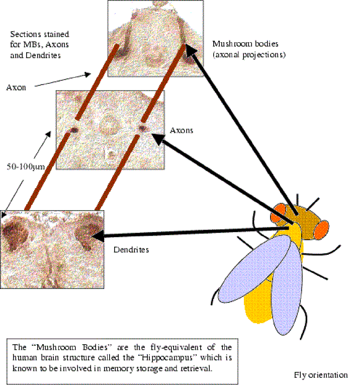
We utilize a well-established, olfactory conditioning protocol to teach Drosophila melanogaster fruit flies to avoid certain odors contingent upon negative reinforcement by electric shock. Following behavioral conditioning and ascertaining acquisition of information, the flies are fixed, their heads sectioned and stained (immunochemically) for the distribution of tau, or microtubule-associated-protein-2 (MAP-2) in the mushroom bodies (an area of the fly’s brain essential for information correlation and memory formation). We are interested in determining whether, as predicted by our model, the distribution of tau and/or MAP-2 will change as a result of memory encoding.
5.3 Why Use Drosophila?
The Drosophila melanogaster fruit fly has long being favored by experimental biologists for numerous reasons including its relatively simple genetic makeup (genome) and quick generation time, powerful classical and molecular genetics and their ability to learn and remember a variety of tasks. However, Drosophila is simply the ideal system for our research for a different reason. In order to track redistribution of tau and MAP-2 in the neurons we must be able to differentiate between the various parts of the neuron such as the dendrites, axons, axonal projections and somata. In humans and other mammals, the neuronal organization is such, that multiple neurons and neuronal types are involved in a given process forming an extensive complex network of axons and dendrites. As a result, it is very difficult to locate specific parts of individual neurons and stain selectively to track changes in distribution of a particular protein. In Drosophila on the other hand, mushroom body neurons represent a highly ordered, tightly packed bundle (see figures 5 and 8).
The mushroom bodies are bilateral clusters of about 2500 neurons, situated in the dorsal and posterior cortex of the Drosophila brain (figure 5). The dendrites of all mushroom body neurons aggregate to form a distinctive structure just ventral to the cell bodies where inputs arrive conveying sensory information. The axons of these neurons bundle together (fasciculate) and project to the anterior of the brain. There, they bifurcate, with some axonal processes extending medially and others projecting dorsally[46] (figure 5). Flies that lack mushroom bodies are able to smell, but totally unable to learn the olfactory associative learning task[47, 48].
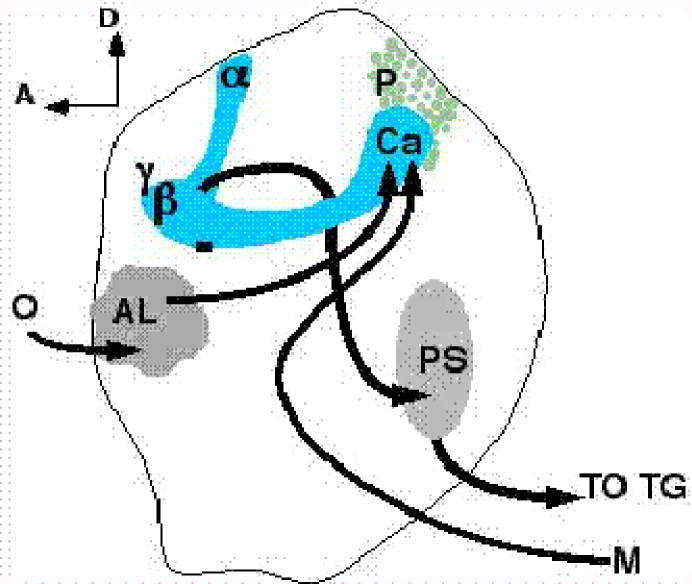
Therefore, the Drosophila system enables us to target a particular set of neurons easily identifiable and well described in their properties. This is essential for our analysis of bulk movement of microtubule-associated proteins within neurons.
5.4 Conditioning Protocol
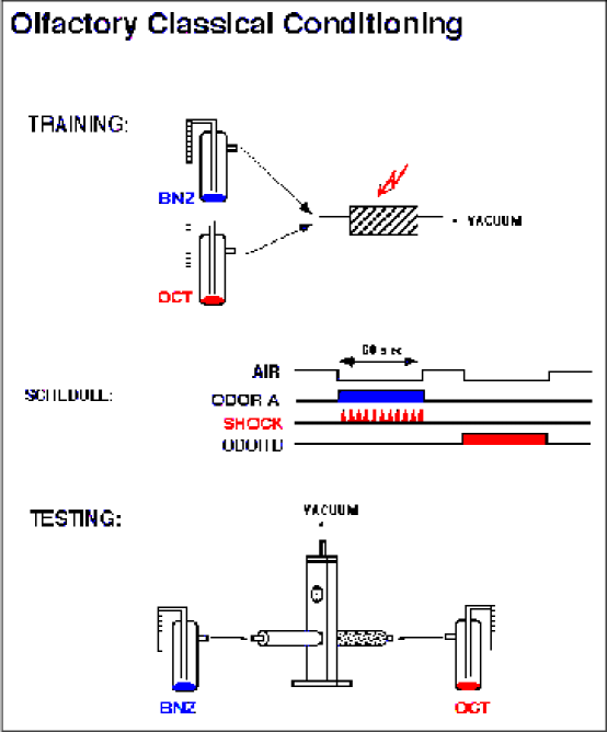
A qualitative description of our protocol[46, 47, 48, 49] for training wild type Drosophila Melanogaster fruit flies follows. Drosophilae are naturally attracted or repulsed by different odors with a variety of affinities. We use the standard, negatively reinforced associative learning paradigm which couples olfactory cues with electric shock to condition flies. We used two equally aversive odorants: 3-Octanol (OCT) and Benzaldehyde (BNZ). The training apparatus consists of a training chamber and a selection maze. The maze is normalized by adjusting the concentration of odorant. Once normalized, wild type, naive (i.e. untrained) flies choose to enter one of two identical tubes smelling of OCT and BNZ respectively, with a probability of 50% since they avoid both odors equally.
Training, or rather conditioning, of the flies takes place as follows. A batch of wild type, naive flies (numbering between 50 and 60) are collected under light anesthesia (using ) and 12-24 hours later are left in the dark for one to two hours. The entire procedure of conditioning the flies takes place in a temperature- and humidity-controlled darkroom. This is done in order to isolate the effects of olfactory stimulation from visual stimulation. Once the flies have been acclimated to the darkroom, they are inserted into training chamber A whose walls are electrified by a signal generator set to 92.0V. The flies receive twelve electrical shocks (of duration 1.25sec each) for one minute. During this time, the chamber is filled with air containing OCT. The flies are given 30 seconds to rest while the air is being cleared of odorants and are then given the opposite (control) odorant (in this case BNZ) for another minute in the absence of electrical shocks. A rest period of 30 seconds follows after which the flies are tested for acquisition of information. They are inserted into the selection maze and given the choice of entering a chamber smelling of OCT or an identical one smelling of BNZ. For control and consistency purposes, the experiment is done simultaneously in apparatus B with the shock-associated and control smells reversed while everything else remains identical. We define a ”trained” fly as one that has chosen to go into the chamber filled with the control odor after given the choice for 90 seconds. The procedure is illustrated in figure 6 below.
It is observed that following training, a good percentage of the flies choose to avoid the smell that was present when they received the electrical shocks. The percentage is calculated as a normalized performance index (PI) where
.
Typical PI values for our experiments have been between 75 and 90 giving us confidence that the flies have truly learned to associate the stimuli. This procedure alters the probability of response of the flies to the stimuli. Re-testing the flies that made the correct choice producing a PI of 90 will not result in 100% of the flies avoiding the shock associated odor, but rather will result in a distribution producing a PI of 90 again. Therefore, the behavioral changes of the flies parallel the probabilistic response of neuronal firing.
5.5 Fixation, Sectioning and Staining
Once the flies have made their choice, those that made the ’correct’ choice are immediately killed by submersion into a fixative solution without subjecting them to anesthesia. An equal number of naive flies that have not been exposed to the training apparatus but are otherwise identical to the trained ones are also fixed. We used three different fixing protocols of different fixation strength each[49]. Following fixation the flies were dehydrated through a series of ethanol baths (0-100%) and Methylbenzoate preparing them for embedding in Paraffin, decapitating and sectioning. It is clear that this experimental approach is likely to capture differences in the distribution of tau and MAP-2 between trained and naive flies and consequently provides us with a way of testing whether their distribution is affected by training as predicted by the quantum brain hypothesis and also suggested by recent studies in rodents[28, 29, 30]. There are however, a number of complications. In order for the distribution of microtubule associated proteins to be seen in the microscope, one must bring it up from the background by immunochemical methods which use in situ chemical staining reactions to indicate the localization of the proteins within the cell. This involves obtaining antibodies (which are also proteins) that selectively and specifically attach to tau or MAP-2. Following standard immunochemical procedures, the antibodies become linked to chromogenic (staining) substances that allow visualization of the distribution of the protein under investigation in the tissue of interest.
Note that there is an important underlying assumption here: the fixative solution is required to fix the tissue as it was at the instant of death so that all proteins in the neurons are permanently bound to their last location before death. It further assumes that embedding in Paraffin will not affect the binding of MAPs to MTs or the structure of MAPs themselves. Such changes may alter the structure of MAPs in ways that will make them no longer identifiable by the antibodies. Historically, fixation complications have been circumvented by using a variety of fixatives and fixation conditions with good results despite the lack of complete theoretical understanding as to the exact action of the fixatives.
Furthermore, due to resolution and staining limitations, it is not possible to directly test the GSM (guitar string model) unless there is sufficient relocation of MAPs. At least in this modus operandi, we have had no choice but to assume that the training has been sufficiently intensive so that the result of encoding this memory was to dramatically change the MAP-2 distribution in a large number of neurons.
5.6 Results
We have been successful in our initial experiments to train flies and test their learning and memory for up to 6hrs. This is an essential point as we are not certain of the exact timing of the proposed redistribution of microtubule associated proteins. However, our initial attempts to localize MAP-2 within the fly brain have been unsuccessful. It must be noted that in our experiments, we used monoclonal antibodies which only recognize one binding site at the target protein and if that site is ”buried” in the secondary structure of the protein, the antibody will not bind. A solution to this problem is to use polyclonal antibodies, which have a number of binding sites on their target protein. We are currently in the process of trying a number of anti-MAP-2 polyclonal antibodies to select the one that best reveals the MAP-2 distribution in the fly brain.
A more interesting interpretation of these initial results is that the antibody binding sites on MAP-2 are ”masked” due to its interaction with the MTs, but upon training and during the proposed re-distribution, these sites may become available for detection. We are in the process of addressing this possibility by training flies and fixing them at different times (0 to 360 minutes) past training, to investigate whether these proposed sites become available which would be evidenced by immunological staining.
5.7 Experiment 2 Basics
Tau is another microtubule associated protein. In humans, it plays a role similar to MAP-2 and it seems that tau is of paramount importance in keeping axonal MTs parallel and aligned.
We have obtained transgenic flies that will express human tau-protein in their mushroom bodies. This is important as tau has long being implicated in the encoding of human memory and it has recently been shown that mutations in the human NC-17 tau gene are one of the causes of Alzheimer’s Disease (AD)[20]. In fact, earlier theoretical research by this group has led us to assert this prior to it becoming well accepted. We had claimed that subneural abnormalities such as Neurofibrillary Tangles (NFTs) and abnormally phosphorylated tau are the main causes of AD symptoms, rather then the other way around. NFTs are axonal MTs that have lost their structural integrity due to the inability of mutated or hyperphosphorylated tau to hold them in parallel and as a result have been tangled up and are unable to function properly.
5.8 Motivation, Relevance to Alzheimer’s Disease
By inducing flies to express the human tau-gene specifically in their mushroom bodies, we anticipate that we will in fact be replacing, at least to some extent, the MAP the fly actually uses to hold its MTs together by human tau-protein. We do not know a priori what to expect, as the flies can exhibit an increase or decrease in learning performance, or there might be no overt phenotype. In the case that there is observable phenotype in their learning, we will be able to deduce something about the role that MAPs in general play in memory encoding. We already know that the introduced tau gene is not lethal and a preliminary investigation does not indicate anomalies in general feeding, mating, or circadian behaviors of the flies. Furthermore, assuming that tau will play a similar role as it does in humans, we will be able to test whether overproduction of tau in older flies makes them susceptible to ”dementia” in the form of a neurodegenerative disease such as Alzheimer’s. In the case that the flies exhibit learning deficits, we will examine their brains for histological hallmarks such as NFTs. Alternatively, the introduction of human tau may reduce or eliminate the observed age dependent decline in the learning capability of fruit flies.
5.9 Protocol
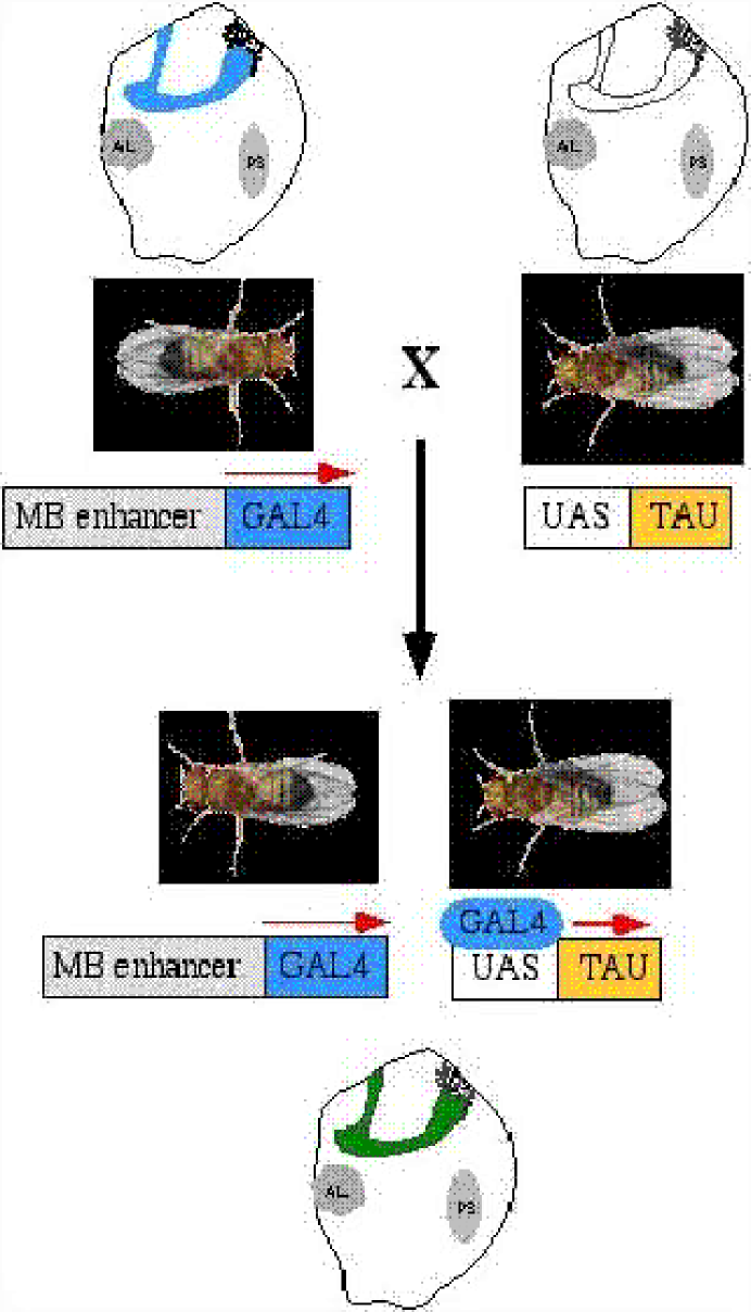
How does one go about persuading a fly to create a human protein in its brain? This process is called directed gene expression and uses genetic engineering to force the expression of genes in specific tissues, even if they are alien to the organism. This method also allows turning gene expression on or off at specific times. The main idea is to have two genetically manipulated lines the first of which contains a gene of choice (human tau in our case) fused to and under the direction of an upstream activating sequence (UAS) activated only by the presence of its unique, selective and specific activator protein GAL4 in the same cell. To generate lines expressing GAL4, the GAL4 gene is inserted randomly into the fly’s genome in front of various genes expressed in specific tissues at specific developmental times (temporal control) due to the action of their native enhancers dictating this expression pattern. The GAL4 transgene ”usurps” these native enhancers, resulting in its tissue and temporal specific expression. A GAL4 target gene (UAS-tau) will remain silent in the absence of GAL4. To activate the target gene, the flies carrying the UAS-tau are crossed to flies expressing GAL4 and thus in their progeny, the UAS-tau transgene will be expressed in the tissue and temporal specific pattern specified by the GAL4. This is illustrated in figure 7 below.
5.10 Fixation, Sectioning and Results
This experiment is currently underway. To ascertain that we have flies expressing human tau in their mushroom bodies we must first test whether the GAL4 ”driver” line directs expression in the mushroom bodies as advertised. To do this, we cross flies that contain the GAL4 gene with flies that contain another gene whose activity can be readily monitored (reporter gene) by histological methods. We have used the bacterial beta-galactosidase gene (UAS-LACZ).
Flies that are the progeny of GAL4xLACZ will have beta-galactosidase activity in their mushroom bodies visualized as blue pigment. This provides us with a simple test of where the actual tau-gene is going to be expressed once activated in a GAL4xtau cross.
The sectioning procedure employed here is different from the one for experiment 1. The naive fly heads are cryo-sectioned by freezing to and an embedding gel is used instead of chemical fixation which would destroy the activity of the reporter gene. The staining is provided by the activity of the reporter LACZ gene which converts a colorless substrate into a blue precipitate within the tissue where the reporter is expressed. The results of this preliminary experiment are encouraging as it is seen beyond doubt that the mushroom bodies as well as certain other sections of the fly brain do indeed express LACZ indicated by the blue color in the sections. This is illustrated in figure 8.
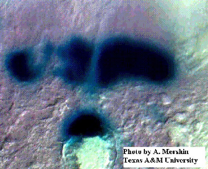
The next steps of this experiment are to first investigate the learning phenotype of the GAL4xtau flies and then proceed with the sectioning and staining methods described for experiment 1 using anti-tau antibodies to visualize potential changes in the distribution of tau immediately after training and at later times to assess changes due to memory formation. Figure 9 shows our preliminary immunohistochemical results where the dark staining corresponds to expression of tau. The flies used for this were naive, i.e. have not been exposed to the training apparatus.
6 SUMMARY, DISCUSSION AND CONCLUSION
6.1 Summary
This review gives an overview of the new field of ’quantum physics motivated neurobiology’. A biological model of memory was presented that stressed the role of the cytoskeleton in encoding memory. The quantum brain hypothesis was outlined and some phenomenological aspects discussed. Some fundamental aspects of cell and molecular neurobiology were presented, concentrating on microtubules and microtubule-associated-proteins. The reasons why we believe quantum coherence is a realistic possibility even at the scale of MTs and even at temperatures of order were outlined in Section 3. Quantum computation was discussed and a brief example of entangled states of qubits was presented in Section 4. In Section 5, two experiments targeted at indirectly testing the phenomenological predictions derived from our quantum theory of brain were described and some preliminary results presented.
6.2 Discussion
It cannot be stressed enough that this line of research is suffering from a total lack of in situ, direct quantum mechanical experiments. Recently published[34] and some yet unpublished efforts by Zioutas et. al. concentrate on showing that MTs are indeed ferroelectric. They use detection of hypothetical ferro-to-paraelectric state phase transitions due to temperature changes to investigate the conjectured ferroelectric properties of MTs. They employ a novel approach where they try to detect electromagnetic radiation (of order some GHz) emitted by MTs as a result of the expected phase transition.
The theoretical aspects of this work have been criticized by some physicists claiming that quantum coherence is impossible in the C biological environment[12]. It has been argued that if one sets up a wavefunction describing a whole neuron including its membrane and the surrounding ions it will decohere with decoherence times no longer than s due to collisions between ions and molecules as well as long range Coulomb interactions. This certainly seems plausible and biological manifestation of quantum mechanical effects will not be observed since the biological dynamical timescale is of order . This argument is flawed in assuming that the entire neuron must be in a coherent state of ”firing-and-not-firing” and thus for a typical axon up to order Na+ ions must be in a superposition of being in-and-out of the axon and in coherence with each other and the membrane. The firing or not of the neuron is an emergent property and it seems unlikely that quantum mechanics will apply directly to the generation and propagation of APs let alone the bulk dynamics of extracellular ions. Our model does not suggest superposition of ions and membrane.
Further, it has been argued[12] that even MTs will have difficulty sustaining coherence and an estimate of the decoherence time is given to be of order . The dominant decoherence mechanism for these considerations is assumed to be Coulomb interactions with closest neighboring ions. Screening effects are not considered by arguing that the closest ions will be the ones doing the screening and they are also the ones effecting decoherence. This, it is claimed, does not allow information propagation by the mechanisms suggested. However, as described in Section 3, the particular ordered arrangement of molecules inside and outside the MT as well as the presence of the K-code may well protect MTs from such decohering mechanisms[5]. It is an ongoing effort[6] to further unveil how these properties will affect decoherence-time estimates. Decoherence may well be effected by strong electromagnetic interactions resulting from neurotransmitter binding and this is exactly the kind of decoherence mechanism which should be most favored by biologists since neurotransmitter action and consequent AP propagation along the membrane must somehow ”reset” the system to where it can receive the next ”information package”. It remains to be seen if by further theoretical analysis and biophysical experimentation, the decoherence time can be conclusively brought up to match the dynamical time scale.
A natural extension of the quantum hypothesis is that there is place for quantum coherent effects in other, non-neural cells. In fact anything with a cytoskeleton-like structure, any protein whose function depends on electron mobility (and this includes all known proteins) can be treated as a fundamentally quantum mechanical object. Whether there are observable emergent properties depends on the system at hand but it seems that the difference between neural and ordinary cells is made by the characteristically ordered and long MTs in the axons and dendrites. It remains to be seen what role quantum mechanics is going to play in the molecular biology of the future.
6.3 Conclusion
This review is an account of the initial steps of what we expect to be a long process of theoretical and experimental research in this field. What we have achieved so far is to design and conduct the first ever experiments capable of indirectly testing some of the predictions of the quantum brain hypothesis. It cannot be stressed enough that the experiments described here can only tell us whether the quantum hypothesis, at least in its present formulation, is wrong. Even if all of the theoretical predictions are shown to hold in the laboratory, these results can have other, more conventional, interpretations. For instance, if we are able to show a definite redistribution of MAPs as a result of learning, it can be argued that this changes neural cells in ways which do not directly depend on quantum coherence e.g. axonal transport can be altered thus affecting synaptic weight and effectively training the neuron. As discussed earlier, in order to discover at which level the transition from quantum to classical takes place, direct quantum mechanical experiments on the MT system are needed but those seem to be quite far ahead in the future.
What we have succeeded in doing is to show that the quantum hypothesis is experimentally falsifiable. We have made the first step in the phenomenology of the quantum brain and this has opened up the way for more experiments and will hopefully make this hypothesis more attractive to mainstream theoretical and experimental physicists and biologists.
It certainly is an astonishing premise that neurobiological processes taking place in a fly’s brain are fundamentally tied to quantum events and this brings us full circle to the long conjectured connection between quantum physics and human consciousness.
7 Acknowledgments
The work of DVN supported in part by grant no. DE–FG–0395ER40917.
References
- [1] Penrose, R. ”The Emperor’s New Mind”. (Oxford University Press, Oxford 1989)
- [2] Penrose, R. ”Shadows of the Mind”. (Oxford University Press, Oxford 1994)
- [3] Hameroff, S.R. & Penrose R. in Toward a Science of consciousness: the first Tucson discussion and debates. Edited by Hameroff S. R., Kaszniak, A.W. & Scott. A.C. (MIT Press, Cambridge), 507-539 (1996)
- [4] Nanopoulos D.V. hep-ph/9505374 from XV Brazilian National Meeting on Particles and Fields, (1994) and Physics Without Frontiers Four Seas Conference (1995)
- [5] Mavromatos, N.E. & Nanopoulos, D.V., in Advances in Structural Biology 5 283-318 (JAI Press Inc, London 1998) Malhotra S.K., Tuszynski J.A. Eds. Also in quant-ph/9802063
- [6] Sataric M.V., Tuszynski J.A. & Zakula R.B. Phys. Rev. E48 589 (1993)
- [7] Frölich, H. Int J. Quantum Chem. 2, 641-649 (1968)
- [8] Frölich, H. Nature 316, 349-351 (1970)
- [9] Frölich H.& Kremer F. Coherent excitations in biological systems (Springer-Verlag, New York) (1983)
- [10] Mavromatos, N.E. & Nanopoulos, D.V. Int. J. Mod. Phys. B11 ****** (1997)
- [11] Mavromatos, N.E. & Nanopoulos, D.V. Int. J. Mod. Phys. B12 517-542 (1998)
- [12] Tegmark, M. quant-ph/9507009 submitted to Phys.Rev. E (1995)
- [13] Edelman, G., Gall, W.E. & Cowan W.M. Synaptic Function (John Wiley & Sons, New York) (1987)
- [14] Voronin L.V. Synaptic modifications & Memory An electrophysiological Analysis (Springer-Verlag, New York) (1993)
- [15] Haas H.L. & Buzsaki G. Synaptic Plasticity in the Hippocampus, (Springer-Verlag, New York) (1988)
- [16] Dermietzel, R. Brain Research Reviews xxx C 97000 123-133 (1997)
- [17] Fitzgerald, R. Physics Today 17-19 March (1999)
- [18] Ventura D. & Martinez T. quant-ph/9807059 submitted to IEEE Transactions on Neural Networks (1998)
- [19] Hameroff, S. R. & Penrose, R. Scale in conscious experience: Is the brain too important to be left to specialists to study? 243-274 (Mahwah, Erlbaum) (1995)
- [20] Vogel, G. Science, 280, 5 June (1998)
- [21] Arriagada P. et. al. Neurology, 42 631 (1992)
- [22] Franks,N.P. & Lieb W.R. Nature 300 487-493 (1982)
- [23] Franks,N.P. & Lieb W.R. Nature 316 349-351 (1985)
- [24] Hameroff, S.R. & Watt, R.C. Anesth..Analg. 62 936-940 (1983)
- [25] Koruga, D. L. Ann. NY Acad. Sci. 466, 953-955 (1986)
- [26] Clark, I. J.Biochemistry and Bioenergetics 41 59-61 (1996)
- [27] Martin, K.C., Casadio, A., Zhu, H., Yaping, E., Rose, J.C., Chen, M., Bailey, C.H. & Kandel, E.R., Cell 91, 927-938, Dec. (1997).
- [28] Woolf, N.J. Neurobiology of Learning and Memory 66 258-266 (1996)
- [29] Woolf, N.J. Progress in Neurobiology 55, 59-77 (1998)
- [30] Woolf, N.J., Zinnerman, M.D. & Johnson, G.V. Brain Res. 821(1), 241-249 (1999)
- [31] Mavromatos N.E., Nanopoulos D.V., Samaras I. & Zioutas K. in Adnances in Structural Biology 5 127-134 (JAI Press Inc., London 1998)
- [32] Kandel, E.R., Schwartz J.H., Jessel T.M. Essentials of neural science and behavior (Appleton & Lange, Norwalk, Connecticut 1995)
- [33] Fukunaga, K., Muller, D. & Miyamoto, E. Neurochem. Int. 28, 343-358 (1996)
- [34] Sackett D., based on a presentation at the workshop Biophysics of the Cytoskeleton. Banff, August 18-22 1997, Canada
- [35] Del Giuduce, E., Preparata, G., Vitiello, G. Phys. Rev. Lett. 61 1085 (1988)
- [36] Del Giuduce, E, Doglia, S., Milani, M., Vitiello, G. Nucl. Phys. B251(FS13) 376 (1985 ibid B275 (FS 17) 185 (1986)
- [37] Jibu, M., Hagan, S., Hameroff, S.R., Pronbram, K., Yasue, K. Biosystems 32 195(1994)
- [38] Hameroff, S., R. Am.J.Clin.Med. 2 149 (1978)
- [39] Nogales, E., Wolf, G. & Downing, G., Nature 39, 124-134 (1998)
- [40] Shor, P. SIAM Journal of Computing, 26 no. 5 1474-1483 (1997)
- [41] Grover, L. Proceedings of the 28th Annual ACM Symposium on the Theory of Computing (ACM, New York) 212-219 (1996)
- [42] Sharf, Y. & Cory, D. G. quant-ph/0004030 (2000)
- [43] Preskill., J. Physics Today June (1999)
- [44] Steane, A.M. Phys. Rev. Lett.,77 793 (1996)
- [45] Sakurai, J. J. Modern Quantum Mechanics Revised Edition (Addison-Wesley, New York 1995)
- [46] Davis, R.L., Neuron, 11 1-14 (1993)
- [47] Davis, R.L., Physiological Reviews, 76(2) 299-317; (1996)
- [48] Skoulakis, E.M.C. & Davis, R.L. Neuron, 12 931-944 Nov. (1996)
- [49] Tully T. & Quinn, W. J. Comp.Physiol. 157, 263-277 (1985)
- [50] Hirokawa, N. The Neuronal Cytoskeleton Burgoyne R.D. (Wiley-Liss, New York.) 5-74 (1991)
- [51] Amos, L.A. & Klug, A. J. Cell Sci. 14 523-550. (1974)