Kinetics of viral self-assembly: the role of ss RNA antenna
Abstract
A big class of viruses self-assemble from a large number of identical capsid proteins with long flexible N-terminal tails and ss RNA. We study the role of the strong Coulomb interaction of positive N-terminal tails with ss RNA in the kinetics of the in vitro virus self-assembly. Capsid proteins stick to unassembled chain of ss RNA (which we call ”antenna”) and slide on it towards the assembly site. We show that at excess of capsid proteins such one-dimensional diffusion accelerates self-assembly more than ten times. On the other hand at excess of ss RNA, antenna slows self-assembly down. Several experiments are proposed to verify the role of ss RNA antenna.
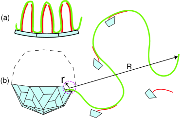
Viruses self-assemble in host cells from identical capsid proteins (CPs) and their genome which in many cases is a long single stranded (ss) RNA. Icosahedral viruses are formed from 60T CPs for only certain triangulation number T such as 1, 3, 4, or 7, etc Book . The Coulomb interaction between CP and ss RNA plays an important role in their self-assembly Brancroft ; adolph ; Bruinsma1 ; Bruinsma . Two recent papers Belyi ; Hu emphasized that CPs of a big class of T = 3 and T= 4 viruses have long flexible N-terminal tails. They explored the role played in the energetics of the virus structure by the Coulomb interaction between the brush of positive N-terminal tails rooted at the inner surface of the capsid and the negative ss RNA molecule (see Fig. 1a). It was shown Hu that virus particles are most stable when the total length of ss RNA is close to the total length of the tails. For such a structure the absolute value of the total (negative) charge of ss RNA is approximately two times larger than the charge of the capsid. This conclusion agrees with available structural data. (Similar result was obtained earlier Bruinsma1 assuming that the positive charge of CP is smeared on the inner surface of the capsid).
In this paper we continue to deal with electrostatic interaction of N-terminal tails and ss RNA, but switch our attention from the thermodynamics to the kinetics of in vitro self-assembly. Most of papers on in vitro kinetics study self-assembly of an empty capsid at much higher than biological concentrations of salt, where the Coulomb repulsion of capsid proteins is screened and hydrophobic interactions dominate Zlotnick00 ; Casini . In Ref. Casini one can clearly discriminate the initial nucleation ”lag phase”, followed by the ”growth phase”, where the average mass of the assembled particles linearly grows with time. The recent study of the kinetics of self-assembly with ss RNA genome emphasizes that CPs stick to ss RNA before the assembly Vogt95 ; Zlotnick04 , so that a virus is assembled actually from the linear CP-RNA complex. Not much is known about the nucleation and growth phases of such assembly.
The goal of this paper is to understand the role of the large length of ss RNA in kinetics of self-assembly at biological salt concentrations. We assume that after nucleation (for example, at one end of ss RNA) the capsid growth is limited by CP diffusion. We calculate the acceleration of self-assembly, which originates from the fact that due to the Coulomb interaction of N-terminal tails with ss RNA, CPs stick to ss RNA and slide on it to the assembly site. In this case, ss RNA plays the role of a large antenna capturing CPs from the solution and leading them to the assembly site. Figure 1b illustrates this process. We show below that for a T=3 virus this mechanism can accelerate self-assembly by approximately 15 times.
We consider a dilute solution of virus CPs with molecules of its ss RNA genome. For the most of this paper we assume that concentrations of the protein , where is the concentration of ss RNA and is the number of proteins in the assembled virus (for T=3 viruses ). In this case there are enough proteins in the system in order to assemble the virus around each ss RNA molecule and changes weakly in the course of assembly. Viruses, however, self-assemble only when the concentration of CP is larger than some threshold concentration Bruinsma1 , which is similar to the critical micelle concentration for the self-assembly of surfactant molecules colloidal . The critical concentration can be estimated as
| (1) |
where is the CP volume, is the absolute value of the electrostatic adsorption energy of the CP N-terminal tail to ss RNA, and is the absolute value of the CP-CP attraction energy in the capsid (per CP). Both and can be of the order of , so that the critical concentration can be very small. In this paper we always assume that . As shown in Ref. Hu , in a partially assembled capsid, CP sticks to a piece of ss RNA of the length equal to the tail length (Fig. 1a). A partially assembled capsid with CPs encapsulates the length of ss RNA . To continue this process the next th CP should attach itself to the partially assembled capsid at the site, where ss RNA goes out of the capsid (see Fig. 1b) and this CP gets more nearest neighbors. We call this slowly moving site ”the assembly site”. It has the size of the order of the size of CP (see Fig. 1b).
CPs diffuse to the assembly site through the bulk water. For one can neglect the dissociation flux from the assembly site. In this case the net rate of assembly (the number of CP joining the capsid per unit time) is equal to the rate at which diffusing CP find the absorbing sphere with the radius . It is equal to the Smoluchowski three-dimensional reaction rate Smoluchowski
| (2) |
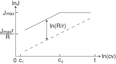
where is the diffusion coefficient of CP in water. The rate as a function of CP concentration is plotted in Fig. 2 by the dashed straight line.
Our main idea is that the long chain of yet unassembled ss RNA outside of the capsid provides an additional route for the diffusion of CPs to the assembly site, in analogy to the well-known faster-than-diffusion locating of the specific site on DNA for a protein Riggs ; BPJ . The dramatic enhancement of the assembly rate is achieved because, due to the Boltzmann factor , the three dimensional concentration of CP on unassembled chain of ss RNA is larger than the bulk concentration . This concentration can be estimated using the cylinder with cross-section build around RNA as the axis: it is equal to the number of CPs per unit length of ss RNA divided by . At large distances the one-dimensional flux of CP sliding on the ss RNA should be balanced by the three dimensional diffusion flux of CP to the ss RNA. This balance determines the radius of the sphere around the assembly site at which two fluxes match each other and the crossover between three-dimensional and one-dimensional diffusions of CP takes place. The ss RNA coil inside this radius is called antenna.
The maximum possible antenna size is the characteristic size of the unassembled portion of ss RNA with length . (Here we assume the ss RNA is a flexible Gaussian coil with the persistence length nm, where nm is the monomer size, and do not account for the excluded volume interaction.) In the case when , the whole ss RNA adsorbs CPs arriving by three-dimensional diffusion and provides a path of fast one-dimensional diffusion to the assembly site (See Fig. 1). As a result, in this case the size replaces the protein size in Eq. (2) leading to a much faster rate
| (3) |
which is shown in Fig. 2 by the part of the solid line parallel to the dashed one. Equation (3) is correct until CPs adsorbed on the unassembled chain of ss RNA are still sparse and do not block each other’s diffusion on ss RNA. Let us use the notation for the concentration , where the antenna becomes saturated by CPs and the dependence of the self-assembly rate on saturates roughly speaking at the level , which is the Smoluchowski rate at (see the solid line in Fig. 2). It was shown in Ref. BPJ that if
| (4) |
We see that the largest enhancement of the self-assembly rate can be achieved in the range of relatively small CP concentrations . For a typical T=3 virus the ss RNA genome consists of 3000 bases, so that the length nm and nm. Using nm, we arrive at the acceleration factor . One can calculate the assembly time limited by diffusion. As we said above for , the concentration of proteins can be regarded as a constant. Thus, the assembly time with the help of antenna is given by
| (5) |
while according to Eq. (2), the assembly time without antenna is simply . Since nm, we can neglect the difference between and , and arrive at the assembly time with the help of antenna times shorter than .
Strictly speaking, these estimates are correct only for self-assembly with a homopolymeric ss RNA or a synthetic negative polyelectrolyte Brancroft . For these cases, a small additional acceleration by a factor 2 or 3 can be provided by the excluded volume effect. On the other hand, the native ss RNA is more compact than gaussian one due to hydrogen bonds forming hairpins and thus the estimated acceleration rate can be reduced by a factor between 2 or 3. Above we for simplicity replaced by its maximum value . The actual calculation of the antenna size can follow the logic of the scaling estimate for the search rate of the specific site on DNA by a protein in Ref. BPJ . In our case, the assembly site plays the role of the target site (diffusion sink) for the protein, the unassembled chain of ss RNA plays the role of DNA and the Coulomb attraction energy of N-terminal tails to the unassembled ss RNA is analogous to the non-specific binding energy of diffusing protein on DNA. One may argue that the virus self-assembly problem is different, because ss RNA plays a dual role. It is not only an antenna for the sliding CPs, but ss RNA itself also moves to the assembly site, where it gets packed inside the capsid (each newly assembled CP consumes the length of ss RNA). However, for a small concentration in the range , where the unassembled ss RNA chain is weakly covered by CPs, the velocity of ss RNA drift in the direction of assembly site is much smaller than the average velocity of CP drift along ss RNA. Thus, for the calculation of the assembly rate at a given length of the unassembled ss RNA chain we can use the approximation of static ss RNA. This brings us back to the problem of proteins searching for the specific site on DNA BPJ . Note that this means that the idea of self-assembly from the prepared linear ss RNA-protein complex Zlotnick04 ; Vogt95 is literally correct only at .
It is shown in Ref. BPJ that for a flexible ss RNA, the antenna size reads , where , and is the one-dimensional diffusion coefficient of the protein sliding on ss RNA. This result remains correct as long as the antenna size is smaller than the ss RNA coil size . The energy of adsorption of the N-terminal tail with approximately 10 positive charges on ss RNA can be as large as . For we get nm, while nm. Thus, a simple estimate leads to the antenna length somewhat smaller than .
There are, however, two reasons why may easily reach its maximum value . First, some viruses self-assemble from dimers Casini ; Zlotnick04 . Naturally dimers with their two positive tails bind to ss RNA with the twice larger energy . This easily makes . ii) The theory of Ref. BPJ assumes that a sliding protein molecule has only one positive patch, where it can be attached to a double helix DNA. Even if two distant along the chain pieces of DNA come close in the three-dimensional space, such protein can not simultaneously bind both pieces and, therefore, can not crawl between them without desorbing to water and losing the binding energy . For a globular protein this is quite a natural assumption. On the other hand, for CP attached to ss RNA by a flexible N-terminal tail, the tail can easily cross over (crawl) between the two adjacent pieces of the same ss RNA molecule losing only small fraction of the energy . This should lead to faster protein diffusion on ss RNA and may easily push up to .
Let us discuss ideas of three in vitro experiments, which can verify the role of ss RNA antenna in virus self-assembly. In the first experiment, one breaks ss RNA molecule into short pieces of approximately equal length. It was shown Rein ; Vogt that the assembly is possible even when , because in order to glue CPs short ss RNA should bind two N-terminal tails of neighboring proteins in the capsid. Virus assembly from short ss RNA pieces goes consecutively through two different diffusion limited stages. In the first stage, capsid fragments (CFs) made of proteins self-assemble on each short ss RNA molecule. According to Eq. 5, the time necessary for this stage is proportional to and is much shorter than the assembly time with the intact ss RNA. The second stage, where CFs aggregate to form the whole capsid takes much larger time ( stands for short). In order to calculate we assume that when two CFs with CPs each collide, they can relatively fast rearrange their ss RNA and CPs in order to make one bigger CF with CPs. We also assume that at any time all CFs are approximately of the same size . Then the concentration of such CFs is , where is the concentration of original intact ss RNA. Therefore, the time required for doubling of a CF can be estimated from Eq. (2)
| (6) |
where and are diffusion coefficient and effective radius of a CF with CPs. Since the diffusion coefficient is inversely proportional to the droplet radius, the product , (where is the water viscosity), is the same constant as for a single protein. One collision of droplets transfers CPs to the growing CF. Therefore, the average time needed to add one CP to the growing CF does not depend on . In other words, the number of CP per CF increases at a constant rate. The assembly ends when reaches . Therefore, the assembly time is given by
| (7) |
Above equation shows the assembly time depends on which stands for the concentration of CP involved in the CF aggregation. However has no dependence on . Comparing Eqs. 5 and 7, we obtain that at
| (8) |
We see that the virus assembly time with short ss RNA pieces is much larger than that for the intact ss RNA. This happens due to the breaking of big antenna of the original ss RNA.
In the second experiment, we return to the intact ss RNA and discuss what happens when we vary relative concentrations of CP and ss RNA , for example, keeping and changing . Until now we assumed that , i.e. we have marginally more proteins than it is necessary to assemble a virus at every ss RNA. If the assembly time is practically the same as that at and is given by Eq. 5. Let us now consider much larger , for which . Here situation changes dramatically. There are two assembly stages. In the first stage, a CF is assembled with part of each ss RNA molecule, leaving the rest of the ss RNA molecule as a tail. This assembly uses up all the proteins and stops, when all CFs are still much smaller than the complete capsid and their ss RNA tails are long (see, for example, Fig. 1b). This state is essentially a kinetic trap. If energies and are much larger than , CFs on different ss RNA molecules can not exchange CPs trough the solution or via collision of their ss RNA tails. They can grow only via CF-CF collisions, while merging on one ss RNA and releasing the other empty one. We explained above, at (CPs are in excess), CFs without RNA tails produce a capsid during time given by Eq. 7. On the other hand, at , only occupied by CP ss RNA molecules take part in the aggregation and in order to get the assembly time, in Eq. 7 should be replaced by , which does not depend on . However, due to the long ss RNA tail, a CF diffuses slower than it does without a tail. The time grows substantially with decreasing , because with more ss RNA, the initial CFs have fewer CPs and longer ss RNA tails. This time saturates at , where and each CF has only one protein and the longest ss RNA tail. Thus, a long antenna accelerates assembly at and decelerates it at . This behavior of is schematically plotted in Fig. 3.
In the third experiment we can combine the first two and break ss RNA into pieces at several different values of . At a CF gets a shorter tail of ss RNA and larger mobility, so that assembly is faster than for intact ss RNA. When , the assembly time grows according to Eq. (7) with decreasing (increasing ). This is because the smaller the ss RNA concentration, the harder for the CFs to collide with each other and form larger CFs. In other words, kinetics is determined only by CPs already assembled in CFs and their number decreases with growing . We illustrate such nontrivial role of broken ss RNA in Fig. 3.
Now let us give some numerical estimates for , and for the in vitro assembly. Using the radius of CP nm, we obtain nM and M from Eqs. (1) and (4). For nM and the diffusion coefficient , the assembly time is about 10 min. At excess of CP, ss RNA antenna reduces it to min. At excess of ss RNA roughly speaking increases to . One can make even larger using much longer than native ss RNA.
In conclusion, we studied the role played by unassembled tail of ss RNA, which we call antenna. We showed that one-dimensional diffusion accelerates the virus self-assembly more than ten times when proteins are in excess with respect to RNA. On the other hand when RNA is in excess long tail of ss RNA slows down the assembly. We discussed several experiments which can verify the role of antenna. Although in this paper we focus on viruses for which CPs have long positive N-terminal tails, our idea can be also applied to the case where a CP binds to ss RNA by its positive patch. Our ideas are applicable beyond icosahedral viruses, for example, to the assembly of immature retro-viruses such as RSV or HIV Rein ; Vogt95 ; Vogt .
We are grateful to V. A. Belyi, R. Bruinsma, A. Yu. Grosberg, T. T. Nguyen, A. Rein, Iu. Rouzina, V. M. Vogt and A. Zlotnick for useful discussions.
References
- (1) S. J. Flint, L. W. Enquist, V. R. Racaniello and A. M. Skalka, Principles of Virology 2nd ed (ASM Press, Washington D.C., 2004)
- (2) J. B. Brancroft, E. Heibert and C. E. Bracker, Virology 39, 924 (1969).
- (3) K. W. Adolph and P. J. Butler, Philos Trans R. Soc Lond B (Biol Sci.) 276, 113 (1976).
- (4) P. van der Schoot and R. Bruinsma, Phys. Rev. E 71, 061928 (2005).
- (5) R. F. Bruinsma, Eur. Phys. J. E, Soft Matter. 19, 303 (2006).
- (6) V. A. Belyi and M. Muthukumar, PNAS 103, 17177 (2006).
- (7) Tao Hu, Rui Zhang, B. I. Shklovskii, q-bio.BM/0610009
- (8) A. Zlotnick, R. Aldrich, J. M. Johnson, P. Ceres and M. J. Young, Virology, 277, 450 (2000)
- (9) G. L. Casini, D. Graham, D. Heine, R. L. Garsea and D. T. Wu, Virology, 325, 320 (2004).
- (10) S. Campbell and V. M. Vogt, J. Virol. 69, 6487 (1995).
- (11) J. M. Johnson, D. A. Willis, M. J. Young, and A. Zlotnick, J. Mol. Biol. 335, 455 (2004).
- (12) D. F. Evans and H. Wennerstrom, The Colloidal Domain 2nd ed (Wiley-VCH, New York, 1999).
- (13) M. V. Smoluchowski, Z. Phys. Chem. 92, 129 (1917).
- (14) O. G. Berg, R. B. Winter and P. H. von Hippel, Biochemistry, 20, 6929 (1981); R. F. Bruinsma, Physica A, 313, 211 (2002); S. E. Halford, J. F. Marko, Nucleic Acids Res. 32, 3040 (2004); M. Slutsky, L. A. Mirny, Biophys. J. 87, 4021 (2004);
- (15) Tao Hu, A. Yu. Grosberg and B. I. Shklovskii, Biophys. J. 90, 2731 (2006).
- (16) S. Campbell and A. Rein, J. Virol. 73, 2270 (1999).
- (17) M. Y. Ma and V. M. Vogt, J. Virol. 76, 5452 (2002); 78, 52 (2004).