Kinesin as an electrostatic machine
Kinesin as an electrostatic machine
Abstract
Kinesin and related motor proteins utilize ATP fuel to propel themselves along the external surface of microtubules in a processive and directional fashion. We show that the observed step-like motion is possible through time varying charge distributions furnished by the ATP hydrolysis circle while the static charge configuration on the microtuble provides the guide for motion. Thus, while the chemical hydrolysis energy induces appropriate local conformational changes, the motor translational energy is fundamentally electrostatic. Numerical simulations of the mechanical equations of motion show that processivity and directionality are direct consequences of the ATP-dependent electrostatic interaction between the different charge distributions of kinesin and microtubule.
I Introduction
The kinesin family is a set of motor proteins that move on the surface of microtubules and shuttle various cargo molecules to different parts of the cellTuz (1)-parallel (9). The energy for kinesin transport motion is provided by hydrolysis of ATP molecules that are in its vicinity and attach to a specific site of the protein. Despite much progress in the area, the precise, microscopic fashion of ATP action is not known. Kinesin motion is step-like with one step for each ATP hydrolysis while processivity and directionality depend on specific neck properties of each particular type of the family. These features of motor protein motion led several years ago in the introduction of mechanical ratchet models that could provide some insight on the phenomenology of individual Reimann (10, 12, 13, 14, 15) and collective julicher (16, 17) motion. Although many issues and especially those related to the random aspects of the walk where addressed successfully by these models, fundamental questions on the energetics and nature of forces that enable the walk have not been discussed. In the present work we focus on the latter; specifically we use electrostatic information of the microtubule and kinesin and evaluate the full electric force that binds the latter on the former. We then show that the actual kinesin walk is caused by charge distribution changes on kinesin enabled by the action of ATP. The walk that is a combination of conformational changes accompanied by kinesin charge reshuffling is found to be fundamentally governed by electrostatic forces.
II Kinesin electrostatic model
Kinesins walk on microtubules; the latter are made by
alpha and beta of tubulin proteins that
are highly charged. Molecular modeling shows that, in the presence of
water, tubulins have additionally a permanent dipole moment that points
towards the cylindrical symmetry axis of the microtubule with
a non-zero component parallel to the protofilaments towards their minus
endTuz (1). Electrostatically thus a microtubule has negative surface
charge accompanied by a positive charge distribution in its immediate interior
with a slanted polarization vector that is larger in the beta than the
alpha subunit (Figure 1). This symmetry breaking induced by the tubulin dipole moment direction
plays a key role in the specificity of the attachment of a kinesin head onto a
beta tubulin subunitdecoration (2).
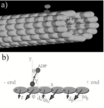
Structural analysis of kinesin and related proteins suggests that there are three charge domains, one in each head region and a third one in the central neck linker domain. The head charge distribution depends strongly on the ATP or ADP presence while the neck charge is specific to the protein. For modeling purposes it is sufficient to consider kinesin and similar motor proteins as a body containing three charges located on the apexes of a triangle with angle subtending from the neck charge to the two head charges (Figure 1b) These charge distributions interact with tubulin multipole fields and determine at each instant of time the dynamic state of the molecule.
In our analysis of kinesin walk to be detailed below, we place motor proteins and microtubules in an overdamped medium with thermal energy and motor drag force per rotational degree of freedom. For the sake of simplicity, we assume a relative electric permitivity equal to 80 everywhere and a Debye length above . Although Debye-Hückel theory predicts , the interaction range may be enhanced by the channeling of the electric field along the interior of the proteins, which is not accesible by diffusing ions or water molecules. Then it is reasonable to take of the order of the size of the kinesin. We take consecutive tubulin unit distance to be , per or subunit. The lateral space between two protofilaments is while the pitch of between consecutive protofilamens is also included. For a processive plus-ended kinesin and its non-processive-minus-ended chimera ncd we use for the head-to-head distance the value of while we consider the heads to be charged with in the absence of nucleotides, with ATP attachment and with ADP. These head charge values are consistent with the charges involved in hydrolysis reaction
| (1) |
The value of the central motor charge changes for different proteins; as a result we consider different motors with charges in the range .
For the electrostatic distribution of microtubules we assign negative surface charge per tubulin subunit while include a positive charge distribution in the interior leading to dipole moment magnitude of Tuz (1), or (). Finally, we use a dipolar tilt with length and angle values equal to , and , , for the and subunits respectively (Figure 1b).
The microtubule-induced kinesin interaction potential is given by
| (2) |
where is the position vector labeling the charges on kinesin (), while is the location of the N microtubule charges on the and subunits. is the inverse of the Debye length, which we take around , and is the excluding volume radius as described in Ref. Levin (18). A regime with a greater and lower Debye length is also operative. We considered a flat microtubule with five protofilaments; due to the rapid decay of the force out from protein volumes, we include in total the closest tubulin charges to kinesin. For the simplest case when neck and head charges are aligned we have and the protein reduces to a triply charged rigid rod.
We have tested the model for several values of smaller than ; we have found that the model is not fully opperational for . We have also tested the model considering that the angle has some elasticity as well. We found that in case the angle is very soft, the raising head rapidly loses its power to pull the motor forward. For the purposes of the model, the motor core and the neck need to be stiff enough to maintain the values of in the range discussed above even at maximum load conditions. In this simpler rod-like configuration the polar angle and the azimuthal angle are sufficient for describing the motor rotation. We can describe the motion using the following overdamped equations of motion for kinesin:
| (3) |
and
| (4) |
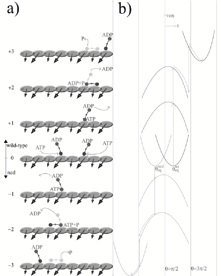
where is the drag coefficient, the head-to-head distance and the total microtubule electrostatic potential of Eq. (2) at the Cartesian location . The environment is simulated through the thermal forces and ; for each we have and . In order to integrate Eqns. (3, 4) we need to perform at each instant of time the Cartesian-to-polar transformation . For the rotation we consider the attached head to coincide with the origin of the coordinate system; the latter is shifted by each time a step is completed. The simple Larmor-like rotation of the protein for becomes a more complex rigid body rotation for . Extensive numerical simulations with the model described previously for the kinesin-microtubule complex led to the following quantitative picture for the motion. In equilibrium (parked) state the nucleotide-free head is positively charged and thus attached to the beta subunit of the negative microtubule surface while the other head containing ADP is negatively charged and tethered through the neck. Due to the slanted microtubule dipole moment, the kinesin axis is tilted but in a direction determined by the central charge sign; for wild type kinesin points to the fast-growing-end while for ncd points to the minus end ( Figure 2).
All experimental data agree that after nucleotide entry in the head pocket, the binding of the attached head to the microtubule and the ADP binding to the tethered head become unstable, signaling the onset of a cascade of events leading to the walk. In our model this critical juncture occurs because in the absence of nucleotide the positively charged head becomes negative acquiring charges after ATP binding. Although the entry of a negatively charged molecule in the head pocket is necessary for the commencement of the walk process, the release of the attached head is not immediate, since a head-tubulin chemical bond needs to be broken. ATP hydrolysis energy thus is used for a local conformational change that captures the negative charge after dephosphorylation. Although charged ADP nucleotides in the medium compete with ATP entering also in the pocket, they do not hydrolyze and, as a result, cannot induce the local conformation that will trap them. In our electrostatic model thus hydrolysis energy induces primarily a specific local conformational change, being the power stroke electrostatic in nature.
III Kinesin dynamics
Based on this picture, the protein parked configuration is a result of the microtubule-empty head attraction, microtubule-tethered head repulsion while the kinesin angle wrt microtubule is due to the tubulin dipolar tilts. This stable configuration is maintained during a random and [ATP]-load-dependent dwell time until a new ATP binds into the attached head. When this occurs, the local charge changes and the ADP trapped in the tethered head experiences an additional repulsion, becoming unstable and eventually opening the pocket and exiting. After ADP expulsion the tethered head becomes positive, the attached head charge is negative and the protein becomes electrostatically unstable. As a result the tethered head collapses onto the microtubule (”falling processes”), while the attached head is forced to detach from the surface (”rising process”) and a new, shifted, park state is reached.
Every kinesin step thus includes falling of the leading head onto the microtubule followed by rising of the trailing head ( Figure 2). We performed extensive 3D numerical simulations and found that the electrostatic force field generated by the microtubule is able to drive these two sequential processes while keeping the motor faithful to a given protofilament. We note that, energetically, the parked state represents a potential energy minimum for the interaction between free head charge and microtubule. When ADP is released and the electrostatic charge distribution of the motor changes this minimum becomes a potential maximum, i.e. the repulsion of the tethered head turns into attraction.
For a null-charged-neck case, even though the motor is tilted in the parked state, there is no motion directionality since the falling of the tethered head does not have a preferred collapsing side. The directionality features of the motor enter through the charge distributions of the neck region, which is responsible of the potential shift between stable and unstable equilibrium points. Specifically, when the central charge is positive, not only the protein is more processive due to the attraction with the negatively charged tubulin surfaces, but it also walks towards the plus-end of the microtubule; this is precisely the case with wild-type kinesin. In ncd, on the other hand, the central charge is negative, the protein is non-processive and walks in the opposite direction towards the minus end. Additionally, in proteins with no charge in the neck region, the walk is known not to be deterministic but randomMutante (3). We note further that our simulations are compatible with the experiments in reference parallel (9) whereby the falling motor direction is parallel to the protofilament axis, with some variations and latteral collapses leading to protofilament changes due to thermal fluctuations. The lateral periodicity of the microtubule lattice is while the axial one only ; these values determine crucially the motion after the destabilization of the parked state since the electric force is stronger in the axial direction. We find that this electrostatic model captures fully the directionality and processivity features of the known motors.
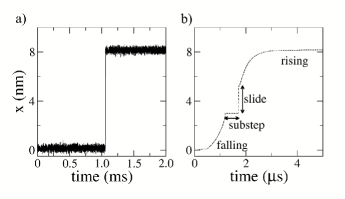
Two different time-scales characterize the actual kinesin motion. While dwell times between two steps, modulated by ATP concentration and the external load are of the order of millisecondsVisscher (4), the action of the step itself lasts only some microseconds substepsno (6); this is the reason for the stepping appearance of the trajectories. In the relatively short period of the step time, several processes must occur. First, falling of the leading head after ADP release, a process activated by ATP binding on the attached head. Subsequently the two heads remain attached onto the microtubule for a time not longer than 30 microseconds substepsno (6). Recent measurements were able to track bead-movements at the microsecond scale substepsno (6). The recording of a single step, in our interpretation, reveals two different regimes that may be separated by a quick stage at which both kinesin heads are attached to the microtubule. Kinesin crytallization data show that the separation between heads is smaller than 8 nmcristalina (7), i.e. slightly smaller than the protofilament spatial period and, as a result, after falling the leading head cannot reach the beta subunit location before the trailing head detaches. Thus, sliding may take place while the trailing head rises to reach the new parked state. This aspect is portrayed in Figure 3 in a sequential form although in practice both sliding and rising could occur simultaneously.
In order to address motor protein processivity
we consider the two sets of chemical reactions that are activated by ATP binding at the trailing head. The first group is related to a cascade of rapid reactions involving
ATP hydrolysis, dephosphorylation and subsequent head detachment
with an estimated time 500
for eachchemistry (8).
Secondly, ATP activates ADP release in the other head, although
the typical time of this process is not known. There is a
competition between these two reactions; if hydrolysis and
detachment occur faster, the whole motor detaches from the microtubule
and processivity ceases. If, on the other hand, ADP release and falling is the fastest
process, then a small substep occurs while both heads are attached.
Processivity thus is related to the competition of these two processes.
In the case of non-processive ncd, for instance, the existence of a
negative central charge has the effect of slowing
down the falling process while
speeding up the rising one resulting, thus, in a non-processive motor. This effect has been tested experimentally in Ref. Vale (19).
We point out that when ATP enters into the attached head, the system becomes electrostatically unstable. However, detachment of the head occurs some time later triggered by the phosphate release. This fact implies that the chemical bonding that binds the motor to the microtubule, while stable in absence of ATP, becomes unstable when the nucleotide arrives. The time delay between ATP entry and bond dissociation appears to be crucial for motor processivity. In the Figure 2, we have assumed that both possesive and non-possesive motors may perform the full detachment-attachment cycle, although, in the case of ncd, the chemical bond may be actually broken before the falling head reaches the next binding site. The time duration of a single kinesin step is very small compared to the parked dwell times, and, as a result, global observables such as the mean velocity or randomness can be predicted using only chemical kinetics. This has been shown in reference Ciudad (20) where a kinetic approximation was shown to be sufficient in fitting the measured data, even in the presence of an external load. However, our present approach shows, that thermal fluctuations alone may not produce directly the long displacements in a time-scale necessary for the walk.
IV Conclusions
In this work we attempted to analyze the complex motion of molecular motor proteins from a mesoscopic point of view placing emphasis on the fundamental interactions that enable the motion. We found that the motion is primarily driven by the electrostatic interaction between the charged microtubule surface and the fluctuating motor head charges. The nonequilibrium aspect of the walk is provided by the ATP hydrolysis cycle that furnishes appropriate charge motor distributions that make the walk possible. We made several assumptions in this work, most of which have been motivated by the current status of knowledge in the area. We considered the motor as a relatively rigid one; this assumption is presently well founded, however it may be lifted through an improved motor model that includes additionally protein eleasticity. Furtheremore, our model does not address at all the structual changes in the head pockets effected by ATP hydrolysis, the mode of the local energy relase as well as the mechanism for the ADP detachment from the thethered head. These issues that may involve complex conformational changes as well as possibly charge or energy transfer processes must be addressed through a more foundamental, microscopic model. Finally, it is crucial to evaluate the range of Debye screening in the vicinity of a highly charged surface such as the microtubule. While our motor model remains fully operational to a Debye length range of approximately , one certainly needs to address the complex nature of the electrostatic shielding in the vicinity of the microtubule and assess the true range of the electrostatic forces involved in the walk. We note that our model provides a consistent dynamical picture for the kinesin walk that is based on two premises, viz. (a) that the ATP hydrolysis energy is used for a head local conformational change that captures locally charges and (b) the motor motion is driven by electrostatic forces. The emerging qualitative and quantitative picture for the walk is fully compatible with all known experimental data, while, furthermore, it is testable experimentally. If electrostatics, charge transfer as well as capture indeed link the chemistry of ATP and the mechanics of kinesin, this may hold true for other ATP-dependent processes as well.
Acknowledgements.
We thank Marta Ibañes for discussions. This work was supported by the Ministerio de Educación y Ciencia (Spain) under Project No. , Grant No. , by grant of the Generalitat de Catalunya and Grant ”Pythagoras II” of the Ministry of Education of Greece and the European Union.References
- (1) J. A. Tuszynski, T. Luchko, E. J. Carpenter and E. Crawford. Electrostatic Properties of Tubulin and Their Consequences for Microtubules. Journal of Computational and Theoretical Nanoscience, 1:1–6, 2005.
- (2) M. Thormählen, A. Marx, S. A. Müller, Y. H. Song, E. M. Mandelkow, U. Aebi and E. Mandelkow. Interaction of Monomeric and Dimeric Kinesin with Microtubules. Journal of Molecular Biology, 275:795–809, 1998.
- (3) S. A. Endow and H. Higuchi. A mutant of the motor protein kinesin that moves in both directions on microtubules. Nature, 406:913–916, 2000.
- (4) K. Visscher, M. J. Schnitzer and S. M. Block. Single kinesin molecules studied with a molecular force clamp. Nature, 400:184–189, 1999.
- (5) A. A. Kasprzak and A. Hajdo. Directionality of kinesin motors. Acta Biochimica Polonica, 49(4):813–821, 2002.
- (6) N. J. Carter and R. A. Cross. Mechanics of the kinesin step. Nature, 435(19):308–312, 2005.
- (7) F. Kozielski, S. Sack, A. Marx, M. Thormählen, E. Schönbrunn, V. Biou, A. Thompson, E. M. Mandelkow and E. Mandelkow. The Crystal Structure of Dimeric Kinesin and Implications for Microtubule-Dependent Motility. Cell, 91:985–994, 1997.
- (8) R.A. Cross. The kinetic mechanism of kinesin. TRENDS in Biochemical Sciences, 29(6):301-309, 2004.
- (9) S. Ray, E. Meyhfer, R. A. Milligan and J. Howard. Kinesin Follows the Microtubule’s Protofilament Axis. The Journal of Cell Biology, 121(5):1083–1093, 1993.
- (10) P. Reimann. Brownian motors: noisy transport far from equilibrium. Physics Reports, 361:57–265 , 2002 and references therein.
- (11) G. P. Tsironis and K. Lindenberg. . Adv. in Structural Bio., 5(eds):271–281 , 1998.
- (12) G. Stratopoulos, T. Dialynas and G. P. Tsironis. Directional Newtonian motion and reversals of molecular motors. Physics Letters A, 252:151–156, 1999.
- (13) M. E. Fisher and A. B. Kolomeisky. Simple mechanochemistry describes the dynamics of kinesin molecules. PNAS, 98:7748–7753, 2001.
- (14) M. Bier. Processive Motor Protein as an Overdamped Brownian Stepper. Physical Review Letters, 91(14):148104, 2003.
- (15) A. Ciudad, A. M. Lacasta and J. M. Sancho. Physical analysis of a processive molecular motor: The conventional kinesin. Physical Review E, 72:031918, 2005.
- (16) F. Jülicher, A. Ajdari and J. Prost. Modeling molecular motors. Review of Modern Physics, 69:1269–1282, 1997.
- (17) K. Nishinari, Y. Okada, A. Schadschneider and D. Chowdhury. Intracellular Transport of Single-Headed Molecular Motors KIF1A. Physical Review Letters, 95:118101, 2005.
- (18) Yan Levin. Electrostatic correlations: from plasma to biology. Reports on progress in physics, 65:1577–1632, 2002.
- (19) K. S. Thorn, J. A. Ubersax and R. D. Vale. Engineering the Processive Run Length of the Kinesin Motor. The Journal of Cell Biology, 151(5):1093–1100, 2000.
- (20) A. Ciudad and J. M. Sancho. External mechanical force as an inhibition process in kinesin’s motion. Biochemical Journal, 390:345–349, 2005.