Focal adhesions as mechanosensors: the two-spring model
Abstract
Adhesion-dependent cells actively sense the mechanical properties of their environment through mechanotransductory processes at focal adhesions, which are integrin-based contacts connecting the extracellular matrix to the cytoskeleton. Here we present first steps towards a quantitative understanding of focal adhesions as mechanosensors. It has been shown experimentally that high levels of force are related to growth of and signaling at focal adhesions. In particular, activation of the small GTPase Rho through focal adhesions leads to the formation of stress fibers. Here we discuss one way in which force might regulate the internal state of focal adhesions, namely by modulating the internal rupture dynamics of focal adhesions. A simple two-spring model shows that the stiffer the environment, the more efficient cellular force is built up at focal adhesions by molecular motors interacting with the actin filaments.
I Introduction
During recent years, tremendous progress has been made in regard to a quantitative understanding of the metabolic, signal transduction and genetic networks characteristic of biological systems Kitano (2002); Alon (2003); Alm and Arkin (2003). Although network approaches capture many of the essential aspects of simple organisms, for higher organisms a quantitative and systems-level understanding also has to include structural aspects, including the spatial organisation and mechanical properties of cells. In particular, modelling tissues and organs requires a quantitative understanding of the roles played by cytoskeleton, membranes, and the extracellular matrix (ECM).
One field which cannot be understood completely without considering biochemical and structural aspects on an equal footing is cell adhesion, which is an essential element of many physiological situations, including development, tissue maintenance, wound healing, angiogenesis, and cell migration Gumbiner (1996). In general, most cell types require anchorage to the ECM to proliferate. Moreover, cell adhesion also determines how cells interpret soluble ligands like hormones and growth factors Stupack and Cheresh (2002); Guo and Giancotti (2004). The behaviour of adhering cells is strongly influenced by the chemical, topographical and mechanical properties of the surfaces they attach to Curtis and Riehle (2001). During recent years, experiments with elastic substrates have shown that elastic properties of the extracellular evironment are also highly relevant for cellular decision making Pelham and Wang (1997); Lo et al. (2000); Wong et al. (2003); Engler et al. (2004); Georges and Janmey (2005).
A growing body of evidence now suggests that the essential link between the mechanical properties of the extracellular environment and cellular decision making are mechanotransductory processes at integrin-based cell-matrix contacts Chicurel et al. (1998); Galbraith and Sheetz (1998); Geiger et al. (2001); Katsumi et al. (2004). For cells spreading on flat substrates, cell-matrix contacts initially form as focal complexes close to the lamellipodium. Depending on the presence of appropriate signals, focal complexes can mature into focal adhesions which are connected to actin stress fibers. Focal adhesions have a twofold purpose. As they connect the actin cytoskeleton with the ECM, they guarantee structural integrity. Equally important, they are also strong signaling centers. In fact more than 50 different kinds of proteins are known to localize to the cytoplasmic plaque of focal adhesions, many of which are known signaling molecules. Therefore focal adhesions provide an excellent opportunity to study the interplay between biochemical and structural aspects in biological systems.
The details of the mechanosensory processes at focal adhesions are still elusive. It has been shown some time ago that application of force on integrin-based contacts between cells and ligand-coated beads leads to contact reinforcement and mechanotransduction Wang et al. (1993); Choquet et al. (1997). Recently, force reconstruction at single focal adhesions on compliant substrates showed that the internal forces exerted at focal adhesions correlate with their sizes Balaban et al. (2001); Tan et al. (2003). In a complementary study, it was shown that force exerted externally by a micropipette leads to growth of those focal adhesions which are tensed Riveline et al. (2001). Other recent experiments imply both a membrane-independent stretch response of the protein network connected to focal adhesions Sawada and Sheetz (2002) as well as some role for stretch-activated ion channels Munevar et al. (2004). In fact it is very likely that several force-mediated mechanisms work in parallel at focal adhesions, including changes in integrin and extracellular ligand densities, rearrangements in the cytoplasmic plaque, stretch-activated ion channels and opening of cryptic binding sites in focal adhesion molecules Bershadsky et al. (2003). Recently a quantitative model has been introduced which explains anisotropic growth of focal adhesions under force by density variations in the sheared layer of integrins Nicolas and Safran (2004); Nicolas et al. (2004). Other theoretical efforts have modelled force-mediated growth as strain relaxation due to incorporation of new material, phase transitions due to force-mediated coupling between neighboring receptors and force-mediated release of a soluble signal. However, a systems-level description of focal adhesions as mechanosensors has not been presented yet.
In this contribution, we discuss several modelling efforts which in the future might be integrated into such a systems-level understanding of focal adhesions. Such a description will have to integrate the effects of extracellular elasticity, molecular motor activity, and signal transduction. We start with a discussion of integrin signaling at focal adhesion and how it relates to the spatial and temporal organization of cells. Next we describe a simple model for the stochastic rupture dynamics of adhesion clusters under force, which quantitatively demonstrates that the internal state of adhesion clusters can be regulated by force. Finally we introduce a new model (two-spring model), which shows in a quantitative way how extracellular elasticity might modulate the build-up of intracellular force at focal adhesions.
II Integrin signaling at focal adhesions
Although physical concepts like force and elasticity are essential to understand active mechanosensing at focal adhesions, the biochemical aspects of these systems are equally important and far from understood. Focal adhesions are based on heterodimeric transmembrane-receptors from the integrin family, which connect the ECM with the actin cytoskeleton. Integrins are large allosteric machines which are activated both by biochemical and mechanical cues and which transmit both inside-out and outside-in signals Hynes (2002). For mammals, 24 integrin variants are known, which bind to different subsets of ECM-ligands. For example, the main integrin-receptors for fibronectin and vitronectin are and , respectively. Interestingly, cancer cells switch their integrins: they loose integrins like , which mediate adhesion, and upregulate integrins like , which promote migration and survival in new environments Guo and Giancotti (2004). The whole complexity of the integrin systems becomes apparent when one considers the interaction with the the cytoplasmic plaque and the signaling to the cytoskeleton Geiger et al. (2001).
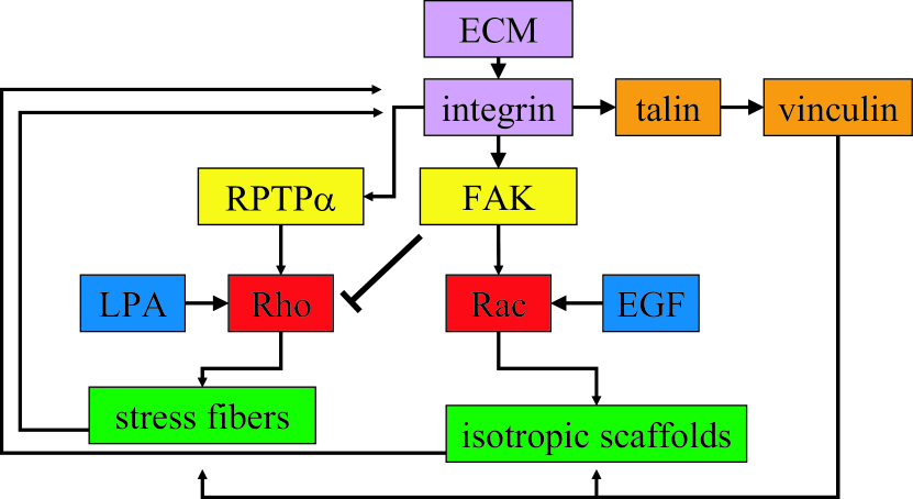
In Fig. 1, we schematically depict some of the aspects which are known to be important in this regard. The scheme deliberately focuses on three important downstream targets of integrin signaling to the actin cytoskeleton DeMali et al. (2003). Focal adhesion kinase (FAK) is a protein tyrosine kinase which has been shown to be a key component of mechanosensing at focal adhesions Wang et al. (2001). It is activated by integrin ligation and one of its main downstream targets is the small GTPase Rac, which leads to reorganization of the actin cytoskeleton into an isotropic network structure. At the same time, FAK-activation downregulates another small GTPase, Rho, mainly through activation of p190RhoGAP. Rho promotes the reorganization of the actin cytoskeleton into stress fibers and it often has an antagonistic role to Rac. Both small GTPases belong to the Rho-family and are also activated by pathways involved in cell survivial (epidermal growth factor (EGF) and lysophosphatidic acid (LPA) in the cases of Rac and Rho, respectively). Upregulation of Rac and downregulation of Rho is typcial for phases of spreading and cell migration, when focal complexes and lamellipodia are more prominent than focal adhesions and stress fibers. However, the initial dip in Rho-activity is often followed by long-term activation, albeit in a ligand-specific and cell-type-specific manner Bershadsky et al. (2003). This typically corresponds to the phase of mature adhesion, which is discussed here. Although experimental findings are conflicting, there is good evidence that the receptor-like protein tyrosine phosphatase RPTP- activates Rho through the tyrosine kinase Fyn and a RhoGEF which has not been identified yet von Wichert et al. (2003). Irrespective of the detailed mechanism, Rho-activation has been shown to be an essential part for the force-mediated stabilization of focal adhesions Riveline et al. (2001). The main issue here is that Rho-mediated activation of myosin II molecular motor activity as well as formation of stress fibers is essential for maturation of focal adhesions, thus providing positive feedback to growing adhesions. Rac-mediated organization of the actin cytoskeleton into isotropic networks might provide positive feedback for the growth of focal complexes, but possibly in a force-independent way.
A third major player in focal adhesions is talin, one of the four proteins known to link the integrins directly with the actin cytoskeleton. Talin is essential for early focal adhesion reinforcement under force Jiang et al. (2003) and leads to recruitment of vinculin, which also stabilizes focal adhesions. Both talin and vinculin can exist in closed and open conformations, a fact which might be related to the mechanosensor at focal adhesions Bershadsky et al. (2003). They also might act as nucleators for the actin cytoskeleton, thus locally modulating the effects of the small GTPases Rac and Rho. Finally it is interesting to note that the actin cytoskeleton also features crosstalk to the microtubule system. For example, it has been shown that one of the main downstream targets of Rho is mDia Riveline et al. (2001), which might regulate microtubule polymerization. Moreover it has been found that microtubules are targeted into mature focal adhesions, possibly in order to deliver some kind of death signal Krylyshkina et al. (2003).
The scheme presented in Fig. 1 shows that there exists a positive feedback involving integrin ligation, assembly of the cytoplasmic plaque, Rho- and Rac-signaling to the cytoskeleton and reorganization of the cytoskeleton. In the case of Rho-signaling, an essential element of this feedback is generation of stress through myosin II molecular motors and growth of focal adhesions under force. One of the future challenges in this field is a more complete and data-based description of the interplay between signaling at and spatial organization of integrin-based adhesions and the actin cytoskeleton. In order to understand the role of force in the feedback loop between integrins and actin cytoskeleton, physical mechanisms have to be identified by which force affects the state of focal adhesions.
III Rupture dynamics of adhesion clusters under force
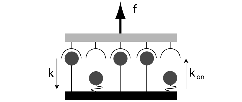
In order to study how force affects adhesion clusters in general, we recently studied a simple model for the stochastic dynamics of parallel bonds under shared constant loading Erdmann and Schwarz (2004b, c). This model is a stochastic version of a classical yet deterministic model which has been introduced by Bell Bell (1978). The model assumes that receptor-ligand bonds have been clustered on opposing surfaces, of which the upper one acts as a rigid transducer which transmits the constant force homogeneously onto the array of bonds. In our model, is a constant, but in future work it might be combined with a growth model for adhesion clusters Nicolas and Safran (2004); Nicolas et al. (2004). At each time, () bonds are closed and bonds are open. Closed bonds are assumed to rupture with a force-dependent rupture rate , where is the unstressed (intrinsic) rupture rate (typically around 1/s) and the internal force scale (typically a few pN) of the adhesion bonds. The exponential dependance between force and rupture rate results from a Kramers-type description of bond rupture as escape over a transition state barrier Evans and Ritchie (1997). The factor results because force is assumed to be shared equally between closed bonds, which holds true when the transducer is connected to a soft spring (in the opposite limit of a stiff spring, all bonds feel the same force and cooperativity is lost). Open bonds are assumed to rebind with a force-independent rebinding rate . A schematic representation of our model is shown in Fig. 2. The model has three dimensionless parameters, namely cluster size , dimensionless total force and dimensionless rebinding rate . With dimensionless time , it leads to the following one-step master equation
| (1) |
where is the probability that bonds are closed at time and the and are the reverse and forward rates between the possible states :
| (2) |
This equation implies , that is, after rupture of the last closed bond, new bonds are allowed to form. However, in many situations of interest, rebinding from the completely dissociated state is prevented by elastic recoil of the transducer. Therefore in the following we use (absorbing boundary at ). For the mean number of closed bonds, , one can derive from Eq. (1)
| (3) |
This suggests to study the following differential equation
| (4) |
as has been done by Bell Bell (1978). However, this deterministic treatment is a good approximation for the first moment of the stochastic model only in the case of large systems. For small systems, stochastic fluctuations in combination with the non-linearity and the absorbing boundary lead to different results.
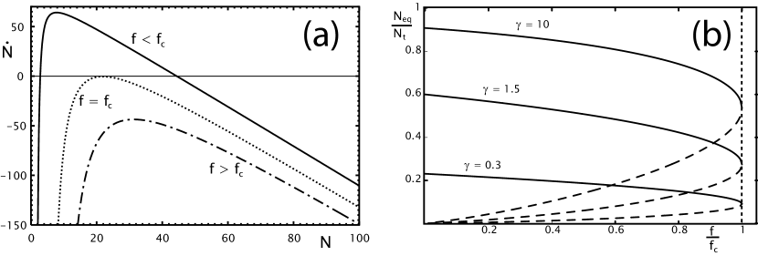
While force destabilizes the cluster, rebinding stabilizes it. We first study this interplay in the framework of the deterministic equation Eq. (4). In Fig. 3a, we plot as a function of for several values of force . This shows that two fixpoints with exist up to a critical force , with the lower one being unstable (a saddle) and the upper one being stable (a node). At , the two fixpoints collapse and stability vanishes in a saddle-node bifurcation. The critical force can be calculated exactly to be
| (5) |
Here the product logarithm is defined as the solution of . For , we have . Thus the critical force vanishes with , because the cluster decays by itself with no rebinding. For , we have . This weak dependence on shows that the single bond force scale set by also determines the force scale on which the cluster as a whole disintegrates. Fig. 3b shows the full bifurcation diagrams for different values of the rebinding rate . The larger rebinding, the larger are the values for the stable steady state. In particular, for we have , that is first increases linearly with and then saturates towards the maximal value .
In conclusion, the bifurcation analysis of the Bell-model shows that force can switch the stability of adhesion clusters. It is tempting to speculate that focal adhesions might be regulated to be close to such a critical state, because then small changes in cytoskeletal loading would result in strongly accelerated cluster dynamics and larger forces on single bonds. This in turn could trigger signaling events, e.g. by exposure of cryptic binding sites. In fact the stress constant at mature focal adhesions recently has been measured to be around nN/ for different cell types and different experimental conditions Balaban et al. (2001); Tan et al. (2003). Using Eq. (5), this idea can be used to estimate the rebinding rate in focal adhesions, which has not been measured yet. Estimating and using the dissocation parameters Hz and pN for activated -integrin binding to fibronectin Li et al. (2003) gives a rebinding rate Hz.
Although the deterministic model gives non-trivial insight into possible mechanisms for switching the state of focal adhesions by force, it neglects fluctuation effects. In particular, cluster lifetime is predicted to be infinite below the critical force . In the stochastic treatment, lifetime is finite for all parameter values due to the possibility that the systems reaches the absorbing boundary at the completely dissociated state. Average cluster lifetime then can be identified with the mean first passage time to reach the state , which can be calculated exactly from the adjoint master equation. For one bond, one simply has , as suggested by Bell Bell (1978). For two bonds, we find
| (6) |
This result generalizes Bell’s suggestion to and already reveals the characteristic structure of the solution for general cluster size : mean cluster lifetime is suppressed exponentially by force and the rebinding correction is a polynomial of power . A detailed analysis shows that although very different for , for the stochastic treatment gives similar results in regard to as the deterministic one.
In order to investigate the effect of fluctuations, we used computer simulations to numerically solve the master equation Eq. (1). This can in fact be done very efficiently by using the Gillespie algorithm for exact stochastic simulations Gillespie (1976). Our computer simulations show that for , single rupture trajectories show a characteristic shape which is not revealed by considering the first moment only. Initially they follow the average value, but then they abruptly move towards the completely dissociated state, while the average value approaches this state in a more gentle way. Therefore the average behaviour results not so much from differently shaped trajectories, but rather from the distribution of the timepoints of abrupt decay. This observation shows the importance of fluctuations and can be understood from the rates given in Eq. (2): once there is a fluctuations to a smaller number of closed bonds, force on the remaining bonds rises and leads to even more increased dissociation. Therefore a positive feedback exists for bond rupture, which for cannot be balanced anymore by rebinding effects.
It is well known that bifurcations often lead to switch-like behaviour in biochemical control systems Tyson et al. (2003). In general, thresholds have evolved for many biological systems, including the cell cycle and the MAPK-cascade. Our model shows that switch-like behaviour can also arise from the mechanical effect of force. Similar mechanisms are very likely to be at work at focal adhesions. In particular, the experimental evidence described above suggests that a certain threshold of force is required to trigger signaling events which eventually lead to regulated growth of focal adhesions. Since the build-up of internal force has to be balanced by the extracellular environment, its mechanical properties modulate the way in which the threshold is reached. Therefore an internal threshold for force is an appealing candidate for the exact mechanism of the mechanosensor at focal adhesions.
IV The two-spring model
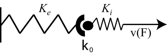
In order to investigate this point in quantitative detail, we now introduce a simple two-spring model for build-up of force at focal adhesions. The model is depicted schematically in Fig. 4. Here the ECM and the force-bearing intracellular structures are represented by harmonic springs with spring constants and , respectively. Since the two springs act in series, the effective spring constant is given by . Therefore the overall stiffness is mainly determined by the softer spring. For the time being we assume that this applies to the extracellular environment. Tension in the actin stress fibers is generated by myosin II molecular motors. For simplicity, we represent their activity by a linearized force-velocity relation
| (7) |
where free velocity is of the order of m/s and stall force is a few pN Howard (2001). As the motors pull, the springs get strained. For the static situation, the energy is stored in the spring. Therefore the stiffer the environment, the less work has to be invested into building up a certain level of force . For the dynamic situation, we have . The dynamics of force generation can be derived by noting that the power invested into the spring is generated by the molecular motors:
| (8) |
with the force-velocity relation from Eq. (7). This equation can be readily integrated:
| (9) |
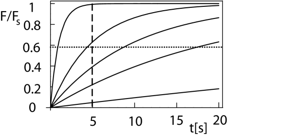
with . If the cell pulls on a material with a bulk modulus of kPa, then the corresponding spring constant on the molecular level can be expected to be of the order of . Thus the typical time scale is seconds. If the bulk modulus is of the order of MPa, then (which is also the range for protein stiffness) and the typical time scale is milliseconds. In Fig. 5, we plot Eq. (9) for different values of the spring constant . All curves eventually saturate at , but the stiffer the environment (the larger ), the faster a given threshold in force (indicated by the horizontal dashed line) can be reached.
Like the general adhesion clusters discussed in the preceding section, focal adhesions are also subject to force-modulated internal dynamics. In the two-spring model from Fig. 4, the internal structure of the focal adhesion is represented by one biomolecular bond with unstressed dissociation rate . In principle one now can apply the concept of rupture under force to the loading history from Eq. (9). In fact recent years have shown that rupture under non-constant force is essential to understand the details of biomolecular bonding Evans (2001). For linear ramps of force, this issue has been addressed theoretically in great details, both for single bonds Evans and Ritchie (1997); Shillcock and Seifert (1998) and adhesion clusters Seifert (2000); Erdmann and Schwarz (2004a). Unfortunately, the differential equation for the probability that one bond with the loading history from Eq. (9) breaks at time can be solved only numerically. Therefore it is instructive to consider two simple limites of this situation. As in the preceding section, we assume that the single bond under constant force has the average lifetime . In the case of large , and the bond effectively experiences constant loading with stall force . In the case of small , loading is approximately linear, with a loading rate . If the dimensionless loading rate , then the bond will decay with its intrinsic rate before the effect of force becomes relevant. The general case will be within these two limites. Since the stall force is of the same order as the internal force scale , the effect of the loading history is expected to change the result by not more than one order of magnitude. For simplicity, we therefore now use the force-independent dissociation rate . Then we deal with a Poisson process with an exponentially decaying probability that the bond breaks at time in a time interval . Using Eq. (9), we then calcuate the average force which has been built up until bond rupture:
| (10) |
We therefore conclude that the level of force reached is essentially determined by the quantity . Since unstressed dissociation constant , stall force and maximal motor velocity are molecular constants, the only relevant quantity in this context is indeed the external stiffness . Using the typical values given above, we find that is of the order of and for soft and stiff springs, respectively. This results in an average force which is larger by a factor of in the stiff environment. Note that this outcome for the average force is somehow weaker than one would expect by naively inspecting Fig. 5 in regard to the level of force reached after some internally determined time (indicated by the vertical dashed line).
The simple two-spring model can now be used to make first quantitative predictions for active mechanosensing at focal adhesions. If a cell is pulling at several focal adhesions with a similar investment of resources, then those contacts will reach the level of force putatively required for activation of the relevant signaling pathways which experience the largest local stiffness in their environment. Growth of contacts in an elastically anisotropic environment might then lead to cell polarization and locomotion in the direction of maximal effective stiffness in the environment, which has been observed experimentally for different adhesion-dependent cell types on elastic substrates Lo et al. (2000); Wong et al. (2003). Recently we have shown that such an effective cell behaviour can be described by an extremum principle in linear elasticity theory Bischofs and Schwarz (2003); Bischofs et al. (2004). Solving the elastic equations for different geometries and boundary conditions of interest, one then can predict non-trivial effects for cell positioning and orientation, in good agreement with numerous experimental observations for cells on elastic substrates and in hydrogels. The two-spring model introduced here also makes interesting predictions regarding the way cells perceive extracellular rigidity. Since , cells can only perceive external stiffness relative to their internal stiffness. This suggests that cells have mechanisms to match their internal with the external stiffness.
V Conclusions
In order to understand mechanotransduction processes in animal tissues and organs quantitatively and on the systems level, one has to investigate the way the mechanical properties of the environment, the regulation of actomyosin contractility and the conversion of physical force into biochemical signals work together at focal adhesions. Here we have presented first quantitative steps in this direction. We first discussed integrin signaling from focal adhesions and how it feeds back to the integrins through the actin cytoskeleton. Next we discussed a model for the rupture dynamics of adhesion clusters under force which showed that force is an important regulator of the internal state of focal adhesions and that switch-like control mechanisms can result from a structural model. Introducing the two-spring model, we then showed how this internal dynamics can in principle be coupled to extracellular elasticity and intracellular force generation. Our treatment shows how biochemical and structural aspects might be coupled at focal adhesions. In order to arrive at a complete and predictive understanding of focal adhesions, future work has to develop new concepts along these lines and to incorporate as much experimental data as possible. In the long run, this effort then might become an important part of the future systems biology of tissues and organs.
Acknowlegdments: This work was supported by the Emmy Noether Program of the German Research Foundation (DFG) and the Center for Modelling and Simulation in the Biosciences (BIOMS) at Heidelberg University.
References
- Alm and Arkin (2003) Alm, E., Arkin, A. P., 2003. Biological networks. Curr. Opin. Struct. Biol. 13, 193–202.
- Alon (2003) Alon, U., 2003. Biological networks: the tinkerer as an engineer. Sience 301, 1866–1867.
- Balaban et al. (2001) Balaban, N. Q., Schwarz, U. S., Riveline, D., Goichberg, P., Tzur, G., Sabanay, I., Mahalu, D., Safran, S., Bershadsky, A., Addadi, L., Geiger, B., 2001. Force and focal adhesion assembly: a close relationship studied using elastic micro-patterned substrates. Nat. Cell Biol. 3, 466–472.
- Bell (1978) Bell, G. I., 1978. Models for the specific adhesion of cells to cells. Science 200, 618–627.
- Bershadsky et al. (2003) Bershadsky, A., Balaban, N. Q., Geiger, B., 2003. Adhesion-dependent cell mechanosensitivity. Annu. Rev. Cell. Dev. Biol. 19, 677–95.
- Bischofs et al. (2004) Bischofs, I. B., Safran, S. A., Schwarz, U. S., 2004. Elastic interactions of active cells with soft materials. Phys. Rev. E 69, 021911.
- Bischofs and Schwarz (2003) Bischofs, I. B., Schwarz, U. S., 2003. Cell organization in soft media due to active mechanosensing. Proc. Natl. Acad. Sci. USA 100, 9274–9279.
- Chicurel et al. (1998) Chicurel, M. E., Chen, C. S., Ingber, D. E., 1998. Cellular control lies in the balance of forces. Curr. Opin. Cell. Biol. 10, 232–239.
- Choquet et al. (1997) Choquet, D., Felsenfeld, D. F., Sheetz, M. P., 1997. Extracellular matrix rigidity causes strengthening of integrin-cytoskeleton linkages. Cell 88, 39–48.
- Curtis and Riehle (2001) Curtis, A., Riehle, M., 2001. Tissue engineering: the biophysical background. Phys. Med. Biol. 46, R47–R65.
- DeMali et al. (2003) DeMali, K. A., Wennerberg, K., Burridge, K., 2003. Integrin signaling to the actin cytoskeleton. Curr. Opin. Cell Biol. 15, 572–582.
- Engler et al. (2004) Engler, A., Bacakova, L., Newman, C., Hategan, A., Griffin, M., Discher, D., 2004. Substrate compliance versus ligand density in cell on gel response. Biophys. J. 86, 617–628.
- Erdmann and Schwarz (2004a) Erdmann, T., Schwarz, U. S., 2004a. Adhesion clusters under shared linear loading: a stochastic analysis. Europhys. Lett. 66, 603–609.
- Erdmann and Schwarz (2004b) Erdmann, T., Schwarz, U. S., 2004b. Stability of adhesion clusters under constant force. Phys. Rev. Lett. 92, 108102.
- Erdmann and Schwarz (2004c) Erdmann, T., Schwarz, U. S., 2004c. Stochastic dynamics of adhesion clusters under shared constant force and with rebinding. J. Chem. Phys. 121, 8997–9017.
- Evans (2001) Evans, E., 2001. Probing the relation between force, lifetime and chemistry in single molecular bonds. Annu. Rev. Biophys. Biomol. Struct. 30, 105–28.
- Evans and Ritchie (1997) Evans, E., Ritchie, K., 1997. Dynamic strength of molecular adhesion bonds. Biophys. J. 72, 1541–1555.
- Galbraith and Sheetz (1998) Galbraith, C. G., Sheetz, M., 1998. Forces on adhesive contacts affect cell function. Curr. Opin. Cell Biol. 10, 566–571.
- Geiger et al. (2001) Geiger, B., Bershadsky, A., Pankov, R., Yamada, K., 2001. Transmembrane crosstalk between the extracellular matrix and the cytoskeleton. Nat. Rev. Mol. Cell Biol. 2, 793–805.
- Georges and Janmey (2005) Georges, P. C., Janmey, P. A., 2005. Cell type-specific response to growth on soft materials. J. Appl. Physiol. 98, 1547–1553.
- Gillespie (1976) Gillespie, D. T., 1976. A general method for numerically simulating the stochastic time evolution of coupled chemical reactions. J. Comput. Phys. 22, 403–434.
- Gumbiner (1996) Gumbiner, B. M., 1996. Cell adhesion: the molecular basis of tissue architecture and morphogenesis. Cell 84, 345–357.
- Guo and Giancotti (2004) Guo, W., Giancotti, F. G., 2004. Integrin signalling during tumour progression. Nat. Rev. Mol. Cell Biol. 5, 816–826.
- Howard (2001) Howard, J., 2001. Mechanics of motor proteins and the cytoskeleton. Sinauer Associates, Sunderland, Massachusetts.
- Hynes (2002) Hynes, R. O., 2002. Integrins: bidirectional, allosteric signaling machines. Cell 110, 673–687.
- Jiang et al. (2003) Jiang, G., Giannone, G., Critchley, D. R., Fukomoto, E., Sheetz, M. P., 2003. Two-piconewton slip bond between fibronectin and the cytoskeleton depends on talin. Nature 424, 334–337.
- Katsumi et al. (2004) Katsumi, A., Orr, A. W., Tzima, E., Schwartz, M. A., 2004. Integrins in mechanotransduction. J. Biol. Chemistry 279, 12001–12004.
- Kitano (2002) Kitano, H., 2002. Systems biology: a brief overview. Sience 295, 1662–1664.
- Krylyshkina et al. (2003) Krylyshkina, O., Anderson, K. I., Kaverina, I., Upmann, I., Manstein, D. J., Small, J. V., Toomre, D. K., 2003. Nanometer targeting of microtubules to focal adhesions. J. Cell. Biol. 161, 853–859.
- Li et al. (2003) Li, F., Redick, S. D., Erickson, H. P., Moy, V. T., 2003. Force measurements of the integrin-fibronectin interaction. Biophys. J. 84, 1252–1262.
- Lo et al. (2000) Lo, C.-M., Wang, H.-B., Dembo, M., Wang, Y.-L., 2000. Cell movement is guided by the rigidity of the substrate. Biophys. J. 79, 144–152.
- Munevar et al. (2004) Munevar, S., Wang, Y.-L., Dembo, M., 2004. Regulation of mechanical interactions between fibroblasts and the substratum by stretch-activated Ca2+ entry. J. Cell Sci. 117, 85–92.
- Nicolas et al. (2004) Nicolas, A., Geiger, B., Safran, S. A., 2004. Cell mechanosensitivity controls the anisotropy of focal adhesions. Proc. Nat. Acad. Sci. USA 101, 12520–12525.
- Nicolas and Safran (2004) Nicolas, A., Safran, S. A., 2004. Elastic deformations of grafted layers with surface stress. Phys. Rev. E 69, 051902.
- Pelham and Wang (1997) Pelham, R. J., Wang, Y.-L., 1997. Cell locomotion and focal adhesions are regulated by substrate flexibility. Proc. Natl. Acad. Sci. USA 94, 13661–13665.
- Riveline et al. (2001) Riveline, D., Zamir, E., Balaban, N. Q., Schwarz, U. S., Geiger, B., Kam, Z., Bershadsky, A. D., 2001. Focal contact as a mechanosensor: externally applied local mechanical force induces growth of focal contacts by a mDia1-dependent and ROCK-independent mechanism. J. Cell Biol. 153, 1175–1185.
- Sawada and Sheetz (2002) Sawada, Y., Sheetz, M. P., 2002. Force transduction by Triton cytoskeletons. J. Cell Biol. 156, 609–615.
- Seifert (2000) Seifert, U., 2000. Rupture of multiple parallel molecular bonds under dynamic loading. Phys. Rev. Lett. 84, 2750–2753.
- Shillcock and Seifert (1998) Shillcock, J., Seifert, U., 1998. Escape from a metastable well under a time-ramped force. Phys. Rev. E 57, 7301–7304.
- Stupack and Cheresh (2002) Stupack, D. G., Cheresh, D. A., 2002. Get a ligand, get a life: integrins, signaling and cell survival. J. Cell Sci. 115, 3729–3738.
- Tan et al. (2003) Tan, J. L., Tien, J., Pirone, D. M., Gray, D. S., Bhadriraju, K., Chen, C. S., 2003. Cells lying on a bed of microneedles: An approach to isolate mechanical force. Proc. Natl. Acad. Sci. USA 100, 1484–1489.
- Tyson et al. (2003) Tyson, J. J., Chen, K. C., Novak, B., 2003. Sniffers, buzzers, toggles and blinkers: dynamics of regulatory and signaling pathways in the cell. Curr. Opin. Cell Biol. 15, 221–231.
- von Wichert et al. (2003) von Wichert, G., Jiang, G., Kostic, A., Vos, K. D., Sap, J., Sheetz, M. P., 2003. RPTP- acts as a transducer of mechanical force on -integrin-cytoskeleton linkages. J. Cell Biol. 161, 143–153.
- Wang et al. (2001) Wang, H.-B., Dembo, M., Hanks, S. K., Wang, Y.-L., 2001. Focal adhesion kinase is involved in mechanosensing during fibroblast migration. Proc. Natl. Acad. Sci. USA 98, 11295–11300.
- Wang et al. (1993) Wang, N., Butler, J. P., Ingber, D. E., 1993. Mechanotransduction across the cell surface and through the cytoskeleton. Science 260, 1124–1127.
- Wong et al. (2003) Wong, J. Y., Velasco, A., Rajagopalan, P., Pham, Q., 2003. Directed movement of vascular smooth muscle cells on gradient-compliant hydrogels. Langmuir 19, 1908–1913.