The prion-like folding behavior in aggregated proteins
Abstract
We investigate the folding behavior of protein sequences by numerically studying all sequences with maximally compact lattice model through exhaustive enumeration. We get the prion-like behavior of protein folding. Individual proteins remaining stable in the isolated native state may change their conformations when they aggregate. We observe the folding properties as the interfacial interaction strength changes, and find that the strength must be strong enough before the propagation of the most stable structures happens.
pacs:
87.10.+e, 87.14.Ee, 87.15.-vI Introduction
The biological function of the protein is tightly related to its conformation. The loss of biological activity of the proteins, such as in the case of insoluble protein plaques consisting of amyloid fibrils in organs 1 ; 2 , may arise from the aggregation of misfolded protein which frequently cause various diseases. These diseases include prion diseases, Alzheimer’s disease and Parkinson’s disease 3 . For the prion diseases, the prion protein is regarded as the origin of some brain-attacking diseases which is known as spongiform encephalopathies. The structures of normal form of prion protein (PrPC) have been obtained 4 ; 5 ; 6 . Both the PrPC and the corresponding misfolded form (PrPSc) have identical sequences. The only difference is in their conformations which are considered to be responsible for the aggregation and disease 7 . Current experiment methodologies encounter difficulties in obtaining atomic details of seed formation and conversion from PrPC to PrPSc 8 . Large challenges are still in existence for people to theoretically investigate the mechanism of seed formation and propagation at atomic level.
The importance of protein folding has been recognized for a long time 9 ; 10 ; 11 . It is widely believed that for most single domain proteins, the native structure is the global free-energy minimum state, and the amino-acid sequence alone encodes sufficient information to determine its 3-D structure 11 . A great deal of researches have been performed to get the general properties of protein folding and interpret the basis of misfolding diseases. Li et al. 12 introduced the HP lattice model and presented a meaningful interpretation on the structure selection of nature proteins by proposing a concept of designability. Protein refolding to an alternative form has been observed by Harrison et al. 13 using the lattice model in a propagatable manner. Under conditions where the normal native state is marginally stable or unstable, two chains refold from monomeric minimum (the normal native state) to an alternative multimeric minimum in energy, comprising a single refolded conformation that can propagate itself to other protein chains. Harrison et al. treated both 2-D and 3-D HP models to investigate the dimer formation and found that the structures in the homodimeric native state re-arrange so that they are very different in conformation from those at the monomeric native state 14 . Giugliarelli et al. showed how the average inter-amino acid interaction affects the properties of both single and interacting proteins in a highly ordered aggregation, but only in 2-D lattice model. They found the propensity to structural changes of aggregated protein, namely, the prion-like behavior of protein 15 . A Monte Carlo simulation has been used by Bratko and Blanch to examine the competition between intramolecular interactions which is responsible for the native protein structure, and intermolecular association which results in the aggregation of misfolded chains 16 . These works enhanced our understanding on the diseases caused by protein misfolding and aggregating. However, the folding behavior of all sequences is mostly studied within two-dimensional models.
As we have known that the lattice model used to be a valuable model for the designability, it is therefore interesting to investigate the behavior of protein misfolding and aggregating together with the changes of designability and stability on the basis of lattice model. In this paper we apply the 3-D HP model to elucidate the mechanism of protein aggregation diseases such as prion diseases. In Sec. II, we introduced our model that consists of a stack of twenty-seven toy bricks, each of them stands for a cubic lattice. This imitates 27 aggregated proteins interacting with each other through interface. In Sec. III, we discussed the effect of interaction between proteins on the structural stability. Our results show the conversion of most stable structures between isolated state and aggregated state. The change of most stable structures is strongly correlated with the strength of interfacial interaction.
II “Toy Brick” Model
Both two- and three-dimensional lattice models which describe the protein folding into the native structure as free energy minimization are NP-hard 17 . It is significant to introduce simplified models to achieve some essential properties of protein folding. The HP lattice model introduced first by Dill 18 has helped one in understanding essential properties of protein folding and evolution. That model and its extended ones are widely used up to date 19 ; 20 ; 21 , and we have investigated the medium effects on the selection of sequences folding into stable proteins with the simple HP model and got some meaningful results 22 .
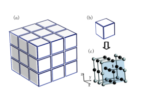
In order to investigate the folding behavior of aggregated protein (multimer), we reconstruct the original HP model by putting a number of individual model proteins (monomers) together, namely, building the cubic toy bricks layer and layer in order. Each monomer has identical sequence and structure but is in different orientation. In this paper, we study the multimer which is stacked by ordered monomers(Fig. 1 (a)). As is shown in Fig. 1 (c), each monomer is figured as a cube formed by a chain of 27 beads occupying the discrete sites of a lattice in a self-avoiding way, with two types of beads of polar (P) and hydrophobic (H) amino-acids respectively. These monomers interact with each other through the contact amino-acids at the surfaces of adjacent monomers respectively. In this model, the total energy of the aggregated protein includes the pair contact energy of the amino acids inside each monomer and the interfacial potential (the additional energy) caused by the contact of amino acids from two adjacent monomers. In our case, 54 interfaces between ordered monomers should be considered. Thus, the energy of the multimer is given by
| (1) |
where the former part represents the total energy of 27 isolated monomers, and i, j denote for the successive labels of residues in a sequence, for the position (of the residue) on the lattice sites, and for H (P) corresponding to hydrophobic (polar) residue respectively. Here the delta notation is adopted, i.e., if a=b and if . As the hydrophobic force drives the protein to fold into a compact shape with more hydrophobic residues inside as possible 23 , the H-H contacts are more favorite in this model, which can be characterized by choosing , , and as adopted in Ref. 12 . The second term in Eq. (1) is introduced to describe the interfacial energy caused by the contact of model proteins. The sum will be done over all the 54 pairs of surfaces. The denotes for the strength of the interfacial interaction and it ranges from 0.1 to 0.9. The is the pair interfacial energy of two contact faces and which belong to two adjacent monomers respectively.
For calculation convenience, we label the six faces of model protein with for a given structure, of which the normal directions correspond to the six directions along , and -z respectively (Fig.1(c)). For each face there are still four states related by rotation of that are specified by second label 0,, and . Then we can easily denote each face of the protein with brackets: , where is the face label and is the rotation label. When the structure and sequence of the protein are fixed, we can write down 24 pairs of kets and bras, , . The interfacial energy between model proteins can be represented as: , it denotes the contact energy between -th rotated state of -th face of a monomer and -th rotated state of -th face of the adjacent monomer, and the stands for the -th rotated state of -th face. The second term of energy in Eq. (1) is then given by:
| (2) |
Both and can be represented by matrixes with entities being either or . In terms of these matrixes, we can easily get the contact energy of two faces by accounting the pair contact energies of corresponding matrix entities, namely,
| (3) |
Here and stands for the aforementioned matrixes corresponding to and . To avoid the ambiguous in defining the matrices of and , we make the following appointment. The matrix is determined by the face of the cubic by taking right hand and head along positive directions of two axis respectively, Additional appoint is that we look toward inside the cubic to define a ket while toward outside to define a bra. For example, in case as show in Fig. 1(c), the -th face contact with -th face of another monomer, we write down the and as:
| (4) |
Then the contact energy of these two faces is evaluated as
| (5) |
clearly, in our definition. To minimize the total energy of the aggregated proteins, the optimal configuration is adjusted by rotating the bra to an appropriate such that the inner product take the smallest magnitude.
III Results and Discussion
It has been noticed by Li et al. 12 that some structures can be designed by a large number of sequences, while some ones can be designed by only few sequences. To elucidate this difference, they introduced the designability of a structure which is measured by the number of sequences that take this structure as their unique lowest energy state. In conclusion, structures differ drastically according to their designability, i.e., highly designable structures emerge with a number of associated sequences much larger than the average ones. Additionally, the energy gap represents the minimum energy a particular sequence needs to change from its ground-state structure into an alternative compact structure. And the average energy gap for a given structure is evaluated by averaging the gaps over all the sequences which design that structure. In the single-monomer HP model, the structures with large designability have much larger average gap than those with small designability, and there is an apparent jump around in the average energy gap. This feature was first noticed by Li et al.12 , thus these highly designable structures are thermodynamically more stable and possess protein-like secondary structures into which the sequences fold faster than the other structures.
In our model, the designability and the energy gap for the aggregated protein are similarly defined, with the energy of single protein replaced by the total energy of the multimer. Considering that the different orientation of the monomers contributes differently to the total energy of the multimer in spite of the identical sequence and structure of each monomer, the designability () in this case denotes for the number of sequences that take the individual structure as the unique lowest energy state of multimer. Similarly, the energy gap is the minimum energy for the multimer to covert from its ground-state conformation (including 27 particularly oriented monomers) into an alternative form .
In our simulation, we search the maximal compact cubic structure of the monomer with various designability first. Based on this, we calculate the total energy of the multimer by stacking 27 monomers one by one to form the aggregated proteins. At each step the state with minimal energy will be preserved. After all monomers are on their positions, we regulate the orientation of each monomer again to search the minimal energy of multimer. After the exhaustive enumeration of all possible sequences, we find that many structures with high average gap in isolated case are no more highly designable in aggregated case when . There are 2,197,634 prion-like sequences taking one structure as the native state(unique energy minimum) in isolated case, but taking another structure when aggregated. While 1,905,960 sequences take the same structures as their native states both in isolated and aggregated cases.
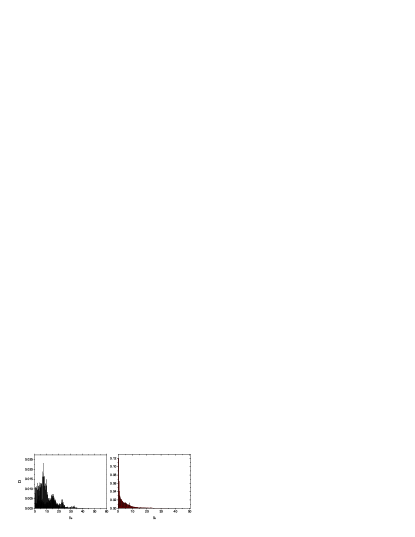
By investigating the sequences of these two cases, we find that the normal sequences possess much more proportion of sequences with larger energy gap, though more prion-like sequences there are. Fig. 2 shows the percentage of total sequences versus the energy gap respectively. The distribution of normal sequences (left) has a peak at , in the right figure plotted for prion-like sequences, the distribution decreases monotonously with the increase of . In this simplified model, the energy gap is considered to be a good parameter to indicate the stability of a sequence at its native state. The larger the energy gap is, the more stability the structures have. Consequently, minority sequences are prion-like. Actually in the nature there should be few prion-like sequences and most protein sequences are stable in their native states. We analyze some of the prion-like sequences by assuming that the decrease of energy can drive the proteins in a multimer to re-fold to new lower energy states. When the isolated native monomers aggregate to form a multimer, some replacement of isolated native structure by new PrPSc-like structure will reduce the energy of the multimer. In this case, all monomers in the multimer will prefer to change their structures for staying much lower total energy level.
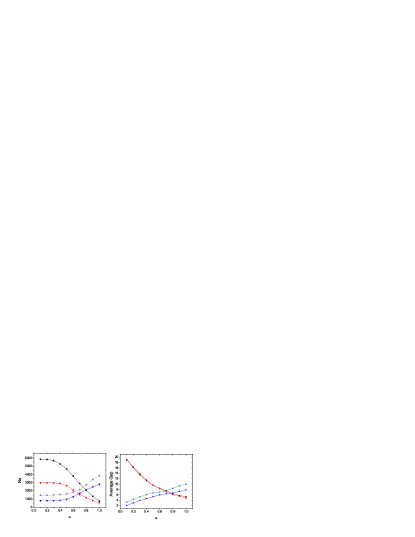
In our simulation, we obtain that the average gaps of the structures with high designability diminish with increasing from 0.1 to 0.9, while the average gaps of most structures with low designability increase. Additionally, a number of new structures which have no sequences taking them as native state in isolated case emerge. In isolated case, there is an abrupt jump in the average energy gap which separates the structures into two groups, the highly designable and lowly designable. These two groups are mixed around in aggregated case (Fig. 3). This implies that the stable structures in isolated case may become unstable and the new stable structures emerge only when the strength of interaction is large enough. This may be consistent with the result in Ref. 24 that most misfolding diseases have a broad incubation period before they cause symptoms and some patients will not be injured. After the seed formation of the misfolding protein, it maybe still take some time for misfolded proteins to get strong enough as condition changes. If the strength stays below the transition point, the injury will not be induced. We investigate some particular structures. The designability and average gaps of highest designable and largest average gap structures in isolated case diminish continuously with increasing , but some other structures (lowly designable in isolated case) enhance their designability and average gaps when aggregate (Fig. 4).

We compare the change of average gap versus the designability in isolated and aggregated cases respectively. In the latter case with , our simulation shows that the average gap increases almost continuously with the increasing of Ns, and there is no abrupt jump in average gap (Fig. 5). Thus the structures can’t be distinguished by the designability and the average gap obviously. The largest average gap reaches 9.945 which is much larger than that in isolated case. Considering an individual sequence, there are two sequences whose gaps reach the value of 55.4 in the aggregated case (2.6 in isolated case) respectively. There are no sequences with energy gap larger than 70.2, the interfacial energy counterpart of these gaps. As is shown in Ref 12 , there are 60 highly designable structures which are distinguished by large average gap from other ones in isolated case. When the proteins aggregate, the average gap increases with designability Ns continuously. But there are large difference in the structures. We find the structures with largest average gap or the highest designability are no more the ones in isolated case, which implies that the most stable structures change when the proteins aggregate. This is similar to the situation that prion-like protein differs in their conformations, PrPC and PrPSc, in isolated and aggregated cases respectively 13 , which is thought to deduce the diseases.


When , 36.7 percent of sequences take one structure as their unique ground states, which is much larger than that in the isolated case (4.75). In the aggregated case, the highest designability Ns is of 3831. Hence there must be much more sequences which average over the other structures and make some lowly designable structures possessing more sequences. As is shown in Fig. 6, in aggregated (left) case, there are much more structures with large Ns, i.e., there are 1497 structures whose Ns are larger than 1400. In the situation of , the distribution of structures with Ns will reach a maximum when Ns=113 (Fig. 6), and it is noticeable that the distributions are same when we change the from 0.000001 to 1. In comparison with the isolated case, there are much less structures that take for individual small designability : in aggregated case the largest number of structures gets 111 at , which is 1109 with Ns being 2 in isolated case. The sequences are much more averaged in all structures in aggregated case, but there are still some structures which are occupied by large number of sequences, and particularly with large average gap. The average gap changes from 0.65 () to 9.95 (), and the largest designability Ns is 3831 with average gap being 7.85.
In summary, by 3-D HP lattice model we observed that there are some sequences which do have prion-like behavior when aggregated. It has been known that the thermodynamic stability of proteins in solution is affected by a variety of factors including temperature, hydrostatic pressure, and presence of additives, such as salts and cosolvent species, which implies that the interfacial interactions between isolated proteins are ubiquitous. If the concentration of protein is sufficiently high, these isolated proteins have a tendency to aggregate reaching a lower energy state. Our calculation showed that a few proteins will propagate the aggregated normal form to abnormal conformation to get more stable multimer (with lower energy), namely, the prion-like behavior. Particularly, we found that the most stable structures are no more the ones in isolated case when the proteins aggregate, such as the structures with largest average gap or the highest designability. Furthermore, we obtained that the average gaps of the structures with high designability diminish with increasing , while the average gaps of most structures with low designability increase. Thus there is no obvious jump in average gap between lowly and highly designable structure for the aggregated proteins. We expect this result can give some hints on the study of misfolding diseases. Since this is a simplified model, further understandings about protein aggregation and misfolding diseases are expected. A more realistic protein-like model with the various interaction being close to the real proteins is in progress.
Acknowledgements.
This work is supported by NSFC No.10225419 and the Key Project of Chinese Ministry of Education No.02046.References
- (1) G. Glenner, N. Engl. J. Med. 302, 1283 (1980).
- (2) C. C. F. Blake and L. C. Serpell, Structure 4, 989 (1996).
- (3) E.McKintosh, S.J.Tabrizi, and J.Collinge,Journal of NeuroVirology 9, 183(2003).
- (4) R. Riek, S. Hornemann, G. Wider, M. Billeter, R. Glockshuber, and K. Wuthrich, Nature 382, 180 (1996).
- (5) T. L. James, H. Liu, N. B. Ulyanov, S. Farr-Jones,et al. Proc. Natl. Acad. Sci. 94, 10086(1997).
- (6) S. B. Prusiner, Proc. Natl. Acad. Sci. 95, 13363(1998).
- (7) C. A. Ross and M. A. Poirier, Nature Medicine 10, S10 (2004).
- (8) M. Buyong,N.Ruth, Protein Science 11, 2335(2002).
- (9) L. Pauling, R. B. Corey, Proc. Natl. Acad. Sci. 37, 235 (1951).
- (10) L. Pauling, R. B. Corey, Proc. Natl. Acad. Sci. 37, 251 (1951).
- (11) C. Anfinsen, Science 181, 223 (1973).
- (12) H. Li, R. Helling, C. Tang, and N. S. Wingreen, Science 273, 666 (1996).
- (13) P.M.Harrison, H.S.Chan, S.B.Prusiner, and F.E.Cohen, Protein Science 10, 879(2001).
- (14) P.M.Harrison,H.S.Chan,S.B.Prusiner,and F.E.Cohen, J. Mol. Bio. 286, 593(1999).
- (15) G. Giugliarelli, C. Micheletti, J.M. Banavar, A. Maritan, J Chem Phys 113, 5072 (2000).
- (16) D. Bratko and H. W. Blanch, J Chem Phys 114, 561 (2001).
- (17) AS. Fraenkel, Bull Math Biol. 55, 1199 (1993).
- (18) K. A. Dill, Biochemistry 24, 1501 (1985).
- (19) J. R. Banavar,M. Cieplak,and A. Maritan, Phys. Rev. Lett. 93, 238101(2004).
- (20) G.Tiana, B.E. Shakhnovich, N.V.Dokholyan, and E.I. Shakhnovich, Proc. Natl. Acad. Sci. 101, 2846 (2004).
- (21) G. Salvi and P. De Los Rios, Phys. Rev. Lett. 91, 258102 (2003).
- (22) Y.Q.Li, Y.Y.Ji, J.W.Mao and X.W.Tang, q-bio.BM /0408024.
- (23) W. Kauzmann, Adv. Protein Chem. 14, 1 (1959).
- (24) D.N. Irani,M. Otto,T. Weber, Current Treatment Options in Infectious Diseases 5, 477(2003).