Magnetic trapping and Zeeman relaxation of imidogen (NH )
Abstract
Imidogen (NH) radicals are magnetically trapped and their Zeeman relaxation and energy transport collision cross sections with helium are measured. Continuous buffer-gas loading of the trap is direct from a room-temperature molecular beam. The Zeeman relaxation (inelastic) cross section of magnetically trapped electronic, vibrational and rotational ground state imidogen in collisions with is measured to be at 710 mK. The NH-He energy transport cross section is also measured, indicating a ratio of diffusive to inelastic cross sections of , in agreement with recent theory Krems et al. (2003).
Driven by the promise of new physics and applications, the field of cold molecular physics has undergone tremendous growth in the past decade Doyle et al. (2004); Bethlem and Meijer (2003). In particular, polar molecules have been proposed DeMille (2002); André et al. (2006) as qubits for quantum information processing and for studies of highly correlated condensed matter systems Micheli et al. (2006). Cold molecules are also very promising candidates for fundamental physics measurements, such as the search for the electric dipole moment of the electron Kozlov and Labzowsky (1995) and time variation of the electron-proton mass ratio Schiller and Korobov (2005). Chemistry with cold molecules may be possible to observe in a new quantum regime where the large deBroglie wavelength and long interaction times of reactants can reopen chemical reaction pathways through tunneling Balakrishnan and Dalgarno (2001); Krems (2005). A key to realizing these new phenomena is the production of cold, trapped, high-density samples of polar molecules.
The complexity inherent in molecules, compared to atoms, makes working with them difficult; the rich internal structure of molecules opens decay modes that, for example, could make evaporative cooling difficult to achieve Bohn et al. (2002). Preparation of cold, dense samples of trapped molecules should provide a pathway to measure cold molecule collisions, critical to elucidating the suitability of molecules for evaporative Avdeenkov et al. (2006); Dhont et al. (2005); Kajita (2006) and sympathetic cooling Lara et al. (2006). So far, several methods have been used to create (ultra-)cold molecules. Production of ultracold molecules from ultracold atoms has been realized through photo and Feshbach association Sage et al. (2005); Donley et al. (2002). Direct cooling or slowing of initially hot polar molecules has also been demonstrated using Stark deceleration Bethlem et al. (1999); Bethlem and Meijer (2003), optical slowing Fulton et al. (2006), billiard-like collisions Elioff et al. (2003), and buffer-gas cooling Weinstein et al. (1998a). Buffer gas cooling uses cryogenic helium gas to cool hot molecules (or atoms). When done in the presence of a magnetic trapping field this can cause the molecules to fall into the conservative trap potential. The first successful trapping of polar molecules was accomplished in our group with buffer-gas loading of CaH, and we know of several other laboratories using buffer-gas loading of magnetic traps Bakker et al. (2006); deCarvalho et al. (2005); Silveira et al. (2001). Other molecules (VO Weinstein et al. (1998b), CaF Maussang et al. (2005) and CrH Stoll ) have also been studied with buffer-gas loading but in all those cases there were loss mechanisms that prevented trapping for extended periods of time.
Despite great progress in the field of cold molecules, no technique has yet realized trapped densities sufficiently high to observe polar molecule-molecule collisions. However, the buffer gas method allowed studies of cold atom-polar molecule spin relaxation of trapped molecules (He-CaH and CaF Weinstein et al. (1998a); Maussang et al. (2005)). These measurements in combination with theory began to uncover the fundamental processes of cold molecule collisions in traps. CaH has a ground state, perhaps the simplest type of magnetically trappable molecule. Although this provided important information on the nature of cold molecule collisions, it was incomplete as there are a variety of molecules. For example, molecules carry with them new internal dynamics, such as the spin-spin interaction, which allows for direct coupling of the rotation during a collision (unlike the case) Krems and Dalgarno (2004). The current vigorous pursuit of high density samples of cold molecules has led several groups to the imidogen (NH) radical. Cold, trapped imidogen is being studied theoretically Krems et al. (2003); Cybulski et al. (2005); Lara et al. (2006); Kajita (2006) and pursued experimentally van de Meerakker et al. (2006); Egorov et al. (2004); Lewandowski et al. (2006), and a scheme for continuous loading of imidogen into a magnetic trap has been proposed for Stark deceleration van de Meerakker et al. (2003).
In this Letter, we demonstrate magnetic trapping of ground state () imidogen radicals and make a direct measurement of the spin relaxation rate in collisions with 3He. Imidogen is continuously loaded directly from a room-temperature molecular beam into a magnetic trap via buffer-gas cooling. More than molecules are loaded into the trap and are observed for longer than 1 s, with lifetimes exceeding 200 ms. The energy transport collision rate is also measured, allowing the determination of the ratio of the diffusive to inelastic cross sections in this system, found to be .
We chose to study imidogen (NH) due to its internal structure, predicted collisional properties, and for technical reasons. The ground state has a magnetic moment and 1.38 Debye electric dipole moment. The imidogen radicals can be produced in an ammonia discharge Ubachs et al. (1984) and detected in absorption or fluorescence on the transition (336 nm, ns). It has been predicted Krems et al. (2003) that molecules with relatively large rotational splitting and weak spin-spin interaction (such as the imidogen radical) will be less likely to undergo collision induced Zeeman transitions. Another important feature of the NH system is that imidogen can be produced in a high flux beam with a room temperature discharge source Egorov et al. (2004). This provides a new challenge - the introduction of this beam into our very low temperature trapping region. This was a key experimental challenge that was met with success and can be applied to numerous other species.
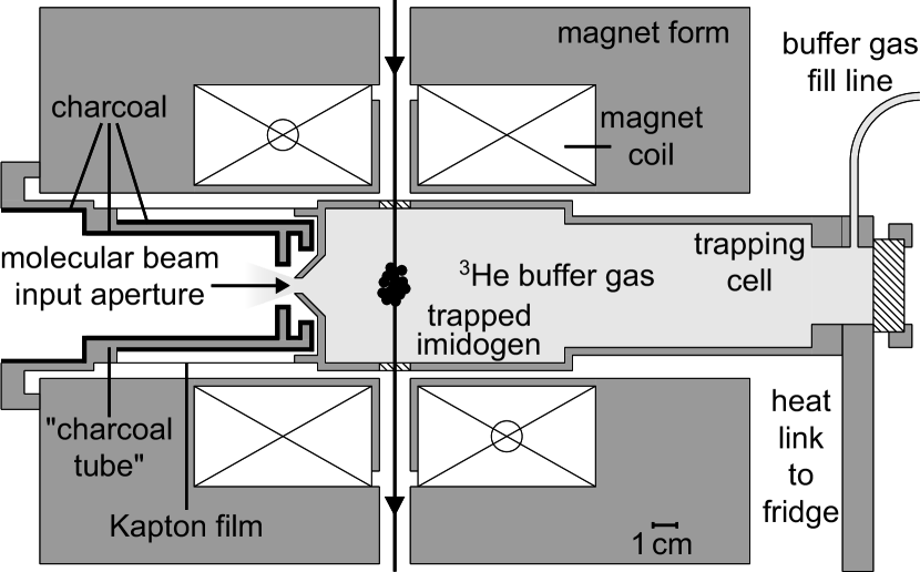
Our apparatus is centered around a cold cell made from copper 111Alloy C10100, annealed in forming gas. and is thermally disconnected from the 4 K magnet surrounding it (see Fig. 1). The maximum trap depth available is 3.9 Tesla. Windows at the magnet midplane allow optical access for laser beams and a fluorescence collection lens. The cell is thermally connected to a 3He refrigerator, giving the cell a base temperature of 450 mK.
Buffer gas enters the cell through a fill line and exits out a 3 mm diameter opening in the side of the cell that faces the discharge source (the “molecular beam input aperture” shown in Fig. 1). Helium is continuously supplied to the cell in order to maintain a constant helium atom density in the trapping region.
The constant flow of helium buffer gas out of the cell aperture poses a significant technical problem. The helium gas can scatter imidogen radicals out of the incident NH beam before they enter the trapping cell. In order to maintain sufficient vacuum just outside the cell, a charcoal coated copper tube (“charcoal tube”) is used to pump away the escaping helium. The charcoal tube is held at 4 K so as to act as a low-profile high-speed vacuum pump. This eliminates any significant scattering of imidogen radicals.
The helium buffer gas density in the cell is determined by the rate at which we flow helium into the cell and the conductance out of the molecular beam input aperture. The conductance of the molecular beam input aperture was measured using a fast ion gauge, agreeing with our calculated theoretical value. This system allows both experimental control and absolute knowledge of the buffer-gas density to better than 20%.
The imidogen radicals in the trapping region are detected using laser induced fluorescence (LIF) excited on the transition. The light source is a CW dye laser frequency doubled in BBO in an external buildup cavity. The excitation laser beam enters the cell and passes through the trap center along a diameter before exiting. Fluorescence from is collected by a midplane lens perpendicular to both the laser and molecular beam axes and imaged to the face of a photomultiplier tube (PMT). In this way, time-resolved narrow-band spectra of trapped imidogen radicals are gathered. As the spherical quadrupole field of the trap magnet is strongly inhomogeneous and the LIF line frequency is field-dependent, laser frequency maps directly to distance from the trap center.
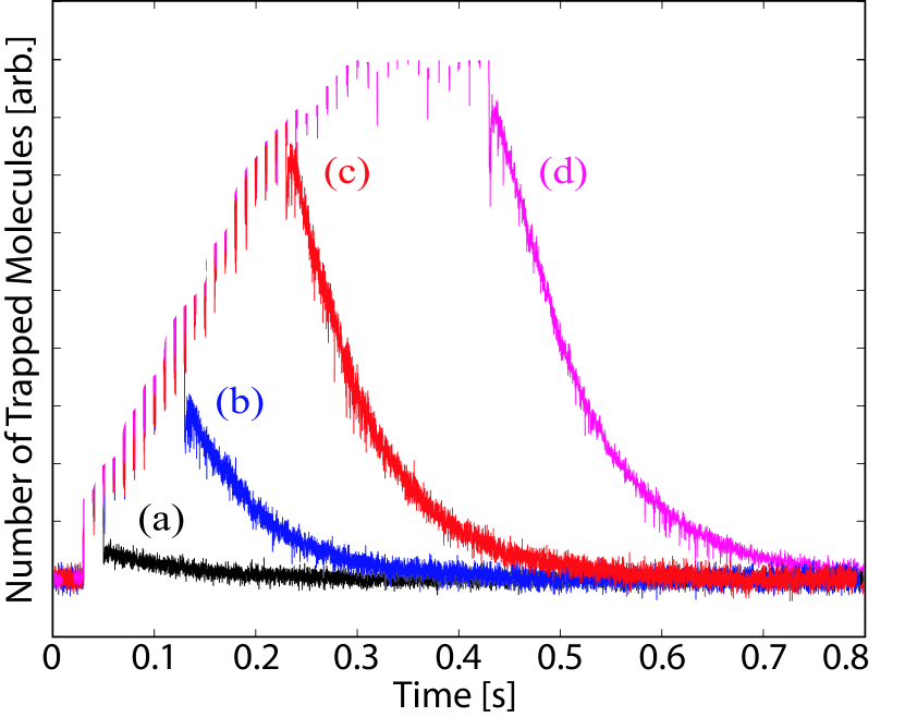
Figure 2 shows a series of time profiles of the number of trapped imidogen molecules for several different loading times. This signal is simply the PMT count rate versus time using an LIF excitation frequency set to the peak of the trapped imidogen spectral feature. As this is a fluorescence experiment, the absolute number of molecules is difficult to determine accurately. By calculating the efficiency of our detection system, excitation probability, and detected molecule fraction we can, however, put a lower bound on our initial number of trapped imidogen radicals of . The data shown in Fig. 2 was taken with 4He, so the temperature of the buffer-gas cell was elevated to 730 mK (in order to maintain sufficient 4He density) at a trap depth of 3.3 Tesla. Each trace corresponds to a different duration of loading from the molecular beam source - the tail ends of all four traces fit well to a single-exponential decay with time constant of ms. For loading times less than the molecular trap lifetime, the number of trapped molecules increases nearly in proportion with with loading time (see curves (a), (b) and (c)). This demonstrates one of the key features of the continuous molecular beam loading technique: as the lifetime of molecules in the trap increases, the loading time can be increased resulting in more trapped molecules. Ablation sources for buffer-gas loading experiments have to this point shown insignificant increases in numbers trapped from multiple loading pulses, likely due to the violent nature of the ablation plume in the trapping region. Fig. 2(c,d) shows that as the loading time exceeds the trap lifetime the signal height saturates and there is no longer any significant increase in the number of trapped molecules, as expected.
Trapping is spectroscopically verified by tuning the LIF laser to be resonant with high-field-seeking (HFS) molecules and comparing time profiles to low-field seekers (LFS). The HFS molecules quickly leave the trap and are undetectable after 10 ms while the lifetimes of the LFS molecules are enhanced to more than 200 ms at our lowest loading temperature. The spatial sensitivity of our fluorescence collection precludes fitting the spectra for temperature, but we have previously demonstrated rotational and translational thermalization of imidogen to the buffer gas temperature Egorov et al. (2004). Finally, as described below, only trapped molecules produce the lifetimes vs. trap depths that we observe.
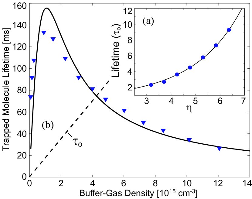
In the absence of collisional losses, the trap lifetime will increase significantly as trap depth increases and the magnetic trapping field holds molecules in the center of the cell. With background buffer gas present, there is an additional lifetime lengthening factor as collisions with helium atoms slow motion of trapped molecules to the walls, essentially enforcing diffusive motion. Fig. 3(a) (inset) shows the measured trap lifetime as a function of , where is the trap depth. The lifetime is plotted in units of the field-free diffusion lifetime in the cell, , given by Hasted (1972)
| (1) |
where is the 3He buffer-gas density, is the thermal average of the diffusion cross section, and are the internal radius and length of the cell, is the reduced mass of the NH-3He system and is the first root of the Bessel function of order zero. For the data in Fig. 3(a) the buffer gas density was cm-3 and the temperature was 690 mK. The solid curve is a fit of a numerical solution to the diffusion equation including the trap potential Weinstein (2001) and the only fitting parameter is the cross section, yielding a measurement of cm2. The quoted uncertainty is systematic and dominated by uncertainty in the buffer-gas density. Multiplying this by the average relative velocity of NH-3He gives the rate coefficient for energy transport, . This is in good agreement with the 0.5 K energy transport collision rate of calculated by Krems et al. in Ref. Krems et al. (2003).
Figure 3(b) shows the measured trap lifetime for different buffer-gas densities. The peak lifetime in this study is somewhat low since cell was heated to 710 mK in order to maintain a constant temperature throughout the full buffer-gas density range (a technical artifact of the helium gas supply system). Trapped imidogen lifetimes for the base temperature of the cell exceed 200 ms. As the density of the 3He buffer gas is increased from to around cm-3 the lifetime of the trapped molecules increases due to the diffusion effect mentioned above. However, as the 3He density is increased past cm-3 it is seen that the lifetime decreases. This decrease is due to collision-induced Zeeman relaxation of the imidogen radicals as they collide with 3He. The time profiles still show good single-exponential decay behavior for these higher buffer-gas densities.
To extract a rate constant for these inelastic collisions, we fit a model curve to the data in Fig. 3. After being loaded, the time profile of the molecule number in the trap can be modeled as where is the buffer gas density, is the initial number of imidogen radicals after loading, and and are fitting parameters corresponding to diffusion enhancement of the lifetime and inelastic collision loss, respectively. This can be rewritten as a single exponential with a time constant given by , which gives the form for the curve fitted to the data in Fig. 3. The constant is the collision induced Zeeman relaxation rate at 710 mK and is found to be , corresponding to an inelastic cross section of and therefore a ratio of diffusive to inelastic cross sections of . The uncertainties are systematic and are dominated by uncertainty in the absolute buffer-gas density. This inelastic collision rate is an order of magnitude higher than the 500 mK value of for 100 Gauss given by Krems et al. Krems et al. (2003) and may be larger due to the strong temperature dependence of the predicted shape resonance. Furthermore, the rate we measure is averaged over the magnetic field range of our trap, and inelastic collision cross sections are predicted to become strongly field-dependent just below this collision energy Krems et al. (2003); Cybulski et al. (2005).
In summary, we have demonstrated magnetic trapping of ground state imidogen molecules using buffer gas loading from a molecular beam. The energy transport (diffusion) and inelastic collision rate constants for NH-3He have been measured
resulting in a ratio of elastic to inelastic collision rates of , which indicates that imidogen radicals should be amenable to the pumping out of the buffer gas and thermal isolation. It is particularly interesting to note that we are able to trap imidogen near the peak of the narrow shape resonance predicted by Krems et al. in Ref. Krems et al. (2003). A factor of two decrease (or increase) in collision energy would decrease the inelastic rate by more than a factor of ten, resulting in a increase in to nearly . This bodes very well for both increasing the number of trapped molecules and perhaps for sympathetic cooling of large numbers of imidogen radicals and other hydrides to the ultracold regime.
Acknowledgements.
We thank Katsunari Enomoto and Michael Gottselig for their valuable assistance in the design and fabrication of the apparatus and also greatly acknowledge Colin Connolly for his construction of the 3He refrigerator used in these experiments. The authors are grateful to B. Friedrich and R. Krems for discussions and careful reading of the manuscript and G. C. Groenenboom for helpful discussions. This work was supported by the U.S. Department of Energy under Contract No. DE-FG02-02ER15316 and the U.S. Army Research Office.References
- Krems et al. (2003) R. V. Krems, H. R. Sadeghpour, A. Dalgarno, D. Zgid, J. Kłos, and G. Chałasiński, Phys. Rev. A 68, 051401(R) (2003).
- Doyle et al. (2004) J. Doyle, B. Friedrich, R. V. Krems, and F. Masnou-Seeuws, Eur. Phys. J. D 31, 149 (2004).
- Bethlem and Meijer (2003) H. L. Bethlem and G. Meijer, Int. Rev. Phys. Chem. 22, 73 (2003).
- DeMille (2002) D. DeMille, Phys. Rev. Lett. 88, 067901 (2002).
- André et al. (2006) A. André, D. DeMille, J. M. Doyle, M. D. Lukin, S. E. Maxwell, P. Rabl, R. J. Schoelkopf, and P. Zoller, Nature Physics 2, 636 (2006).
- Micheli et al. (2006) A. Micheli, G. K. Brennen, and P. Zoller, Nature Physics 2, 341 (2006).
- Kozlov and Labzowsky (1995) M. G. Kozlov and L. N. Labzowsky, J. Phys. B: At. Mol. Opt. Phys. 28, 1933 (1995).
- Schiller and Korobov (2005) S. Schiller and V. Korobov, Phys. Rev. A 71, 032505 (2005).
- Balakrishnan and Dalgarno (2001) N. Balakrishnan and A. Dalgarno, Chem. Phys. Lett. 341, 652 (2001).
- Krems (2005) R. V. Krems, Int. Rev. Phys. Chem. 24, 99 (2005).
- Bohn et al. (2002) J. L. Bohn, A. V. Avdeenkov, and M. P. Deskevich, Phys. Rev. Lett. 89, 203202 (2002).
- Avdeenkov et al. (2006) A. V. Avdeenkov, M. Kajita, and J. L. Bohn, Phys. Rev. A 73, 022707 (2006).
- Dhont et al. (2005) G. S. F. Dhont, J. H. van Lenthe, G. C. Groenenboom, and A. van der Avoird, J. Chem. Phys. 123, 184302 (2005).
- Kajita (2006) M. Kajita, Phys. Rev. A 74, 032710 (2006).
- Lara et al. (2006) M. Lara, J. L. Bohn, D. Potter, P. Soldán, and J. M. Hutson, Phys. Rev. Lett. 97, 183201 (2006).
- Sage et al. (2005) J. M. Sage, S. Sainis, T. Bergeman, and D. DeMille, Phys. Rev. Lett. 94, 203001 (2005).
- Donley et al. (2002) E. A. Donley, N. R. Claussen, S. T. Thompson, and C. E. Wieman, Nature 417, 529 (2002).
- Bethlem et al. (1999) H. L. Bethlem, G. Berden, and G. Meijer, Phys. Rev. Lett. 83, 1558 (1999).
- Fulton et al. (2006) R. Fulton, A. I. Bishop, M. N. Schneider, and P. F. Barker, J. Phys. B: At. Mol. Opt. Phys. 39, S1097 (2006).
- Elioff et al. (2003) M. S. Elioff, J. J. Valentini, and D. W. Chandler, Science 302, 1940 (2003).
- Weinstein et al. (1998a) J. D. Weinstein, R. deCarvalho, T. Guillet, B. Friedrich, and J. M. Doyle, Nature 395, 148 (1998a).
- Bakker et al. (2006) J. M. Bakker, M. Stoll, D. R. Weise, O. Vogelsang, G. Meijer, and A. Peters, J. Phys. B 39, S1111 (2006).
- deCarvalho et al. (2005) R. deCarvalho, N. Brahms, B. Newman, J. M. Doyle, D. Kleppner, and T. Greytak, Can. J. Phys. 83, 293 (2005).
- Silveira et al. (2001) D. M. Silveira, O. Pereira, M. Veloso, and C. L. Cesar, Brazilian Journal of Physics 31, 203 (2001).
- Weinstein et al. (1998b) J. D. Weinstein, R. deCarvalho, K. Amar, A. Boca, B. C. Odom, B. Friedrich, and J. M. Doyle, J. Chem. Phys. 109, 2656 (1998b).
- Maussang et al. (2005) K. Maussang, D. Egorov, J. S. Helton, S. V. Nguyen, and J. M. Doyle, Phys. Rev. Lett. 94, 123002 (2005).
- (27) M. Stoll, private communication.
- Krems and Dalgarno (2004) R. V. Krems and A. Dalgarno, J. Chem. Phys. 120, 2296 (2004).
- Cybulski et al. (2005) H. Cybulski, R. V. Krems, H. R. Sadeghpour, A. Dalgarno, J. Kłos, G. C. Groenenboom, A. van der Avoird, D. Zgid, and G. Chałasiński, J. Chem. Phys. 2005, 094307 (2005).
- van de Meerakker et al. (2006) S. Y. T. van de Meerakker, I. Labazan, S. Hoekstra, J. Küpper, and G. Meijer, J. Phys. B: At. Mol. Opt. Phys. 39, S1077 (2006).
- Egorov et al. (2004) D. Egorov, W. C. Campbell, B. Friedrich, S. E. Maxwell, E. Tsikata, L. D. van Buuren, and J. M. Doyle, Eur. Phys. J. D 31, 307 (2004).
- Lewandowski et al. (2006) H. Lewandowski, L. P. Parazzoli, D. Lobser, and C. Romero, in 379. WE-Heraeus-Seminar on Cold Molecules (2006).
- van de Meerakker et al. (2003) S. Y. T. van de Meerakker, B. G. Sartakov, A. P. Mosk, R. T. Jongma, and G. Meijer, Phys. Rev. A 68, 032508 (2003).
- Ubachs et al. (1984) W. Ubachs, J. J. ter Meulen, and A. Dymanus, Can. J. Phys. 62, 1374 (1984).
- Hasted (1972) J. B. Hasted, Physics of Atomic Collisions (American Elsevier Publishing Company, Inc., 1972), chap. 1.6, 2nd ed.
- Weinstein (2001) J. D. Weinstein, Ph.D. thesis, Harvard University (2001).
- Harris et al. (2004) J. G. E. Harris, R. A. Michniak, S. V. Nguyen, W. C. Campbell, D. Egorov, S. E. Maxwell, L. D. van Buuren, and J. M. Doyle, Rev. Sci. Inst. 75, 17 (2004).