A Two Phase Harmonic Model for Left Ventricular Function
Abstract
A minimal model for mechanical motion of the left ventricle is proposed. The model assumes the left ventricle to be a harmonic oscillator with two distinct phases, simulating the systolic and diastolic phases, at which both the amplitude and the elastic constant of the oscillator are different. Taking into account the pressure within the left ventricle, the model shows qualitative agreement with functional parameters of the left ventricle. The model allows for a natural explanation of heart failure with preserved systolic left ventricular function, also termed diastolic heart failure. Specifically, the rise in left ventricular filling pressures following increased left-ventricular wall stiffness is attributed to a mechanism aimed at preserving heart rate and cardiac output.
keywords:
Left Ventricular motion, Harmonic oscillator, Diastolic heart failure, ,
1 Motivation
1.1 General
The left ventricle (LV) is a highly complex mechanical system. Left ventricular function is determined by both internal and external factors such as myocardial contractility, myocardial stiffness, preload, afterload, rhythm and pace, size, shape etc [1]. All these factors combine to regulate left ventricular motion, which may be characterized by three basic distinguishing attributes: (1) the motion is periodic, including a diastolic expansion phase and a systolic contraction phase, (2) the period is divided unequally between the diastolic and the systolic phases, the former being generally twice as long as the latter [2] and, (3) the forces generated by the left ventricle are in accordance with Frank-Starling law of the heart [1], ], which states that the larger the LV volume, the greater the energy of it s contraction and hence diastolic stretch actually increases LV contractility.
Motivated by the fact that LV dysfunction and heart failure are associated with alterations in the mechanical motion of the left ventricle [3, 4, 5] , simulating this motion has become a major challenge. The use of both theoretical tools (such as finite element geometrical construction[6], fluid-dynamics based computation [7, 8], Immersed Boundary method [9] etc.) and experimental tools (such surgically implanted track markers [10, 11], magnetic resonance tagging [12, 13] etc.) have been utilized for such simulation. As a common factor, many of the theories attempt to include an abundance of factors influencing the left ventricle, to achieve quantitative resemblance between theory and experimental data. While some success has been achieved in describing normal LV function, the models are far from adequate in describing LV dysfunction or the effect of cardiac assist devices, and further progress is needed before such models may be utilized for clinical applications [14].
1.2 Diastolic heart failure
Heart failure (HF) is a clinical syndrome that can result from structural or functional cardiac disorders, impairing the ability of the ventricle to fill with or eject blood [15]. The clinical manifestations of HF are dyspnea and fatigue, which may limit exercise tolerance, and fluid retention, which may lead to pulmonary and peripheral oedema. Diastolic heart failure (DHF) is the clinical syndrome of heart failure associated with preserved LV ejection fraction (an index of LV systolic function and contractility, defined as the ratio of LV stroke volume to end diastolic volume). DHF accounts for approximately 40% of heart failure cases [16, 17] and carries a significant morbidity, comparable to that of systolic heart failure [18, 19]. Diastolic abnormalities of the LV may be related to increased myocardial stiffness and impaired relaxation [20, 21, 22], leading to an essentially mechanical dysfunction, resulting in the inability of the left ventricle to fill with blood at low filling pressures during diastole [23, 24].
The mechanisms underlying increased myocardial stiffness can be divided into factors that are intrinsic or extrinsic to the myocardium [25]. Myocardial factors include cellular factors, such as impaired calcium homeostasis and changes in Titin sarcomeric proteins, which act as viscoelastic springs that provide a recoiling force during diastole [26], and extracellular factors, including changes in extracellular matrix morphology (i.e. fibrosis) leading to increased myocardial stiffness [27]. Relaxation is an energy-dependent process, and abnormalities in cellular energy supply and utilization can lead to impaired relaxation [28]. Both abnormalities may result in a physiological state in which diastolic LV pressures are elevated, leading to elevated left atrial and pulmonary venous pressures, exercise intolerance [29, 30] and acute pulmonary oedema [31, 32].
Despite its prevalence and significant morbidity there are currently very few models that can simulate DHF as an integral part of the mechanical motion of the LV [33]. Such simulation would be beneficial for promoting our understanding of the underlying pathophysiology of DHF and simulating treatment modalities, and as such provided much of the motivation for developing a minimal model for LV motion.
1.3 Scope
In this paper we develop a qualitative minimal approach to describe the motion of the LV. Generalizing the simple linear harmonic oscillator, we qualitatively mimic the full ventricular cycle, with all three basic distinguishing attributes described above. Further more, our model naturally describes the mechanics of DHF, and thus provides a baby-step towards a more rigorous model for LV function in DHF.
2 The model
2.1 Preliminaries
As a mathematical introduction, in this section we discuss the simple harmonic oscillator, describe the general solution and show its relation to the properties of the LV. A simple harmonic oscillator (e.g. a mass attached to a spring, an elastic band etc.) is described by Hooke’s law, that is the larger the displacement from equilibrium the stronger the restoring force acting on the body. In the simplest case, the restoring force is proportional to the displacement, and thus Newton’s law is of the form
| (1) |
where is the proportionality (elastic) constant between the force and the displacement, its physical meaning is the amount of force required to stretch the oscillator by one unit length (per unit mass).
The solution of Eq. (1) depends on the sign of . If is negative, the general solution is
| (2) |
where and are constants determined by the initial conditions of the oscillator. Notice that this solution is ”unstable”, that is will increase indefinitely with the time . Thus, this solution is inappropriate for describing an oscillating body.
For a positive , the solution is periodic,
| (3) |
where the radial amplitude and the phase are constants, which are determined by the initial conditions of the oscillator. The frequency of oscillations is given by , and the period (i.e. the duration of a single cycle) is . We notice that if stands for the LV radius, than this solution already possesses two of the LV characteristics described in the introduction. Obviously, the motion is periodic, and the force (given by the right hand side of Eq. 1) is in agreement with Starlings law.
However, the third characteristic of the LV - the unequal division of the cardiac cycle - is unfulfilled by the simple harmonic oscillator. Following, we introduce a generalization to the simple oscillator, based on a physiologic mechanism, which inherently incorporates this feature.
2.2 Basic model
In this section we describe our generalization of the simple harmonic oscillator. We start by examining the physiologic origin of systolic and diastolic LV function.
The transition from diastole to systole stems from excitation-contraction coupling. The trigger for systole is the electrical excitation of cardiac muscle cells (action potential), which raises the free intra-cellular concentration, thus activating cellular contraction. Contraction leads to elevation of ventricular wall tension, elevation of intra-ventricular pressure and eventually ejection of blood from the ventricle, when a positive pressure gradient is achieved. For diastolic relaxation to occur, intra-cellular concentration must be actively reduced, leading to myocardial relaxation [34].
In essence, this can be simplified to a model in which systole begins when the LV wall is abruptly stiffened (isovolumic contraction) leading to LV pressure elevation and systolic contraction, after-which the ventricle is suddenly relaxed (isovolumic relaxation), followed by a pressure decline and diastolic filling. Even a model based on this simplification would require consideration of additional factors such as, e.g. environmental effects, blood flow (rheological) effects, spatial form of the LV, energy considerations etc.). This type of analysis is probably beyond mathematical rigour.
We thus suggest a phenomenological model for LV function. We describe the LV as a cylindrical elastic membrane. The model thus consists of a harmonic oscillator, for which both the elastic constant (and thus the frequency) and the radial amplitude differ between the systolic and the diastolic phase. Since the systolic (diastolic) phase are defined by a decrease (increase) of the LV radius , both the elastic constant and the amplitude will depend on the sign of the derivative, . This will mimic the sudden stiffening and relaxation of the cardiac muscle due to changes in intra-cellular concentration. The force induced by the intra-ventricular pressure, , is given (in the cylindrical approximation) by , where is the internal pressure (divided by the LV wall mass and height). Assuming that in the systolic and diastolic phases the pressures and are approximately constant, we obtain the following equation for the radius of the LV,
| (4) |
where and are the effective elastic constants at the diastole and systole respectively, and and are the amplitudes at the diastole and systole, respectively. Eq. 4 is subjected to the initial conditions
| (5) |
where is the maximal radius of the LV, and to the following conditions:
-
1.
is a continuous function.
-
2.
The motion is periodic; that is, if is the period of the function, then
Let us start by examining each branch of the solution separately, starting with Eq. (4) for the the systolic phase. Demanding that the solution is stable yields the condition . Denoting and taking in account the initial conditions (Eq. (5)), we have the solution
| (6) |
The motion is governed by Eq. (6) as long as the first derivative of the solution is negative. The derivative changes sign (from negative to positive) at , for which . Thus, (where is the minimal LV radius) is the initial condition for the motion in the diastolic phase, i.e. the solution for the diastolic part of Eq. (4) is subjected to the initial conditions,
| (7) |
Denoting , the solution for the diastolic stage is given by
| (8) |
As in the systolic phase, this branch of the solution is valid when is in the interval . Denoting , and we obtain the solution for a single cycle,
| (9) |
From continuity at and periodicity (i.e. ) we obtain the relation
| (10) |
We thus obtain a full solution for the motion of the LV, characterized by the four parameters and . The ratio between (the duration of diastole) and (duration of systole) is . The parameters and may vary and are determined by both the elastic constants and the pressures in the different phases.
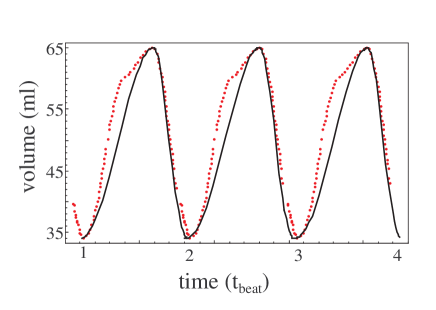
In Fig. 1 we plot the LV volume for several cycles, actual LV data taken from a healthy human subject using non-invasive magnetic resonance imaging technique, adopted from K. Kindberg [13] (dashed line) and a fit with Eq. (9) (solid line). As seen, the model yields fair agreement with the data, and results in the ratio between the diastolic and systolic times, .
3 Diastolic heart failure
As described in Sec. 1.2, diastolic abnormalities in DHF patients include increased LV stiffness and elevated LV filling pressures, leading to the symptoms of heart failure. In this section we intend to answer two questions:
-
1.
Can we simulate the increased LV stiffness within our model?
-
2.
Can this model predict and explain the increase of pressure?
For the first question, the answer seems fairly simple: In the suggested model, LV stiffness is represented by the coefficients and . Thus, in terms of the suggested model, the disease can be simulated by increasing by an amount .
In order to answer the second question we note that in DHF patients there seems to be no significant change in the heart rate, and thus no real change in the duration of the diastole (or systole) [32, 35]. Since the duration of the phases is defined by the frequencies and , from their definition we have that remains constant only if the term remains constant. Thus, if is increased by a portion , then must also increase by . Hence, this model indeed predicts the rise in pressure. In other words, once the diastolic stiffness of the LV wall increases, a compensation is achieved by increasing the pressure, in order to prevent a significant change in heart rate.
4 Summary
In summary, a naive harmonic model for the mechanical motion of the left ventricle was developed. The model consists of a generalized harmonic oscillator, for which both the radial amplitude and the elastic constant depend on the direction of the motion, yielding both the systolic and diastolic phases of the cardiac cycle, and allows for a qualitative simulation of left ventricular motion.
Despite its simplicity, the model yields a natural explanation for diastolic heart failure, in which the left ventricular wall is stiffened and diastolic filling pressures are increased. Our model suggests that the rise in diastolic filling pressures is required in order to maintain ventricular volume when the heart rate is not significantly changed, as is the clinically observed case [32].
One can postulate a correlation between the model and physiological phenomena as follows: Maintaining cardiac output (CO) is essential for normal function. The CO is a product of heart rate and stroke volume. In DHF patients, the stroke volume remains unchanged [36, 35] and hence slowing heart rate should be prevented, or CO will be reduced. However, when our model is considered, if LV volumes are to be maintained despite increased stiffening (again, as is the clinical finding), LV pressures have to be increased or heart rate decreased. It is thus concluded that the increased LV pressures are the result of a forced compensation, aimed at maintaining heart rate and CO when the LV wall is stiffened.
Finally, we note that while the model presented here is very much the minimal model for LV function, additional phenomenological details, such as ventricular pressure- volume loops and atrial parameters may be added in future studies. Such additions would render the model analytically unsolvable, yet numerical solutions may be possible, and may advance our understanding of different physiological phenomena.
References
- [1] Lionel HO, Mechanisms of cardiac contraction and relaxation. In: Zipes DP, Libby P, Bonow RO, Braunwald E, editors. Braunwld’s heart disease 7th edition. Elsevier Saunders, Philadelphia PA, USA 2005, p. 476.
- [2] Tortora GJ, Derrickson B, Principles of anatomy and physiology 11th edition. John Wiley & Sons, Hoboken NJ,USA. 2006, p. 716.
- [3] Colucci WS and Braunwald E, Pathophysiology of heart failure. In: Zipes DP, Libby P, Bonow RO, Braunwald E, editors. Braunwld’s heart disease 7th edition. Elsevier Saunders, Philadelphia PA, USA 2005, p. 509–526.
- [4] Zile MR and Brutsaert DL. New concepts in diastolic dysfunction and diastolic heart failure: Part I: Diagnosis, Prognosis, and Measurements of Diastolic Function. Circulation. 2002;105:1387 -1393.
- [5] Zile MR and Brutsaert DL. New concepts in diastolic dysfunction and diastolic heart failure: Part II: causal mechanisms and treatment. Circulation 2002; 105(12):1503–1508.)
- [6] Hunter PJ, Pullen AJ and Smaill BH, Modeling total heart function, Annu Rev Biomed Eng 2003; 5:147–177.
- [7] Taylor TW, Okino H and Yamaguchi T, Three-Dimensional Analysis of Left Ventricular Ejection Using Computational Fluid Dynamics, ASME J Biomech Eng 1994; 116:127- 130.
- [8] Schoephoerster RT, Silva CL and Ray G, Evaluation of Left Ventricular Function Based on Simulated Systolic Flow Dynamics Computed From Regional Wall Motion, J Biomech 1994; 27:125 -136.
- [9] Peskin, CS and McQueen DM, Fluid Dynamics of the Heart and Its Valves. in: Case Studies in Mathematical Modeling: Ecology, Physiology, and Cell Biology, Othmer, Adler et al., eds., Prentice-Hall Inc., New Jersey 1996, pp. 309 -337.
- [10] Ingels N, Daughters G, Stinson E and Alderman E, Measurement of midwall myocardial dynamics in intact man by radiography of surgically implanted markers. Circulation 1975; 52:859- 867.
- [11] Doss JK, Hillis LD, Curry G, Lewis SE, Dehmer GJ, Parkey RW, Mitchell JH and Willerson JT, A new model for the assessment of regional LV wall motion. Radiology 1982; 143:763- 770.
- [12] Meier D, Ziskin MC, Santamore WP and Bove AA, Kinematics of the beating heart. IEEE Transactions on Biomedical Eng 1980; 27:319- 329.
- [13] Katarina Kindberg, Regional Kinematics of the Heart: Investigation with Marker Tracking and with Phase Contrast Magnetic Resonance Imaging, Thesis, Linkopings universitet, Linkoping, Sweden 2003.
- [14] Saber NR, Wood NB, Gosman AD, Merrifield RD, Yang GZ, Charrier CL, Gatehouse PD and Firmin DN, Progress Towards Patient-Specific Computational Flow Modeling of the Left Heart via Combination of Magnetic Resonance Imaging with Computational Fluid Dynamics. Ann Biomed Eng 2003; 31:42–52.
- [15] Hunt SA, Baker DW, Chin MH, Cinquegrani MP, Feldman AM, Francis GS, Ganiats TG, Goldstein S, Gregoratos G, Jessup ML, Noble RJ, Packer M, Silver MA, Stevenson LW. ACC/AHA guidelines for the evaluation and management of chronic heart failure in the adult: executive summary: a report of the American College of Cardiology/American Heart Association Task Force on Practice Guidelines (Committee to Revise the 1995 Guidelines for the Evaluation and Management of Heart Failure). J Am Coll Cardiol 2001;38:2101–13.
- [16] Senni M and Redfield MM, Heart failure with preserved systolic function. A different natural history? , J Am Coll Cardiol 2001; 38:1277–1282.
- [17] Vasan RS and Levy D, Defining diastolic heart failure: a call for standardized diagnostic criteria, Circulation 2000; 101:2118–2121.
- [18] Vasan RS, Larson MG, Benjamin EJ, Evans JC, Reiss CK and Levy D, Congestive heart failure in subjects with normal versus reduced left ventricular ejection fraction: prevalence and mortality in a population-based cohort, J Am Coll Cardiol 1999; 33:1948–1955.
- [19] Tsutsui M, Tsuchihashi M and Takeshita A, Mortality and re-admission of hospitalized patients with congestive heart failure and preserved versus depressed systolic function, Am J Cardiol 2001:88;530–533.
- [20] Borbely A, van der Velden J, Papp Z, Bronzwaer JGF, Istvan Edes I, Stienen GJM, and Paulus WJ, Cardiomyocyte Stiffness in Diastolic Heart Failure Circulation, Feb 2005; 111: 774–781.
- [21] Kass DA, Bronzwaer JGF, and Paulus WJ, What Mechanisms Underlie Diastolic Dysfunction in Heart Failure? Circ. Res., Jun 2004; 94: 1533–1542.
- [22] Sweitzer NK and Stevenson LW, Diastolic heart failure: miles to go before we sleep, Am J Med 2000; 109(8):683–685.
- [23] Brutsaert DL, Sys SU and Gillebert TC, Diastolic failure: pathophysiology and therapeutic implications, J Am Coll Cardiol 1993; 22:318–325.
- [24] Grossman W, Diastolic dysfunction and congestive heart failure, Circulation 1990;81:III1–III7.
- [25] Zile MR, Brutsaert DL. New concepts in diastolic dysfunction and diastolic heart failure: Part II: causal mechanisms and treatment. Circulation 2002; 105(12):1503–1508.
- [26] Fukuda N, Sasaki D, Ishiwata S, Kurihara S. Length dependence of tension generation in rat skinned cardiac muscle: role of titin in the Frank-Starling mechanism of the heart. Circulation 2001; 104(14):1639–1645.
- [27] Yamamoto K, Masuyama T, Sakata Y, Nishikawa N, Mano T, Yoshida J et al. Myocardial stiffness is determined by ventricular fibrosis, but not by compensatory or excessive hypertrophy in hypertensive heart. Cardiovasc Res 2002; 55(1):76–82.
- [28] Zile MR, Brutsaert DL. New concepts in diastolic dysfunction and diastolic heart failure: Part II: causal mechanisms and treatment. Circulation 2002; 105(12):1503–1508.
- [29] Kitzman DW, Higginbotham MB, Cobb FR, Sheikh KH, and Sullivan MJ, Exercise intolerance in patients with heart failure and preserved left ventricular systolic function: failure of the Frank-Starling mechanism. J Am Coll Cardiol 1991; 17:1065–1072.
- [30] Little WC, Wesley-Farrington DJ, Hoyle J, Brucks S, Robertson S, Kitzman DW and Cheng CP, Effect of Candesartan and Verapamil on Exercise Tolerance in Diastolic Dysfunction. J Cardiovasc Pharmacol 2004; 43(2):288–293 .
- [31] Little WC, Hypertensive pulmonary oedema is due to diastolic dysfunction Eur Heart J 2001; 22: 1961–1964.
- [32] Zile MR, Baicu CF, and Gaasch WH, Diastolic Heart Failure Abnormalities in Active Relaxation, N Engl J Med 2004; 350(19):1953–1955.
- [33] Lemmon JD and Yoganathan AP, Computational Modeling of Left Heart Diastolic Function: Examination of Ventricular Dysfunction. ASME J Biomech Eng 2000; 122:297–303.
- [34] Donald M. Bers, Cardiac excitation - contraction coupling. Nature 2001; 415:198–205.
- [35] Kitzman DW, Little WC, Brubaker PH, Anderson RT, Hundley WG, Marburger CT et al. Pathophysiological characterization of isolated diastolic heart failure in comparison to systolic heart failure. JAMA 2002; 288(17):2144-2150.
- [36] Zile MR, Baicu CF, and Bonnema DD, Diastolic Heart Failure: Definitions and Terminology. Progress in Cardiovascular Diseases, Vol. 47, No. 5 (March/April), 2005: 47;307-313.