Electromagnetically Induced Transparency in 6Li
Abstract
We report electromagnetically induced transparency for the D1 and D2 lines in 6Li in both a vapour cell and an atomic beam. Electromagnetically induced transparency is created using co-propagating mutually coherent laser beams with a frequency difference equal to the hyperfine ground state splitting of 228.2 MHz. The effects of various optical polarization configurations and applied magnetic fields are investigated. In addition, we apply an optical Ramsey spectroscopy technique which further reduces the observed resonance width.
pacs:
32.10.Fn, 32.30.Jc, 32.80.Qk1 Introduction
Electromagnetically Induced Transparency (EIT) is of great interest due to its wide application in lasing without inversion [Zibrov95], control of light propagation (slow light) [Matsko01] and enhanced Kerr nonlinearity [Harris99, Akulshin03]. The concept of a non-absorbing dark state is the key basis of light storage [Lukin03]. Lithium has two stable isotopes, 6Li and 7Li (7.5 and 92.5 natural abundance, respectively). Magnus et al [Magnus05] reported the first study of EIT on the D1 and D2 lines of 7Li in a vapour. In this paper we present EIT in 6Li performed on both the D1 and D2 lines using several nonlinear techniques, paying particular attention to a comparison between the EIT resonances obtained on the D1 and D2 lines.
EIT is a phenomenon in which an opaque atomic medium is turned into a transparent one in the presence of a control laser field [Harris97]. When two Zeeman sub-levels or two hyperfine levels in an atomic ground state are coupled by light to a common excited state, the interference between amplitudes of alternative transition paths can substantially reduce the absorption. The atoms are pumped by the laser light into a coherent superposition of the ground state sublevels which constitutes a non-absorbing state decoupled from the laser field. This phenomenon is also known as coherent population trapping, e.g.,[Arimondo96]. The absorption of the probe exhibits a narrow dip that has a sub-natural linewidth ultimately determined by the relaxation time of the ground state sublevels. The refractive index of the coherent atomic medium reveals extremely steep normal dispersion in the vicinity of the EIT resonance, so that the group velocity of light in such an atomic medium can be dramatically reduced [Schmidt96].

EIT in alkali atoms with half-integer total angular momentum (F=J+I, J=L1/2) have not, to our knowledge, been previously investigated. A schematic diagram of the relevant atomic energy levels is shown in figure 1. The level structure of 6Li is somewhat different to other alkali atoms that have been used to study EIT and other effects related to ground state coherence. There are only three sublevels in the 2 state in comparison with four sublevels for all other alkali n states.
The strongest optical transitions of 6Li are two electric dipole transitions 2 2 (D1 line) and 2 2 (D2 line) which are separated by a fine structure splitting of 10 GHz. Of all the alkali atoms 6Li has the smallest hyperfine splitting in both the ground and excited states. The hyperfine splitting of the 2 ground state, 228.2 MHz [Walls03], is much less than the Doppler width of the D lines near room temperature. But, more interestingly, the hyperfine splitting of the 2 state is smaller than the 5.9 MHz [McAlexander96] natural width of the optical transition. Thus, every atom has comparable probabilities, independent of the atomic velocity, of being excited via different transitions on the D2 line. This unique situation for alkali atoms allows analyzing a role of additional optical transitions, which do not contribute to the preparation of a non-absorbing coherent state responsible for EIT. This study could be useful for optimization of EIT resonances widely used for metrological applications [Knappe05].
The ultimate width of an EIT resonance also depends on the mutual coherence of the probe and control laser fields [Arimondo96]. In the case of Cs and Rb, which have large ground state splitting, several methods can be used to produce two phase-stable laser fields such as high frequency acousto-optical modulators (AOM), two phase-locked lasers, applying current modulation to laser diodes and the use of electro-optic modulators [Wynands99]. High frequency AOMs are expensive while phase locking at precise frequency offset and current modulation at high frequencies are experimentally not simple. However, in the case of lithium the ground state splitting is relatively small and mutually coherent probe and control fields can be easily prepared from the same laser using a low-cost AOM in a double pass configuration, thus eliminating laser linewidth contribution.
2 Experimental Set-up
An external cavity diode laser (ECDL) (Toptica DL 100) is used as the source of the resonant optical field which has an output power of 15 mW and a linewidth of approximately 1 MHz. Using the so-called feed-forward technique, where both the diode current and the grating position are varied, a fine frequency tuning range of over 20 GHz is obtained. The vapour cell consists of a stainless steel crossed tube configuration with a flexible bellow construction and viewports on each end of the 30 cm horizontal arm. Using a thermocoax element the centre part of the vapour cell is heated to 350 ∘C yielding a maximum unsaturated absorption of about 20. At this temperature the Doppler width is 3.5 GHz. Lithium vapour is limited by its mean free path to the centre part of the tube, protecting the viewports from becoming coated with lithium. To further reduce this problem we heat the viewports to 120 ∘C.
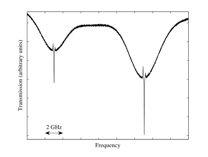
Saturation spectroscopy of lithium vapour provides an excellent frequency reference for our experiments in an atomic beam. Figure 2 shows typical Doppler-free spectra of 6Li with a frequency scan over both the D1 (figure 2 (a)) and D2 (figure 2 (b)) lines. The inset shows a close up of the D2 line whose outer peaks correspond to the F = 3/2 F = 5/2, 3/2, 1/2 and F = 1/2 F = 3/2, 1/2 transitions, respectively. The reduction in absorption of the probe at these outer peaks is due to the saturation of the transition by the pump beam. The enhancement in absorption of the crossover dip is due to compensation of optical hyperfine pumping, which occurs when the pump and probe laser are resonant with both ground state sublevels at the same time. When applying higher intensities the spectroscopy of the D1 line resembles the spectroscopy of the D2 line (inset in figure 2). The width of the crossover peak in the D2 line is 25 MHz. The contrast ratio of the crossover peak with respect to the Doppler broadened resonance is 100.

Figure 3 shows the experimental set-up used for EIT experiments in both the vapour cell and atomic beam. The EIT resonances are usually observed with a bichromatic laser beam consisting of two components with tunable frequency offset changing in the vicinity of the ground state hyperfine splitting. The ECDL (master laser) is used to injection lock a diode laser (slave 1) which injection locks two additional diode lasers (slave 2 and slave 3). The master laser is tuned to either the D1 or D2 line. For the vapour cell experiments the master laser is not actively stabilized since its long-term drift is small compared to the Doppler width. However for the atomic beam experiments the master laser is actively locked to the crossover of the saturated absorption lines. Radiation of slave 3 is used as a pump beam for coupling states between the ground state and the excited levels in the D1 or D2 line. Laser light from slave 2 which is resonant with the F = 3/2 F′ transitions (either D1 or D2 line) is used as a probe beam. The probe laser frequency is scanned over 25 MHz while the frequency of the pump laser is fixed. They are overlapped on a non-polarizing beam splitting cube and expanded to a beam diameter of 5 mm for the vapour cell and 18 mm for the atomic beam experiments.
3 Vapour Cell EIT
By tailoring the optical frequencies present we performed experiments on EIT in both a vapour cell and an atomic beam. To obtain hyperfine ground state coherence we use mutually coherent laser fields whose frequencies are separated by the hyperfine splitting of the ground state. The pump and probe are sent through the vapour cell with intensities of 8 mW/cm2 and 2 mW/cm2 respectively. To improve the signal to noise ratio the pump is amplitude modulated by means of an mechanical chopper (1 kHz) and the probe is detected using a lock-in amplifier. For the results shown here, both beams are linearly polarized and orthogonal to each other.

3.1 Results
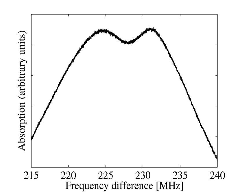
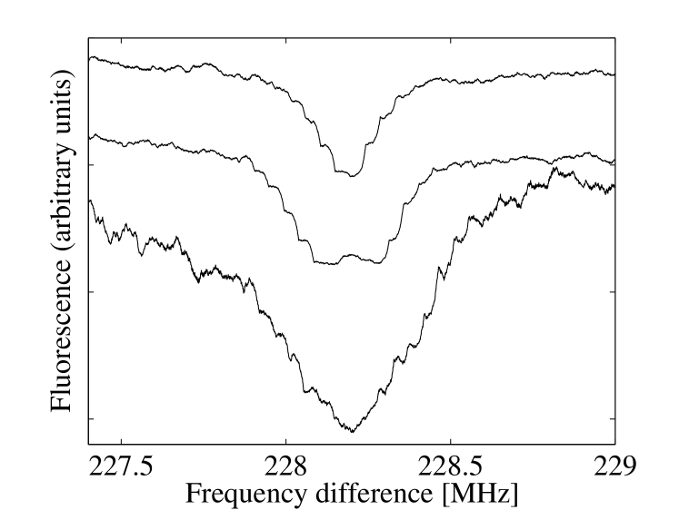
Figure 4 shows absorption plots of the D1 and D2 lines as a function of frequency difference between the collinear pump and probe beams. With no externally applied magnetic field a single absorption dip at a frequency difference of 228 MHz is observed (figure 4 (a) and 4 (c)). The width of these resonances is approximately 4 MHz which is less than the natural width of 5.9 MHz. The amplitudes for the D2 line are notably weaker than for the D1 line. This is due to destructive excitations via cycling transitions which do not contribute to the preparation of dark coherent states. This is in agreement with the results of Stähler et al [Stahler02] and Magnus et al [Magnus05] which show greater contrast in the dark resonance in the D1 line than the D2 line of 85Rb and 7Li, respectively.
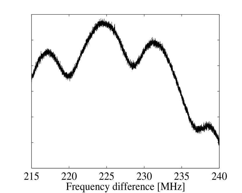
It is difficult to compare the width of the EIT resonances obtained for both lines because any ambient magnetic field can introduce additional broadening. To investigate the possible ambient field broadening an external homogeneous magnetic field (approximately 5 G) was applied perpendicular to the laser beam . The magnetic field removes the degeneracy of the Zeeman levels, splitting the sub-natural EIT resonance (figure 4 (b and d)). The applied magnetic field also introduces a convenient quantization axis. The pump light with linear polarization parallel to the magnetic field can produce transitions, while the probe with orthogonal linear polarization excites the transitions (figure 4 (e)). The m = 0 m′ = 0 type Raman transition, which is magnetic field insensitive in the linear Zeeman approximation, does not exist for 6Li, however, the Raman transitions F = 1/2 (m = 1/2) F = 3/2 (m = -1/2) and F = 1/2 (m = -1/2) F = 1/2 (m = 1/2) are also magnetic field insensitive, because the Zeeman shift of the upper and lower magnetic sub-levels are almost equal. Thus, the EIT dip in absorption, which remains unshifted at 228 MHz frequency difference with increasing magnetic field is due to the above mentioned Raman transitions. The width of this EIT resonance for the D1 line is approximately 3 MHz. Both outer dips are symmetrically shifted by 2 from the unshifted centre dip, where , is the Bohr magneton, is Planck’s constant and is the Landé factor. The width of the outer dips are larger than the width of the unshifted dip due to spatial inhomogeneities in the magnetic field.
We reduced the intensities of the pump and probe lasers but observed no change in the width of the EIT resonances which implies that the width is not limited by power broadening. Although Li-Li collisions are negligible, collisions with residual background gases may cause spin depolarizing collisions of the ground states which possibly contribute to the EIT width. The resolution is further limited by finite interaction (transit) time. The spectroscopy of atoms in a metal vapour cell has the limitations of randomly directed velocities (i.e. Doppler broadening), field inhomogeneities and high collision rates.
4 Atomic Beam EIT
To improve the resolution of EIT resonances experiments were performed using the standard technique of a collimated atomic beam. Isotopically enriched 6Li atoms are evaporated from an oven at a temperature of 450∘ C to produce a thermal atomic beam. At this temperature the 6Li vapour pressure in the oven is expected to be 410-4 Torr. The diameter of the beam at the centre of the probe region is 7 mm. Ultra high vacuum of 210-10 Torr is achieved in an anti-reflection coated glass cell (for 670 nm) which has outer dimensions of 3x3x12 cm3 with a wall thickness of 3 mm. The collimated atomic beam is excited at right angles by resonant laser light in the interaction region. The pump and probe intensities were 1.4 mW/cm2 for both beams. Fluorescence from atoms in the probe region was detected using a photomultiplier tube. The cell was wrapped in -metal to reduce stray magnetic fields.
4.1 Results
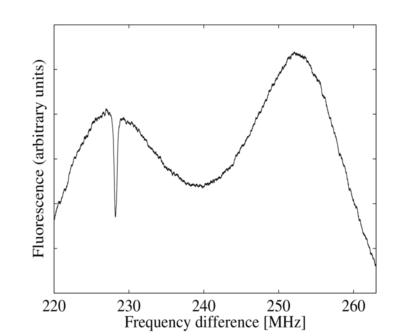
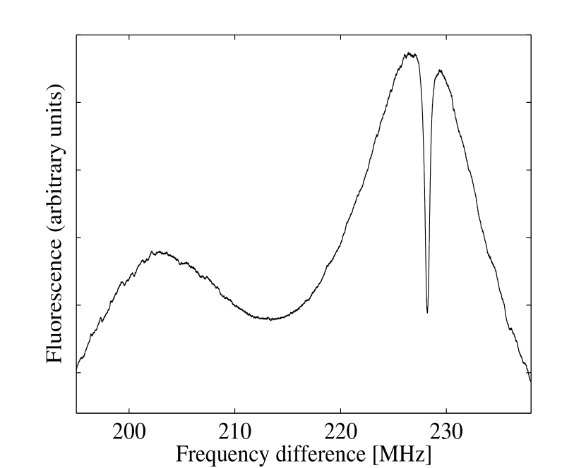
Resonant fluorescence in the collimated atomic beam showed a much narrower transverse Doppler width of 20 MHz compared to 3.5 GHz obtained in the vapour cell. Therefore, the hyperfine splitting of the P1/2 state of 26.1 MHz can be resolved. Figure 5 shows a plot of fluorescence of the D1 line as a function of frequency difference between the pump and probe laser. In this figure the probe laser is scanned over both the F = 3/2 F = 1/2 and F = 3/2 F = 3/2 transitions whereas the pump laser has a fixed frequency tuned to the F = 1/2 F = 1/2 (a) or F = 1/2 F = 3/2 (b) transition. The EIT dip is more pronounced when the fixed laser is tuned to the F = 3/2 state which is due to the larger transition probability. For this reason we performed all of the subsequent experiments with the pump laser tuned to the F = 1/2 F = 3/2 transition.
The EIT resonances obtained on the D1 and D2 lines are shown in
figure 6. The intensity of the resonant fluorescence
on the D2 line is higher, but the contrast of the sub-natural
resonances with respect to the residual Doppler background is
lower compared to the D1 line. In this regard the observations in
the atomic beam and the vapour cell are very similar. However,
better spectral resolution in the atomic beam allows us to
demonstrate that the width of EIT resonances in both cases is also
essentially different. The sub-natural resonances observed in the
atomic beam on both lines under very similar experimental
conditions such as light intensity, polarization and beam
overlapping are shown in figure 6. The curve in
figure 6 (c) represents EIT resonance observed on
the D2 line without applied magnetic field. The width of the
resonance is approximately 560 kHz. The sub-natural resonance on
the D1 line (370 kHz) (figure 6 (b)) reveals a
doublet structure due to residual magnetic field in the
interaction region. This splitting is hidden on the D2 line by the
large width of the EIT resonance. The double structure can be
removed by applying a small magnetic field along the atomic beam
resulting in the 210 kHz wide resonance
(figure 6 (a)) on the D1 line. We believe that the
EIT resonance on the D2 line is wider because of shorter lifetime
of the ground state coherence destructed via the cycling
transition F = 3/2 F = 5/2.

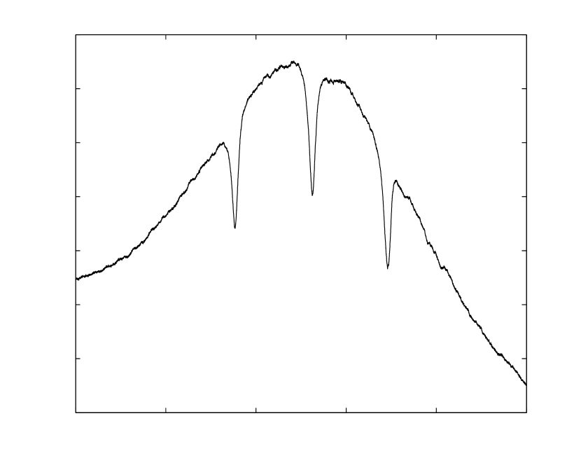
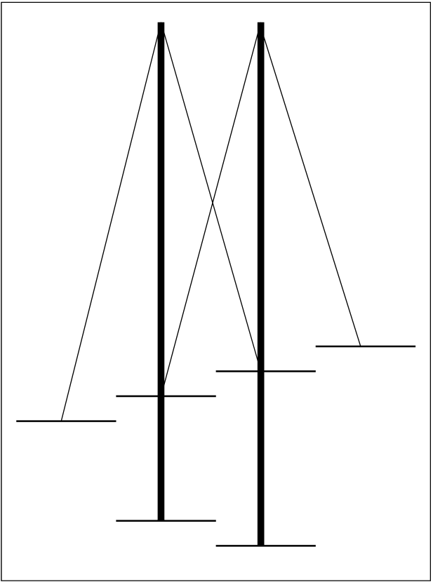
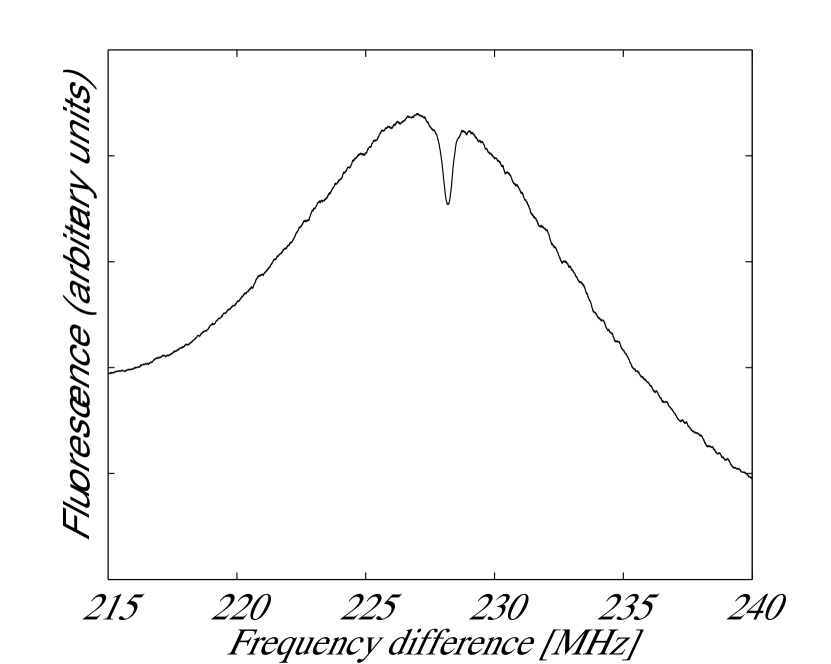

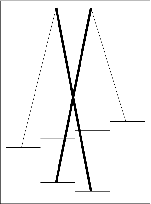
Figure 7 shows several EIT resonances obtained for different polarizations with and without an applied magnetic field ( 2 G) along the atomic beam. In figure 7 (a) and figure 7 (b) the polarization of both lasers is linear and perpendicular to each other. The two outer peaks in figure 7 (b) are both shifted by 2 from the zero field resonance which is consistent with the Raman transitions depicted in figure 7 (c). In figure 7 (d) and figure 7 (e) the polarization of both laser beams are aligned parallel to each other and orthogonal to the magnetic field. Only two -type Raman transitions are possible in this configuration (figure 7 (f)). The two resulting EIT resonances are both shifted by to either side of the zero-magnetic field resonance. The amplitudes are much smaller when parallel polarization light was applied. This can be understood by considering that the atoms will undergo transitions between all Zeeman sublevels, but for parallel polarized pump and probe light, not all of these contribute to coherences (see figure 7 (f)). Similar results were obtained for the D2 line.
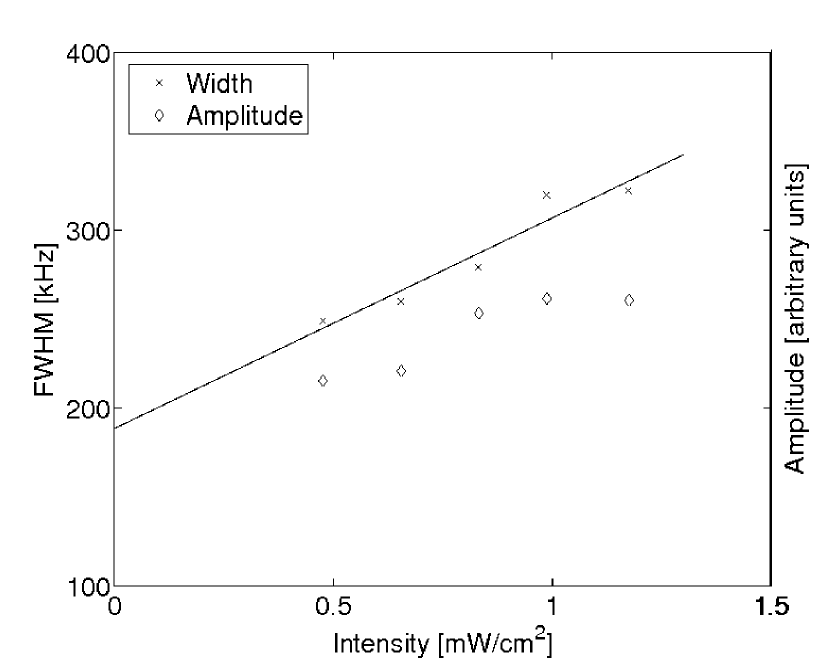
The EIT resonances in the 6Li beam (figures 5 and 7) have a width of 300 kHz for the D1 line. This is a factor of 10 reduction compared to the vapour cell experiments. To get a better understanding of the broadening of our EIT resonances in the atomic beam we investigated the dependence of the width on the laser intensity of the pump laser, as shown in figure 8 (a). In addition, the amplitudes of the EIT resonances are shown. Note that the intensity of the probe laser is kept fixed at 1 mW/cm2. We observe a linear dependence of both the amplitude and the resonance width on the laser intensity. The linear behaviour of the resonance width is expected when power broadening becomes significant [Agapev93]. Extrapolation gives a low-intensity limit of 190 kHz.
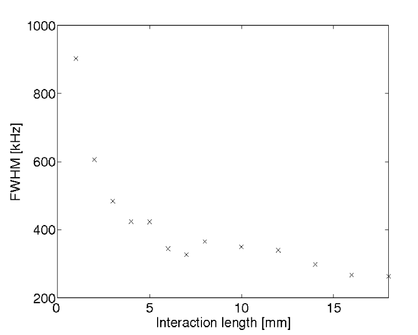
Additional broadening comes from the limited interaction time of the atoms with the light field (transit-time broadening). This was studied by measuring the resonance width for different laser beam cross-sections and the results are shown in figure 8(b). In our work the sum of the laser intensities was kept fixed at 1.7 mW/cm2. A significant increase could be observed for interaction lengths smaller than 5 mm. However, for the beam diameter of 18 mm used in all other experiments the broadening due to the transit time is negligible relative to the contribution of power broadening and magnetic field inhomogeneities.
5 Ramsey Spectroscopy
Further reduction of EIT resonance widths was achieved using Ramsey spectroscopy [Ramsey89]. In this technique atoms or molecules in a beam pass two spatially separated interaction regions. Optical Ramsey fringes were observed on beams of Na and Cs atoms pumped into coherent non-absorbing states [Thomas82, Hemmer93].
Here, a coherent superposition of the and ground states is prepared in a light field. The laser field consists of two co-propagating laser beams which are resonant with the and transitions. After some time the state is probed in a second interaction region consisting of the same two frequencies. Similar to the EIT experiments described previously, the laser resonant with the transition is kept fixed while the other frequency is swept over the transition.

In our experiment the two interaction regions were separated by = 7.7 mm. This was achieved by using only one laser beam in which the complete central vertical portion was blocked. Figure 9 shows a plot of fluorescence as a function of frequency difference. The intensities for both pump and probe were 2 mW/cm2. The polarization of both beams were linear and perpendicular to each other. To amplify the Ramsey fringes the laser light in the first interaction region was chopped, while the fluorescence of the probe region was detected by a photo multiplier tube using a lock-in amplifier. The obtained width (FWHM) of the resonance was narrower than 100 kHz which is consistent with calculations based on a mean atomic velocity 1600 ms-1 and a laser field separation : kHz [Demtroeder03].
6 Summary
We have reported electromagnetically induced transparency in 6Li in both the vapour cell and atomic beam. Resonant light with two frequency components was used to produce coherences between the two hyperfine levels in the 6Li ground state for the first time, allowing fluorescence resonances as narrow as 200 kHz to be observed. The effects of various optical polarization configurations and applied magnetic fields were investigated, and these were readily interpreted with -type Raman transitions. It has been found that the maximum contrast of the sub-natural resonances of suppressed fluorescence occurs for orthogonal linear Zeeman polarizations of the probe and pump components. Magnetic field insensitive fluorescence resonance (in the linear approximation) has been demonstrated despite the fact that the m = 0 m = 0-type Raman transition does not exist for 6Li.
The EIT resonances for the D2 line have been observed in spite of destructive excitation via cycling transition. However, the sub-natural resonances are weaker and broader compared to the D1 line. Additional experiments using the Ramsey separated field technique showed further reduction in the width of the EIT resonances. As an alkali atom with integer nuclear spin, fermionic 6Li presents a potentially interesting system in which to investigate coherence effects such as EIT. Its energy level scheme differs from other atomic species previously investigated, in terms of the number of hyperfine excited states and their small energy splitting. The results reported here are consistent with coherence effects observed using other alkali atoms, such as Rb and Cs, although the smaller hyperfine splitting of 6Li has allowed significant experimental simplification. Our initial atomic coherence investigations on this previously unexplored isotope provide a basis for additional studies.
References
References
- [1]
- [2] \harvarditemAgap’ev1993Agapev93 Agap’ev B D, Gornyi M B, Matisov B G, Rozhdestvenskii Yu V 1993 Usp. Fiz. Nauk 163 1–36
- [3] \harvarditemAkulshin et al2003Akulshin03 Akulshin A M, Cinnnino A, Sidorov A I, McLean R and Hannaford P 2003 Journal of Optics B-Quantum and Semiclassical Optics 5 S479–S485
- [4] \harvarditemArimondo1996Arimondo96 Arimondo E 1996 Prog. Opt. 30 257–354
- [5] \harvarditemDemtröder2003Demtroeder03 Demtröder W 2003 Laser spectroscopy: Basic concepts and Instrumentation vol 3 (New Delhi: Springer-Verlag)
- [6] \harvarditemHarris1997Harris97 Harris S E 1997 Physics Today 50(7) 32
- [7] \harvarditemHarris and Hau1999Harris99 Harris S E and Hau L 1999 Phys. Rev. Lett. 82 4611–4614
- [8] \harvarditemHemmer et al1993Hemmer93 Hemmer P R, Shahriar M S, Lamela-Rivera H, Smith S P, Bernacki B E and Ezekiel S 1993 J. Opt. Soc. Am. B 10 1326
- [9] \harvarditemKnappe et al2005Knappe05 Knappe S, Schwindt P D D, Shah V, Hollberg L, Kitching J, Liew L and Moreland J 2005 Optics Express 13 1249–1253
- [10] \harvarditemLukin2003Lukin03 Lukin M D 2003 Rev. Mod. Phys. 75 457–472
- [11] \harvarditemMagnus et al2005Magnus05 Magnus F, Boatwright A L, Flodin A and Shiell R C 2005 J. Opt. B:Quantum Semiclass. Opt. 7 109–118
- [12] \harvarditemMatsko et al2001Matsko01 Matsko A B, Kocharovskaya O, Rostovtsev Y, Welch G R, Zibrov A S and Scully M O 2001 Advances in Atomic Mol. and Opt. Phys. 46 191-242
- [13] \harvarditemMcAlexander et al1996McAlexander96 McAlexander W I, Abraham E R I, and Hulet R G 1996 Phys. Rev. A 54 R5–R8
- [14] \harvarditemRamsey1989Ramsey89 Ramsey N F 1989 Molecular Beams 2nd Edn, Clarendon, Oxford
- [15] \harvarditemSchmidt et al1996Schmidt96 Schmidt O, Wynands R, Hussein Z and Meschede D 1996 Phys. Rev. A 53 R27 -R30
- [16] \harvarditemStähler et al2002Stahler02 Stähler M, Wynands R, Knappe S, Kitching J, Hollberg L, Taichenachev A and Yudin V Opt. Lett. 27 1472-1474
- [17] \harvarditemThomas et al1982Thomas82 Thomas J E, Hemmer P R, Ezekiel S, Leiby C C Jr., Picard R H and Willis C R 1982 Phys. Rev. Lett. 48 867–870
- [18] \harvarditemWalls et al2003Walls03 Walls J, Ashby R, Clarke J J, Lu B and van Wijngaarden W A 2003 European Physics Journal D 22 159–162
- [19] \harvarditemWynands et al1999Wynands99 Wynands R and Nagel A 1999 Appl. Phys. B 68 1–25
- [20] \harvarditemWijngaarden2005Wijngaarden05 van Wijngaarden W A 2005 Can. J. Phys.83 327
- [21] \harvarditemZibrov et al1995Zibrov95 Zibrov A S, Lukin M D, Nibonov D E, Hollberg L, Scully M O, Velichansky V L and Robinson H G 1995 Phys. Rev. Lett. 75 1499–1502
- [22]