Preliminary study of metabolic radiotherapy with 188Re via small animal imaging
Abstract
188Re is a (Emax = 2.12 MeV) and (155 keV) emitter. Since its chemistry is similar to that of the largely employed tracer, 99mTc, molecules of hyaluronic acid (HA) have been labelled with 188Re to produce a target specific radiopharmaceutical. The radiolabeled compound, i.v. injected in healthy mice, is able to accumulate into the liver after a few minutes. To study the effect of metabolic radiotherapy in mice, we have built a small gamma camera based on a matrix of YAP:Ce crystals, with 0.60.610 mm3 pixels, read out by a R2486 Hamamatsu PSPMT. A high-sensitivity 20 mm thick lead parallel-hole collimator, with hole diameter 1.5 mm and septa of 0.18 mm, is placed in front of the YAP matrix. Preliminary results obtained with various phantoms containing a solution of 188Re and with C57 black mice injected with the 188Re-HA solution are presented. To increase the space resolution and to obtain two orthogonal projections simultaneously we are building in parallel two new cameras to be positioned at 90 degrees. They use a CsI(Tl) matrix with 115 mm3 pixels read out by H8500 Hamamatsu Flat panel PMT.
1 INTRODUCTION
188Re is an attractive therapeutic radioisotope with broad clinical applications. Oncology applications range from palliation of metastatic bone pain to bone marrow ablation and in general to the use of 188Re labelled therapeutic agents to target specific cancerous tissues (see for instance reference [1]). Other therapeutic applications comprise the inihibition of restenosis after Percutanueous Transluminal Coronary Angioplasty (PTCA), radiation synovectomy, and intravasal brachytherapy. In the field of metabolic radiotherapy 188Re shows several favourable characteristics. It can be produced carrier free using a W-Re generator (see for instance reference [2]) and its chemistry is similar to that of 99mTc which is the most used radioisotope in nuclear medicine (medical imaging). The 188W parent has a 69 d half-life, which permits to use the generator for a relatively long period, the equilibrium between parent and daughter setting up within about two days. 188Re decays to 188Os∗ in about 0.7 days via the emission of a -ray with a maximum energy of 2.12 MeV (0.78 MeV average energy), which can be used for destroying cancerous cells. In addition 188Os∗ emits promptly (0.69 ns) a 155 keV -ray (15%), which can be used for imaging. Given the chemical similarity with 99mTc, it can be linked to molecules of hyaluronic acid (HA) which have the function of carrying it to specific sites in the body, e.g. with the production of an accumulation and retention of the drug in the liver [3]. Hence the potential interest for treating liver cancers. On the other hand the radiation could interfere with the labelling process and destroy the molecule used to carry the radioisotope in the body to the target organ, thus reducing the therapeutic effect. Two additional points have to do with the relatively long lifetime and with the rich photon spectrum of 188Re, which extends to high energy, compared to the single 140 keV photon emitted by 99mTc with a half-life of about 6 h. Taking into account the branching ratios (BR) and the lifetimes, the relative 188Re/99mTc counting rate, for an equal number of -moles, is about 9%. This implies that photon detectors developed for imaging with 99mTc are not necessarily sensitive enough. The usual thickness of a low-energy Pb collimator, say 20 mm, will be almost transparent to 188Re photon lines at 300 keV or higher energies, even with BRs depressed by a factor of ten or more, producing background counts which blur the image and worsen the spatial resolution. To study the effect of metabolic radiotherapy in mice, we have therefore built a new small high-sensitivity -camera, following the experience with the YAP-camera [4][5][6], which is used routinely to image small animals (mice) with 99mTc-HA at the Laboratori Nazionali di Legnaro, Italy (e.g. [7][8]).
2 THE GAMMA CAMERA
The gamma camera is based on a matrix of yttrium aluminium perovskite doped with cerium (YAP:Ce or YAlO3:Ce) crystals [9], with friendly mechanical properties (no igroscopicity), fast response (25 ns decay time), high density (5.37 g/cm3) and good X- and -ray absorption. There are 6666 pixels, each 0.60.610 mm3, covering a field-of-view (FOV) of 4040 mm2. The pixels are covered laterally by a special 5 m thick reflective coating which provides also the optical separation between neighboring elements. The scintillator is read out by a R2486 Hamamatsu PositionSensitivePMT [10] with a 76 mm diameter photocathode. The anode consists of 16 plus 16 wires crossing at 90o and connected by two resistive chains, defining the and directions. The wires define an active area with a diameter of about 50 mm. A 20 mm thick lead parallel hexagonal-hole collimator [11], with hole diameter 1.5 mm and septa of 0.18 mm, is placed in front of the YAP matrix. The detector is triggered using the last dynode and the ends of the and resistive chains (x1, x2, y1, y2) are amplified, stretched and read out by a PC using a PCI 6023E card [12]. The coordinates of the photon impact point are then reconstructed by charge division,
| (1) |
and similarly for .
3 CALIBRATIONS
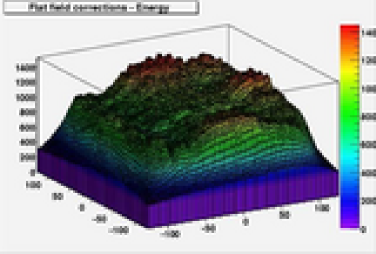
The energy response of the detector to a spatially uniform source (flat field) of 99mTc 140 keV photons prior to energy equalization has been determined using a solution containing 99mTc which covered the whole FOV and was located a few centimeters in front of the collimator surface. The has been arbitrarily divided in pixels and the average energy computed in each pixel. The corrections to the measured energy extracted from this calibration are shown in Fig. 1 and have been used in the following. As shown below with the 188Re spectrum, these corrections improve the energy resolution of the detector. Apart from the energy equalization, no other correction has been applied. With 99mTc the sensitivity of the gamma camera is found to be 210-4 or 7103 cps/mCi, which agrees roughly with calculations.
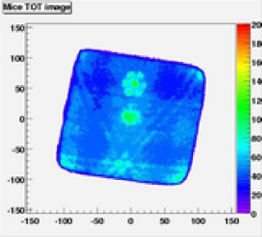
Pointlike 241Am (60 keV photons), 57Co (122 keV photons) and 137Cs (660 keV) sources located in different positions a few millimeters distant from the collimator have been used both to simulate the 188Re energy spectrum and to evaluate the spatial resolution of the detector. In addition 241Am and 57Co, together with 99mTc photons, permit the calibration of energy scale. The overall energy spectrum is presented in Fig. 2 and the cumulative image of the sources is visible in Fig. 3. The 60 keV is well prominent and a 122 keV shoulder is also visible; 137Cs instead produces a broad shoulder at about half of the photon energy. Using appropriate energy cuts the three images can be separated. The intrinsic spatial resolution of the system is quite good, since individual collimator holes can be clearly seen in Fig. 3. The resolution however is worsened in practice by the small thickness of the septa relative to the hole diameter of the present collimators, making them partially transparent to radiation, so the actual effective resolution is 3 mm FWHM at 122 keV.
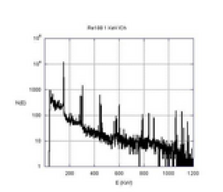
The 188Re photon spectrum measured with a Ge detector is shown in Fig. 4. The 155 keV line is prominent, but many more lines are present at higher energy, in some cases, e.g. at 300 keV, with BRs only a factor of ten lower, or at 800-1200 keV with intensities lower only by a factor of 100 [13].
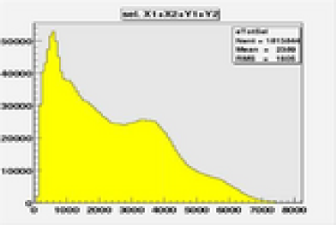
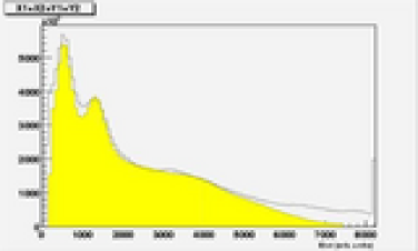
The 188Re spectrum measured with the YAP camera during the imaging of a C57 mouse (see next section) is shown in Fig. 5. The spectrum is shown before and after applying the corrections for energy non-uniformity, and a clear improvement is observed with the shrinking of the 155 keV peak. After the corrections, the energy resolution is E/E 33% 155 keV.
4 FIRST MEASUREMENTS WITH 188Re
The labelling reaction of HA using 188Re was carried out with good yields (65-70%). The radiolabelled compound was purified with a size exclusion chromatographic method before being used for biodistribution studies. Stability studies in rat serum confirmed the maintaining of the Re linked to the polymer and there was no evidence of radio-decomposition after a few hours.
The radiotoxicity of 188Re has been tested “in vitro” and compared with 99mTc, with which no effect is expected. Cells of the M5076 tumor line have been treated with 188Re and 99mTc solutions, and irradiated with X-rays. The number of binucleate cells and of micronuclei in the cells is then counted. M5076 cells turn to out to be highly sensitive to X-rays (0.25-2 Gy)[3]. Activities of 150-300 Ci of 99mTc show no effect, but 188Re -rays, with similar initial activities integrated during 72 h, seem to be quite efficient in inducing DNA damage[14].
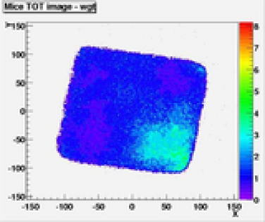
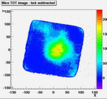
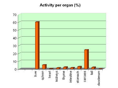
To test the full chain, from the radiolabelling to to the imaging “in vivo”, a C57 black mouse (healthy, female) has been injected with 188Re-HA. After general anesthesia, the solution with an activity of about 250 Ci was injected in the caudal vein. The mouse was positioned along the diagonal of the FOV, with the locus of injection outside it, and was monitored for about three hours. The image collected in the first five minutes shows a large spot close to the locus of injection in the tail (Fig. 6). After 5 mins the activity concentrates roughly in the centre of the body, in a volume which contains the liver (Fig. 7). The activity is slowly decreasing during the 3 h of the measurement. After 3 h the mouse was sacrificed, and the organs were extracted and measured with a microcurimeter (Fig. 8). The liver contains 60% of the residual activity and close by organs another 20%, in agreement with the scintigrafic image (Fig. 7), where individual organs are not resolved. Even with limited resolution the test shows that it is possible to monitor the biodistribution of 188Re in mice, with a potential saving in the number of animals needed for testing the 188Re therapy.
5 CONCLUSIONS
Preliminary results obtained using a new YAP camera in imaging 188Re sources and C57 black mice injected with a 188Re-HA solution have been presented. To increase the space resolution without losing sensitivity, and to obtain different projections simultaneously, we are building two new cameras to be positioned at 90 degrees around the animal. They use a CsI(Tl) matrix with 115 mm3 pixels read out by H8500 Hamamatsu Flat panel PMT [10]. Parallel-hole Pb collimators 20 mm thick, with 1 mm diameter hexagonal holes and 0.2 mm thick septa, will be mounted in front of the scintillators. Also specially made collimators with thicker septa and/or different absorber material will be used. The front-end electronics for the 64 channels of the H8500 has been designed using MPX-08 chips [15]. The system will be mounted on a rotating support in order to produce tomographic images.
References
- [1] Labelling techniques of biomolecules for targeted radiotherapy, IAEA-TECDOC-1359, Vienna July 2003
- [2] http://www.ornl.gov/sci/nuclear_ science_tecnology/nu_med/188info.htm
- [3] A. Antoccia et al., Dati preliminari e danno al DNA del 99mTc-HA in sistemi “in vitro” ed “in vivo”, Suppl. Boll. SIRR VII (2004) 43
- [4] F. de Notaristefani et al., YAP Camera: a small field camera with sub-millimiter spatial resolution, Eur. J. Nucl. Med. 22 (1995) 337
- [5] F. de Notaristefani et al., First Results from a YAP:Ce Gamma Camera for Small Animal Studies, IEEE Trans. Nucl. Sci. 43 (1996) 3264
- [6] F. de Notaristefani, F. Vittori and T. Malatesta, Development of YAP:Ce multi-crystal detectors, Physica Medica 13 (1997) 237
- [7] R. Rossin et al., In vivo Biodistribution Studies on Hyaluronan Butyrate by Means of 99mTc Direct Labelling and YAP Camera, In: Technetium, Rhenium and Other Metals in Chemistry and Nuclear Medicine (M. Nicolini, U. Mazzi Eds.), pp 689-693, SGEditoriali, Padova, 2002.
-
[8]
M.C. Giron et al., YAP-Camera Device for Biodistribution Studies in Mice of 99mTc-Radiotracers, Poster at the 6th Int. Symp. on Technetium in Chemistry and Nuclear Medicine, Brixen, Italy, 2002
http://tc2002.unipd.it/posters_main.htm - [9] CRYTUR Preciosa a.s., Palackeho 175, 511 19 Turnov, Czech Republic.
- [10] http://www.hamamatsu.com/
- [11] http://www.nuclearfields.com/
- [12] http://www.ni.com/
-
[13]
http://www.nndc.bnl.gov/nudat2/
index.jsp - [14] A. Antoccia et al., Study of metabolic radiotherapy with 188Re via small animal imaging, presented at the annual meeting of the Italian Physical society, Brescia 20-25 Settembre 2004 http://www.sif.it
- [15] http://www.novarad.com/