Preprint of:
Alexis I. Bishop, Timo A. Nieminen, Norman R. Heckenberg,
and Halina Rubinsztein-Dunlop
“Optical microrheology using rotating laser-trapped particles”
Physical Review Letters 92(19), 198104 (2004)
Optical microrheology using rotating laser-trapped particles
Abstract
We demonstrate an optical system that can apply and accurately measure the torque exerted by the trapping beam on a rotating birefringent probe particle. This allows the viscosity and surface effects within liquid media to be measured quantitatively on a micron-size scale using a trapped rotating spherical probe particle. We use the system to measure the viscosity inside a prototype cellular structure.
pacs:
87.80.Cc,83.85.Cg,83.85.EiIt is well known that light can transport and transfer angular momentum as well as linear momentum Beth (1936). In recent years, this has been widely applied to rotate microparticles in optical tweezers. Such rotational micromanipulation has been achieved using absorbing particles Friese et al. (1996, 1998a), birefringent particles Friese et al. (1998b); Higurashi et al. (1999), specially fabricated particles Higurashi et al. (1994); Galajda and Ormos (2001), and non-spherical particles using linearly polarized light Bayoudh et al. (2003); Bonin et al. (2002); Santamato et al. (2002); Cheng et al. (2002); Bishop et al. (2003); Galajda and Ormos (2003) or non-axisymmetric beams Paterson et al. (2001); O’Neil and Padgett (2002). Notably, particles rotated in rotationally symmetric beams Friese et al. (1998b); Bishop et al. (2003) spin at a constant speed, indicating that the optical torque driving the particle is balanced by viscous drag due to the surrounding medium. It has been noted that the optical torque can be measured optically Nieminen et al. (2001), allowing direct independent measurement of the drag torque.
For a pure Gaussian beam that is circularly polarised, each photon also has an angular momentum of about the beam axis, equivalent to an angular momentum flux of for the beam, where is the power of an incident beam, and is the angular frequency of the optical field, which can be transferred to an object that absorbs the photon, or changes the polarization state Beth (1936). The reaction torque on a transparent object that changes the degree of circular polarization of the incident light is given by Nieminen et al. (2001)
| (1) |
where is the change in circular polarisation. Thus, by monitoring the change in circular polarization of light passing through an object, the reaction torque on the object is found. We exploit this simple principle as the basis for an optically driven viscometer. The viscosity of a liquid can be determined by measuring the torque required to rotate a sphere immersed in the liquid at a constant angular velocity—a concept we have implemented at the micrometer scale using a system based on optical tweezers. We have rotated birefringent spheres of synthetically-grown vaterite that were trapped three-dimensionally in a circularly polarized optical tweezers trap and measured the frequency of rotation, and the polarization change of light passing through the particle, to determine the viscosity of the surrounding fluid.
At least four distinct techniques have been used previously for microrheology: tracking the diffusion of tracer particles Chen et al. (2003); Mukhopadhyay and Granick (2001), tracking the rotational diffusion of disk-shaped tracer particles Cheng and Mason (2003), measurement of correlations in the Brownian motion of two optically trapped probe particles Starrs and Bartlett (2003), and the measurement of viscous drag acting on an optically trapped probe particle in linear motion Rafał Ługowski and Kawata (2002). The method we use here—direct optical measurement of the rotation of a spherical probe particle in a stationary position—allows highly localized measurements to be made, since the probe particle does not move in the surrounding medium, and, compared with the rotational diffusion measurements by Cheng and Mason using microdisks Cheng and Mason (2003), the use of spherical probe particles greatly simplifies the theoretical analysis of the fluid flow. Also, since the probe particle is rotationally driven by the optical trap, the rotation rate can be readily controlled.
The expression for the drag torque, , on a sphere rotating in a fluid for low Reynolds number flows is well known Landau and Lifshitz (1987), and is given by
| (2) |
where is the viscosity of the fluid, is the radius of the sphere, and is the angular frequency of the rotation. Equating the expressions for torques and gives:
| (3) |
This equation suggests that the viscosity can be determined using knowledge of only a few elementary parameters—the power and the polarization change of the incident beam passing through the particle, and the size and rotation rate of the particle. The power, polarization state, and rotation rate can all be measured optically.
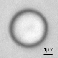
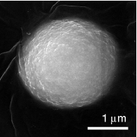
A significant obstacle to implementing this approach has been obtaining spherical, transparent, birefringent particles of suitable size for trapping (approximately 1–10 m in diameter). We have developed a novel technique to grow nearly perfectly spherical crystals of the calcium carbonate mineral vaterite, which has similar birefringence properties to calcite, within the required size regime. We produce the vaterite crystals by adding 6 drops of 0.1 M K2CO3 to a solution of 1.5 ml of 0.1Ṁ CaCl2 plus 4 drops of 0.1 M MgSO4. The solution is strongly agitated by pipetting. The solution initially appears milky, becoming clearer as the crystals form within a few minutes of mixing. The typical mean size is found to be 3 m with a spread of approximately m. Figure 1 shows both an optical image and an electron micrograph of typical crystals used for viscosity measurements. Vaterite is a positive uniaxial birefringent material, with and .
(a)
(b)
A typical optical tweezers arrangement was used as the basis for the experiment, which delivered approximately 300 mW of light at 1064 nm into a focal spot with a diameter of around 0.85 m using a oil-immersion objective of high numerical aperture (). A plate located immediately before the objective converts the linearly polarized light from the laser into a circularly polarized beam which is able to rotate birefringent objects Friese et al. (1998b). Light passing through the probe particle is collected by an oil coupled condenser lens () and then sent to a series of polarization measuring devices. The degree of circular polarization remaining in the beam is determined by passing it through a plate with the fast axis orientated at 45∘ to the axes of a polarizing beamsplitter cube. Photodiodes monitoring the orthogonal outputs of the beamsplitter provide a measure of the degree of circular polarization remaining in the beam, which allows the change in angular momentum, and hence the torque applied to the particle to be known. The photodiodes are calibrated such that they provide a measure of the power at the trap focus. The sum of the two signals gives the total trapping power, and the difference between the signals gives the degree of circular polarization of the beam measured in units of power at the trap focus. The torque applied to the particle is determined by measuring the difference between the initial degree of circular polarization of the beam in the absence of the particle and the final polarization, in accordance with equation (1). The frequency of rotation of the particle can be accurately determined by monitoring the transmitted light through a linear polarizer, as suggested by Nieminen et al. Nieminen et al. (2001). A small amount of light is diverted from the main beam and is passed through a polarizing beamsplitter cube, and the forward direction is monitored using a photodiode. The signal is modulated at twice the rotation rate of the particle, and the depth of the modulation is proportional to the amount of linearly polarized light in the beam. If the particle is thick enough to act as a plate, then circularly polarized light passing through the particle is reversed in handedness and the frequency of the rotation cannot be measured as there is no linear component to the transmitted light. However, as the particles are spherical and are of similar diameter to the beam, the polarization at different radial distances from the rotation axis varies, and there will almost always be some linear component after transmission.
The calculated viscosity depends on the cube of the radius of the particle, and thus it is important to determine the size of the particles accurately. The particles used for viscosity measurements are in the range of 1.5 to 3.5 m in diameter and consequently to obtain the viscosity within 10% requires the diameter to be measured with an accuracy on the order of 90 nm, and it was found that obtaining an accurate measurement of the diameter by direct visualization was problematic. The method of measurement preferred by the authors is to put two spheres of nearly identical size in contact, and measure the distance between centres, which can be found accurately due to the spherical symmetry. The diameter is inferred using elementary geometry, and the error in the measurement by this method is estimated as being nm. The degree of asphericity for typical particles was estimated to be less than 3%.
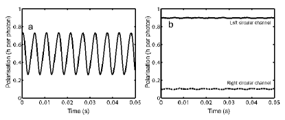
The viscosity of water was measured using the system described. A range of vaterite particles were suspended in distilled water and trapped three dimensionally in a sealed slide/coverslip cell, with a depth of 50 m. Particles that are trapped begin to rotate immediately. However, the rotation may be stopped by aligning the plate located before the objective so that the polarization is made linear. The size of the particles ranged from 1.5 to 3.5 m in diameter, and rotation rates up to 400 Hz (24000 rpm) were observed for powers up to 350 mW at the trap focus. The Reynolds number of the fluid flow around the rotating spheres is quite low, on the order of for rotations up to 1000 Hz, and hence the flow is well within the creeping flow regime required by (2). The signals from the circular polarization and linear polarization monitoring photodiodes were recorded using a 16 bit analogue to digital converter sampling at 10 kHz for a period of 5 seconds. Typical signals recorded during an experiment are reproduced in figure 2. The strong uniform modulation of the linear polarization signal (figure 2(a)) shows that the particle rotates very uniformly, taking a large fraction of the angular momentum carried by the beam. Using a particle of diameter 2.41 m, the viscosity was found to be Pa s, which is in excellent agreement with the established value for the viscosity of water at 23∘C of Pa s CRC Handbook (1974). A series of measurements was made with a range of different sized particles at a range of rotation rates and trapping powers. The variation in viscosity over a range of powers below 100 mW was found to be on the order of 0.7% using a single particle. The variation in inferred viscosity using 11 particles, spanning a range of sizes, was approximately 4.7%. The most significant sources of error arise from the calibration of the trapping power at the focus of the trap (7%), the asphericity of the particles (3%), resulting in a 6% error in the viscosity, and the uncertainty in the measurement of the particle size (1.4%), which contributes to a 4% error in the viscosity. The total error for the viscosity measurement from all error sources is estimated as being 10%.
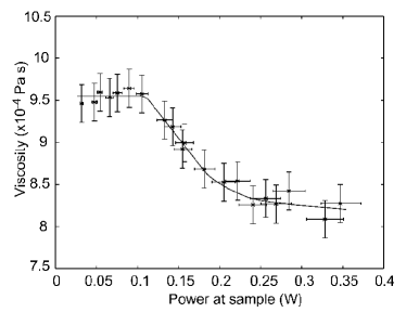
The inferred viscosity was found to be independent of rotation rate, although for trapping powers above approximately 100 mW, the viscosity was observed to decrease with increasing power. This is expected, due to heating of the particle which results from the absorption of light through the sample, and also heating of the surrounding liquid due to absorption of the trapping beam by water, which is on the order of 10 K per W at the focus Liu et al. (1996). Figure 3 demonstrates the behaviour of the measured viscosity as a function of the power at the trapping focus. It was observed that higher trapping powers could be used with smaller particles before the onset of the decrease in viscosity, which is consistent with volume absorption effects. The observed decrease in viscosity with power was initially approximately linear and was approximately 0.13% per mW, which is equivalent to a temperature rise of around 0.06∘C per mW. The total power loss for the trapping beam passing through a particle was measured for a sample of vaterite particles, and was found to be within the range of 0.6% to 1.2%. The reflection loss due to refractive index mismatches at the particle surfaces is approximately 0.6%.
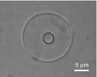
We have also demonstrated the ability to measure the viscosity inside a small confined region, such as a cellular membrane. Hexane filled vesicles were formed by emulsifying hexane, containing a small amount of soy lecithin (1 g/l), with water containing a quantity of vaterite spheres. Spherical membrane structures were formed with diameters up to approximately 20 m which occasionally contained single vaterite crystals. The viscosity inside a 16.7 m diameter vesicle was measured with a vaterite particle of diameter 3.00 m, using the same procedure as outlined earlier, and was found to be Pa s for powers less than 100 mW, which is in good agreement with the established value of viscosity for hexane at 23∘C of Pa s (extrapolated) CRC Handbook (2002). This value is significantly less than that of water (Pa s at 23∘C CRC Handbook (1974). Figure 4 shows the hexane filled vesicle with the vaterite probe particle inside.
The viscous drag torque acting on a spherical probe particle of radius rotating with an angular frequency at the center of a sphere of radius of fluid of viscosity , surrounded by a fluid of viscosity is Landau and Lifshitz (1987)
| (4) |
This can be used to determine the viscosity within a vesicle if the viscosity of the surrounding fluid is known, or to estimate the error if equation (3) is used instead. For other geometries, such as if the probe particle in not in the centre of the vesicle, or is near a plane interface Danov et al. (1998), the effect will be more complicated. However, in most cases the effect of the boundary can be ignored if the probe particle is at least one diameter away from the interface. The effect of nearby boundaries was determined experimentally by measuring the viscous drag torque at varying distances from a solid interface. For rotation about an axis parallel to the interface, no change in the drag torque was observed until the probe particle was within one diameter of the interface. For rotation about an axis normal to the interface, no change in the drag torque was observed until the probe particle was very close to the interface. For the above measurement of the viscosity of hexane, the drag in the vesicle is only 0.4% greater than in pure hexane. The rotation rate of the vesicle is Landau and Lifshitz (1987), equal to for the case above. Small particles in the surrounding fluid near the vesicle were observed to orbit the vesicle at approximately this rate.
The most significant contribution to the error in these measurements is the uncertainty in accurately determining the diameter of the spherical particle. It was not found to be possible to drag a particle through the membrane, and hence the method of size measurement using two spheres in contact could not be used. The diameter was instead directly estimated from an image recorded of the sphere. The error in determining the diameter is estimated as being 5%, which is the main contribution to the total measurement error of 19%.
It is feasible to produce smaller vaterite particles, and the use of these would allow the viscosity to be probed on an even smaller size scale—potentially probing volumes with a capacity of a cubic micron. The ability to functionalise the probe particle surface would enable the selective attachment of the particles to various biological structures, which would allow the torsional response of these structures to be investigated quantitatively. We note that the torque and angular deflection can still be measured accurately in the absence of continuous rotation. The demonstrated ability to measure viscosity within micron-sized volumes makes the system developed here of great value for probing the microrheology of liquid-based materials, such as colloids.
Acknowledgements
This work was partially supported by the Australian Research Council. We wish to thank Professor Rane Curl, University of Michigan, for valuable discussions.
References
- Beth (1936) R. A. Beth, Physical Review 50, 115 (1936).
- Friese et al. (1996) M. E. J. Friese, J. Enger, H. Rubinsztein-Dunlop, and N. R. Heckenberg, Physical Review A 54, 1593 (1996).
- Friese et al. (1998a) M. E. J. Friese, T. A. Nieminen, N. R. Heckenberg, and H. Rubinsztein-Dunlop, Opt. Lett. 23, 1 (1998a).
- Friese et al. (1998b) M. E. J. Friese, T. A. Nieminen, N. R. Heckenberg, and H. Rubinsztein-Dunlop, Nature 394, 348 (1998b), erratum in Nature, 395, 621 (1998).
- Higurashi et al. (1999) E. Higurashi, R. Sawada, and T. Ito, Physical Review E 59, 3676 (1999).
- Higurashi et al. (1994) E. Higurashi, H. Ukita, H. Tanaka, and O. Ohguchi, Appl. Phys. Lett. 64, 2209 (1994).
- Galajda and Ormos (2001) P. Galajda and P. Ormos, Applied Physics Letters 78, 249 (2001).
- Bayoudh et al. (2003) S. Bayoudh, T. A. Nieminen, N. R. Heckenberg, and H. Rubinsztein-Dunlop, Journal of Modern Optics 50, 1581 (2003).
- Bonin et al. (2002) K. D. Bonin, B. Kourmanov, and T. G. Walker, Opt. Express 10, 984 (2002).
- Santamato et al. (2002) E. Santamato, A. Sasso, B. Piccirillo, and A. Vella, Opt. Express 10, 871 (2002).
- Cheng et al. (2002) Z. Cheng, P. M. Chaikin, and T. G. Mason, Phys. Rev. Lett. 89, 108303 (2002).
- Bishop et al. (2003) A. I. Bishop, T. A. Nieminen, N. R. Heckenberg, and H. Rubinsztein-Dunlop, Phys. Rev. A 68, 033802 (2003).
- Galajda and Ormos (2003) P. Galajda and P. Ormos, Opt. Express 11, 446 (2003).
- Paterson et al. (2001) L. Paterson, M. P. MacDonald, J. Arlt, W. Sibbett, P. E. Bryant, and K. Dholakia, Science 292, 912 (2001).
- O’Neil and Padgett (2002) A. T. O’Neil and M. J. Padgett, Opt. Lett. 27, 743 (2002).
- Nieminen et al. (2001) T. A. Nieminen, N. R. Heckenberg, and H. Rubinsztein-Dunlop, Journal of Modern Optics 48, 405 (2001).
- Chen et al. (2003) D. T. Chen, E. R. Weeks, J. C. Crocker, M. F. Islam, R. Verma, J. Gruber, A. J. Levine, T. C. Lubensky, and A. G. Yodh, Phys. Rev. Lett. 90, 108301 (2003).
- Mukhopadhyay and Granick (2001) A. Mukhopadhyay and S. Granick, Current Opinion in Colloid and Interface Science 6, 423 (2001).
- Cheng and Mason (2003) Z. Cheng and T. G. Mason, Phys. Rev. Lett. 90, 018304 (2003).
- Starrs and Bartlett (2003) L. Starrs and P. Bartlett, Faraday Discuss. 123, 323 (2003).
- Rafał Ługowski and Kawata (2002) B. K. Rafał Ługowski and Y. Kawata, Optics Communications 202, 1 (2002).
- Landau and Lifshitz (1987) L. D. Landau and E. M. Lifshitz, Fluid Mechanics, vol. 6 of Course of Theoretical Physics (Butterworth-Heinemann, Oxford, 1987), 2nd ed.
- CRC Handbook (1974) CRC Handbook of Chemistry and Physics, 1974–1975 (CRC Press, 1974), 55th ed.
- Liu et al. (1996) Y. Liu, G. J. Sonek, M. W. Berns, and B. J. Tromberg, Biophysical Journal 71, 2158 (1996).
- CRC Handbook (2002) CRC Handbook of Chemistry and Physics, 2002–2003 (CRC Press, 2002), 83rd ed.
- Danov et al. (1998) K. D. Danov, T. D. Gurkov, H. Raszillier, and F. Durst, Chemical Engineering Science 53, 3413 (1998).