Direct reconstruction of the effective atomic number of materials by the method of multi-energy radiography
Sergey V. Naydenov and Vladimir D. Ryzhikov
Institute of Single Crystals
60 Lenin Avenue, Kharkov, 61001 Ukraine
Craig F. Smith
Lawrence Livermore National Laboratory
PO Box 808, Livermore, CA 94556 USA
A direct method is proposed for reconstruction of the effective atomic number by means of multi-energy radiography of the material. The accuracy of the method is up to 95% . Advantages over conventional radiographic methods, which ensure accuracy of just about 50% , are discussed. A physical model has been constructed, and general expressions have been obtained for description of the effective atomic number in a two-energy monitoring scheme. A universal dependence has been predicted for the effective atomic number as a function of relative (two-energy) radiographic reflex. The established theoretical law is confirmed by the experimental data presented. The proposed development can find multiple applications in non-destructive testing and related fields, including those in the civil sphere as well as anti-terrorist activities.
07.85.-m ,78.20.Bh ,81.70.-q, 81.70.Fy ,87.59.Hp
1. Introduction
Non-destructive testing (NDT) of different unknown materials, objects and media, including complex micro- and macro-structures, is an important field in modern applied physics [1]. Physical methods of NDT find multiple applications in a wide variety of technical areas, including quality control of industrial products, inspection of welded joints and connections, and confirmation of building construction, pipelines, etc. An important role is also played by NDT in radioactive materials control [2], security protection in aviation [3], [4], railway and automobile transport, and customs inspection of goods and luggage. In addition, broad applications related to the use of new NDT technologies in medicine [5], [6], [7], [8], including separate diagnostics of tissues and organs, can be expected. Among the promising directions of NDT, one should note digital (computer) radiography. In it, digital reconstruction of an image of an object examined by X-ray or gamma-radiation is prepared based on the data collected in real-time detection. The most widely used radiation detectors for such applications are solid-state 1D and 2D scintillation systems [9], [10], [11], [12], [13].
The main task of digital radiography, as well as of other NDT methods, is reconstruction of the physical parameters that determine technical characteristics and properties of the controlled objects being examined. This is traditionally carried out by reconstruction of the spatial configuration, observing small details and defects of the image, and determining constitutive components and dimensions (thickness). In recent times, strong interest has been emerging in the development of direct methods for reconstruction of the overall composition of the object, i.e., quantitative determination of the effective atomic number, density, chemical composition, etc. To a large extent, this is due to improvements in technical capabilities for detection and image processing and is driven by the desire for additional detail in the image resulting from the collected data. Often it is not sufficient to know just the geometrical structure of an object to ensure its complete identification for decision-making purposes. For example, fully reliable detection of explosives is not possible without discrimination of materials by their effective atomic number (organics and inorganics), density, content of predominant chemical elements, etc. The importance of such enhanced monitoring cannot be overestimated. It is vitally necessary not only to provide enhanced tools for scientific and technological investigations, but to meet current needs for improved protection against terrorist threats to the safety and health of civil populations. An important milestone on the way to solving this problem is the implementation of two-, and in general, of multi-energy radiography [14], [15], [16], [17].
2. Effective atomic number
Among the parameters determining the constitutive structure of an unknown object or material, one should especially note the effective atomic number . In fact, this measure can provide an initial estimation of the chemical composition of the material. A large generally corresponds to inorganic compounds and metals, while a small is an indicator of organic substances. For many applications, e.g., radioisotope monitoring, cross-section studies of absorption, scattering and attenuation of electromagnetic radiation, testing of multi-component, heterogeneous and composite materials, etc., this parameter is of principal significance. It is very important to obtain its quantitative evaluation from the data provided by radiographic measurements. With the aim of making this possible, in the present work we have developed a physical model for two-energy radiography as a particular case of the multi-energy radiography. We show that a multiplicity of is sufficient for direct determination of . General theoretical expressions are provided for determining the effective atomic number with respect to the photo and pair generation effects.
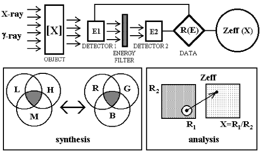
The method proposed here for determination of is substantially different from the approaches used previously. In these procedures, the effective atomic number is comprised of a linear superposition of several reference values, i.e., it is expressed in the form of , where , and corresponds to the multiplicity of the radiography used. Such “synthesis” of is quite similar to the formation of light of arbitrary color from several basic colors (Fig.1). Weights , or the relative content of the components (light, heavy, intermediate), are determined from the data of radiographic measurements. Theoretically, this corresponds to the solution of systems of linear equations of the form (where is the layer thickness of the -th element phantom in the studied object), but with attenuation coefficients that are a priori pre-set at several fixed energy values for the basic elements, each with its selected atomic number and density . The values of depend not only on reflexes , but also on the basis chosen. Therefore, such radiography is, in fact, “indirect”. Erroneous or inaccurate choice of the basis leads to mistakes (artifacts) in determination of . For example, a sample with low content of a heavy element (low ) can be mistakenly identified as a sample with a thick layer of a low element. It is clear that, expanding the basis and increasing the multiplicity, one can substantially reduce the errors. However, even then, errors can be as high as tens of percent. The accuracy of traditional methods in 2- and 3-energy radiography allows distinction of materials with difference in by about 50% . So, it is possible to distinguish iron alloys () from wood () and even from light aluminum alloys (), but it is not possible to reliably discern the difference between water () and organic materials (), iron and many other metals, calcium minerals () and sand (), etc. To distinguish among these materials requires accuracy that should be higher by an order of magnitude (i.e., not worse than 95% ).
For a chemical compound with the known formula of the form (here is the number of atoms of the -th kind with atomic number and atomic mass in the complex molecule ; is the full number of simple components), and are determined from the expressions
| (1) |
where is an index depending upon the chosen absorption mechanism of gamma-quanta. The expression (1) for determination of is derived from the balance of the absorbed energy per each atomic electron of the substance. Absorption over all possible physical channels and by all simple components of the complex compound is accounted for. Parameter is related to the characteristic dependence of the electron coefficient of linear absorption, (here is the mass attenuation coefficient), on the atomic numbers of simple constituents of the substance studied. The choice of values corresponds to the predominant absorption channels at specified radiation energies, i.e. . In the photo effect theory, . In the pair formation theory . For the Compton scattering weakly depending upon atomic properties of the material . In the experiments, such absorption character is also well confirmed. Besides this, the parameter can be considered as a fitting parameter giving the best approximation of the absorption cross-section in a specified energy range and for a specified class of substances. E.g., in the photo effect energy range intermediate values are used. We use for the photo effect , while for the pair formation effect, , i.e.
| (2) |
For the Compton scattering, which normally accompanies one of the former mechanisms, it should be assumed by definition that and
| (3) |
as the Compton effect cross-section does not depend upon properties of the material, but only upon its average electron density. The molar mass . In practice, relative concentrations of the simple components of the material are often also known. Then, as (where ), instead of (1)-(2) we obtain
| (4) |
In Table 1, data are presented on the effective atomic number of selected substances that comprise many materials in practical use. The data were calculated using formulas (1)-(4). It should be noted that the effective atomic number is dependent upon the energy range of the ionizing radiation used. Its value corresponds to the predominant absorption channel. Using elementary inequalities from expressions (1)-(3), one can easily obtain the general relationship valid for chemical compounds:
| (5) |
which is substantiated by the data of Table 1. For homogeneous mixtures (solid, liquid, gaseous), including alloys, these inequalities (5) can be violated at certain concentration ratios of the mixture components. This feature (inversion) can be used, for example, in identification of substitution alloys or composites.
3. Theory and physical model of two-radiography
Let us consider a simple basic model of two-energy radiography used for direct qualitative determination (monitoring) of the effective atomic number of a material. A general scheme of such radiography is presented in Fig. 1. X-ray and gamma-radiation is attenuated exponentially with linear coefficient . The latter depends upon the radiation energy , the density of the material , and its chemical (atomic) composition. is considered as a direct characteristic of the atomic composition. For simplicity, we assume that: 1) the radiation is monochromatic at the two fixed energies and ; 2) its spectrum is not changed when passing through the object, and 3) scattered radiation can be neglected. By the appropriate choice of energy filters, or by using radioactive isotopes as radiation sources and subsequent collimation of the radiation in a sufficiently narrow beam, rather good approximation of these conditions can be realized in the experiment. Corrections for these factors can be also accounted for theoretically. The non-monochromatic character of the emitted and detected radiation, as well as its accumulation due to scattering inside the object (and/or detector), generally lower the monitoring efficiency and relative sensitivity.
Let us write down the (digitized) signal as recorded by detectors in the form
| (6) |
where is the background signal (without an object); is thickness of local cross-section of the object in the direction of ray propagation. One should note that the value of depends upon full conversion efficiency of the system (i.e., the ratio of the energy of the useful electron signal to the energy of the initial photon) and upon the radiation source power. Let us separate the dependence on the effective atomic number in the attenuation coefficient
| (7) |
where the functions define the energy dependence (assumed to be universal) of the actual absorption cross-sections for the photo effect, Compton effect, and pair formation effect, respectively. It should be noted that theoretical determination of for a complex material using formulas (1)-(4) is based just on the absorption structure as given by (7). Let us define the reflex as and go from the system of equation (6) to the following linear equations:
| (8) |
Here, we have introduced the mass attenuation coefficient and the monitoring constants , which depend only upon the radiation energy, but not on the properties of the tested material. It is taken into account that the normal energy ranges of the photo effect () and the pair formation effect (), are, as a rule, sufficiently far from each other on the energy scale. Hereafter, we will consider just the two-channel absorption mechanism, i.e., either of the photo effect/Compton effect type () or pair generation/Compton type (). Monitoring using only the inverse Compton scattering corresponds to . In all cases, it is the relative reflex of 2-radiography that plays the principal role:
| (9) |
As it follows from equations (8), this value is related to the relative attenuation coefficient, . Therefore does not depend upon the geometry (thickness) or the density of the material nor does it depend on its other physico-chemical properties, except its effective atomic number. In the energy range where total absorption coefficient varies monotonically, we have , if , or , if .
Solving the system of equations (8) with respect to unknown variables and leads, after reconstruction of the monitoring constants, to the expression
| (10) |
where ;; is the surface density (its dimensionality ) of the -th reference. Formula (10) involves only the radiography data and the calibration data for measurements with two reference samples with known and fixed thickness . The calibration data are represented by the matrix , where . In calibration, one should account for the solubility conditions of system (8), which are, in fact, conditions on the monitoring feasibility. Hence, it follows that the choice of the radiation range and the references should comply with the requirements ; . There are no limitations imposed on , i.e., there is no limiting relationship between density and thickness of the samples.
Let us present equation (10) in the following more convenient form:
| (11) |
where the new constants are related to the old constants from (10) by certain relationships. As the formulations appear to be rather bulky, we do not write them down explicitly. The dependence (11) is a fraction-rational, non-linear, though monotonic, function. Expression (11) will not be changed under a uniform scaling transformation (). Therefore, only three of the introduced constants are independent. They are determined by calibration using two samples of given composition, i.e., by the matrix . However, there could be another approach to determination of . We use, from the beginning, the functional relationship (11). The fraction-rational function is unambiguously reconstructed from three reference points. Having carried out the measurements for three reference materials with pre-set values of (note that the density and thickness of the samples are completely arbitrary), we obtain three relationships of the form . The constants are then readily determined from these relationships:
| (12) |
where is the relative reflex (9) for radiography of the -th reference () at fixed energies of 2-radiography. Unlike the case of -calibration for expression (10), three, and not two, reference samples are to be used here. This is related to the fact that density and size (dimensions) of the reference samples are not fixed. Obviously, such a calibration procedure is more convenient for experiments and practical applications.
It is convenient to consider parameter in Eq. (11) as fixed. Earlier, it was noted that for energies in the photo effect range, and for the pair formation effect. At the same time, can be considered as one more undetermined constant, especially, if it is not known beforehand which absorption mechanisms prevail, or if the energies are used at which all these mechanisms are essential. Than the value of can be determined numerically by one of the approximation methods. It can minimize in the best way the set of mean square deviations for a certain basis of calibration measurements for substances with known atomic number values (). It should be noted that in the same way it is possible to substantially increase the accuracy of effective atomic number determination in a class of objects with close values, for instance, to increase the accuracy of distinction between organic substances, etc.
4. Discussion and experiment data
The proposed method of “direct” reconstruction of is free from the above-discussed disadvantages related to the monitoring being dependent upon the chosen basis. The “direct” method ensures up to 95% accuracy for determination. In fact, using expressions (6)-(12) to determine the relative sensitivity , we obtain an estimate
| (13) |
where is the minimum detectable change in the effective atomic number, and are the smallest detectable variations of the object thickness. Sensitivity with respect to defect detection for multi-energy radiography corresponds to the sensitivity of conventional radiography (separately for each of the assembly detectors) and is normally of the order of several percent. The numerical factor 2 in the expression (13) is related to the two-energy nature of the monitoring (i.e., sensitivity is assumed to be equal for all detectors, ). Consequently, , if . This requires spatial resolution of pl/mm .
The relationship (13) determines the accuracy of the proposed method. It can be seen that 95% reconstruction of the effective atomic number can be achieved for an unknown material. Below, this conclusion is confirmed by experimental data. As noted before, the reason for such dramatic increase in accuracy is related to the direct reconstruction of from radiography measurements without using a fitting procedure by choosing the “basic” materials. A formal representation (“replacement”) of an arbitrary material by superposition of two specified materials with known absorption coefficients leads to large errors in determination of . The existing materials are too numerous for such rough approximation for absorption of electromagnetic radiation. On the contrary, the direct effective atomic number using only the relative radiographic reflex (9) for the inspected material allows us to avoid this mistake. It is of an order of ( is the order of multiplicity), i.e., for indirect reconstruction of in two-energy radiography the errors can reach 50% . Many industrial radiographic installations using indirect methods have such accuracy, . This is quite sufficient to discern organics from inorganics, e.g., to see the difference between heavy alloys, , and plastics , but is clearly not enough for more accurate measurements in many applications. The direct method of reconstruction opens here many new possibilities.
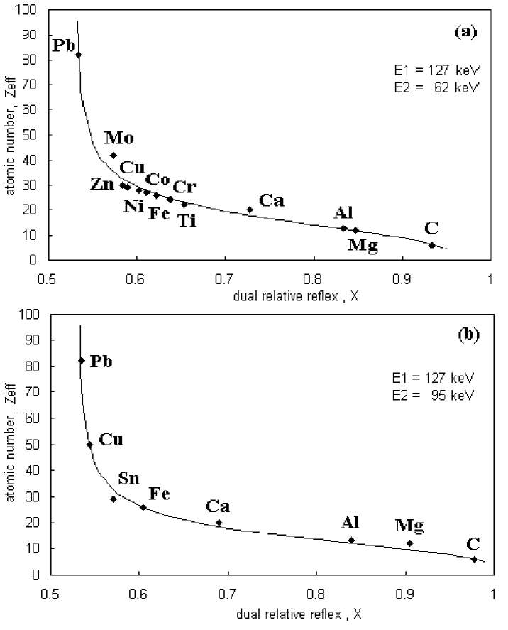
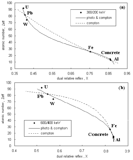
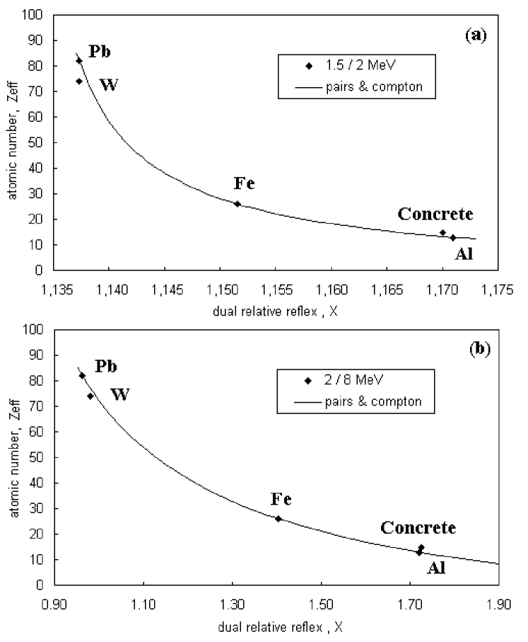
In general, unlike the “synthesis” of , the direct method is based upon “analysis” of the atomic composition. Moreover, in the new approach it is possible to limit oneself to the use of just the 2-radiography. This is important, because passing over to radiography with higher multiplicity is a technically difficult task. To verify the theory, we compared the obtained theoretical dependence (11)-(12) with known experimental data on gamma-radiation absorption in a large range of various materials, starting from carbon () and ending with uranium (). These results are shown in Fig. 2-4. In constructing the theoretical curves, three points were chosen as reference ones (defining a fraction-rational function), which corresponded to materials with large, small and intermediate values. Three characteristic regions of the energy spectrum were considered – i.e., low, middle and high energies, corresponding, respectively, to the photo effect, the Compton effect and the pair production effect. The data presented are in a very good agreement with theory.
The choice of absorption mechanisms depends upon the radiation energies used. The gamma-quanta energy range is mainly determined by the experiment conditions of character of applications. E.g., in security and customs inspection of luggage, medium energies from several tens to several hundreds keV are used. This corresponds to the region of combined effects of photo absorption and Compton scattering of gamma-quanta in the substance (Figs. 2-3). Discarding any of these mechanisms in the medium energy range can lead to substantial errors in determination of the effective atomic number. Thus, in Fig. 3 a theoretical approximation neglecting the photo effect gives much worse results. For inspection of large objects, such as trucks or containers, accelerator energies of several MeV are needed. In this case, for determination of the effective atomic number one should use a model where the pair formation effect and Compton effect are predominant It should be stressed that the proposed method can be used with any choice of the radiation energies. For the data presented in Figs. 2-4, we used only several fixed radiation energy values. We have checked that with any other choice of these energies the principal law (11) remains valid. For the experimental data used, deviations from the theoretical dependence did not exceed 5-10% , which corresponds to 90-95% accuracy in determination.
Detailed analysis of the optimal design for 2-radiography, completion of direct experiments, reconstruction of the atomic composition of different objects and complex materials, micro- and macro-structures, etc. are the subjects of our further studies. The approach presented in this paper allows the most complete extraction of the information on physical properties of the material studied by multi-energy radiography. The number of parameters reconstructed corresponds to the radiography multiplicity. (This is a general principle of multi-energeticity). This direct analytical approach uses only absorption data, showing up the amplitude contrast induced by ionizing radiation of photons (as monochromatic electromagnetic waves). Together with new X-ray analysis methods, including those using synchrotron (coherent) radiation exposing the phase contrast, as well as multi-energy holography [18], [19], [20], micro-focusing and computer tomography [21], [22], etc., the proposed approach to monitoring of materials structure widely broadens the field of NDT possibilities. This direction is similar to new directions in studies of semiconductor and other films. Also, certain prospects are opened for application of NDT in development of various microstructures, as well as in studies of the distribution profile of implanted nano-clusters of alien atoms in the “host” crystal lattice.
5. Summary
Thus, a direct approach has been proposed to reconstruction of the atomic structure of materials by means of multi-energy radiography. The general expressions (10)-(12) obtained for the effective atomic number in 2-monitoring, are closed and convenient for development of computer data processing algorithms. These formulas can be also used for reconstruction of the structure of mixtures, composites, multi-component objects, micro- and macro-structures, systems with variable chemical composition, etc. The validity of a universal law (fraction-rational dependence with calibration over three reference points) in the atomic number radiography is experimentally confirmed in different energy ranges of X-ray and gamma-radiation. An essential feature of multi-energy radiography is direct extraction of additional information on physico-chemical structure of the object under study. This opens new possibilities in materials science, non-destructive testing and other applied fields of science and technology.
Acknowledgements
The research described in this publication was made possible in part by Award No. UE2-2484-KH-02 of the U.S. Civilian Research & Development Foundation for the Independent States of the Former Soviet Union (CRDF).
References
- [1] 15th World Conference on Non-Destructive Testing, Session of Methods and Instrumentation, Rome, Italy, Abstracts Book, p. 1-800 (2000).
- [2] C. Robert-Coutant, V. Moulin, R. Sauze, P. Rizo and J. M. Casagrande, Nucl. Instr. and Meth. A442, 949-956 (2003).
- [3] National Research Council, Airline Passenger Security Screening: New Technologies and Implementation Issues (National Advisory Board, National Academy Press, Washington, D.C. (1996).
- [4] L. Grodzins, Nucl. Instr. and Meth. B36/37, 829 (1991).
- [5] G. T. Barnes, R. A. Sones, M.M. Tesic, D. R. Morgan and J. N. Saunders, Radiology 156, 537 (1985).
- [6] L. A. Feldkamp, S. A. Goldsteine, A. M. Parfitt, G. Jesion, M. Kleerekoper, Journal of Bone and Mineral Research 1, 3-11 (1989).
- [7] F. Inanc, J. N. Gray, T. Jensen, J. Xu, Proceedings of SPIE Conference on Physics of Medical Imaging, Vol. 3336, 830-837 (1998).
- [8] M. Marziani, A. Taibi, A. Tuffanelli, M. Gambaccini, Phys. Med. Biol., 47, 305-313 (2002).
- [9] R. M. Harrison, Nucl. Instr. and Meth. A310, 24-34 (1991), and references therein.
- [10] Rapiscan, Prospects, USA, 2003, http://www.rapiscan.com .
- [11] Heimann, Prospects, Germany, 2003, http://www.heimannsystems.com .
- [12] YXLON International, Prospects, 2002, http://www.yxlon.com .
- [13] Poliscan, Prospects, http://www.isc.kharkov.com/STCRI.
- [14] R. E. Alvarez, A. Macovski, Phys. Med. Biol., 21, 733-744 (1976).
- [15] J. C. G. Coenen, J. G. Maas, “Material classification by dual energy computerized X-ray tomography”, International Symposium on computerized tomography for industrial applications, 120-127 (1994).
- [16] C. Rizescu, C. Beliu and A. Jipa, Nucl. Instr. and Meth. A465, 584-599 (2001), and references therein.
- [17] S. V. Naydenov, V. D. Ryzhikov, B. V. Grinyov, E. K. Lisetskaya, A. D. Opolonin, D. N. Kozin, Los Alamos Archives (http://xxx.lanl.gov), physics/0206014.
- [18] S. W. Wilkins, T. E. Gureyev, D. Gao, A. Pogany, A. W. Stevenson, Nature 384, 335 (1996).
- [19] T. Gog, P. M. Len, G. Materic, D. Bahr, C. S. Fadly, C. Sanches-Hanke, Phys. Rev. Lett. 76, 30-33 (1996).
- [20] P. Cloetens, W. Ludwig, J. Baruchel, D. Van Dyck, J. Van Landuyt, J.P. Guiday, M. Schenker, Appl. Phys. Lett. 75, 2912-2914 (1999).
- [21] M. Van Geet, R. Swennen and M. Wevers, Sed. Geol. 132, 25-26 (2000).
- [22] See, for example, X-ray tomography in Material Science (Hermes Science Publications, Paris, 2000, ed. by J. Baruchel et al.).
Table 1. Effective atomic number of various substances with respect to the photo effect, , pair formation effect, , and Compton scattering, .
| Material | Chemical formula | |||
|---|---|---|---|---|
| Inorganic substances | ||||
| Stainless steel | Fe 66% ; Cr 10% ; Ni 16% ; Ti 8% | 26.57 | 27.58 | 27.80 |
| Black steel | Fe 92% ; C 8% | 25.97 | 25.94 | 25.76 |
| Calcium phosphate; bone tissue (med.) | Ca(PO4)2 | 17.38 | 16.11 | 11.05 |
| Table salt | NaCl | 15.66 | 15.21 | 14.62 |
| Quartz glass; sand | SiO2 | 12.30 | 11.63 | 10.80 |
| Aluminum and light alloys | Al2O3 | 11.70 | 11.23 | 10.65 |
| Glass | Na2SiO3 | 11.49 | 11.02 | 10.51 |
| Water | H2O | 7.98 | 7.89 | 7.22 |
| Air | mixture O2; N2 etc. | 7.6 | 7.4 | 6.9 |
| Organic substances | ||||
| Polyvinyl chloride | (C2H3Cl)n | 15.85 | 14.80 | 11.97 |
| Soft tissue (med.) | CNO-organics; H2O 90% | 7.8 | 7.2 | 6.8 |
| Glucose | C6H12O6 | 7.37 | 7.22 | 6.73 |
| Saccharose | C12H22O11 | 7.38 | 7.18 | 6.71 |
| Cellulose (wood, fabrics) | (C6H10O5)n | 7.31 | 7.14 | 6.68 |
| Organic glass | (C5H8O2)n | 6.96 | 6.76 | 6.24 |
| Polyamide (nylon) | (C6H11NO2)n | 6.85 | 6.70 | 6.18 |
| Polystyrene | (C8H9)n | 5.95 | 5.92 | 5.57 |
| Polyethylene (plastics) | (C2H4)n | 5.94 | 5.86 | 5.29 |
Figure captions
FIG. 1. General scheme of two-energy radiography with reconstruction of the effective atomic number of the material. Synthesis consists in mixing of the fitting basic elements with L – ”light”, M – ”middle” and H – ”heavy” atomic mass. Analysis is unambiguous reconstruction of . “Black-and-white” synthesis corresponds to the two-energy radiography, “three-color” scheme (R – ”red”, G – green, and B – ”blue”) corresponds to 3-radiography, etc. For the direct method (analysis) proposed in this work, 2-radiography is sufficient.
FIG. 2. Dependence of the effective atomic number upon 2-radiography reflexes in the regions of photo effect and Compton scattering. Theoretical dependence (solid line) and experimental points for materials of known composition are indicated. Geometry (dimensions) of samples is arbitrary.
FIG. 3. Dependence of the effective atomic number upon 2-radiography reflexes in the region of intermediate ionizing radiation energies. Theoretical curves are presented modeling the predominant role of one of two possible absorption channels. The best agreement with experimental data is obtained when a mixed absorption mechanism (photo effect/Compton effect) is chosen.
FIG. 4. Dependence of the effective atomic number upon 2-radiography reflexes in the region of Compton scattering and the pair formation effect. When the detected energy ranges are moved apart, sensitivity of the method is not worsened.