Multiple Folding Pathways of the SH3 domain
Abstract
Experimental observations suggest that proteins follow different pathways
under different environmental conditions. We perform molecular dynamics
simulations of a model of the SH3 domain over a broad range of
temperatures, and identify distinct pathways in the folding transition. We
determine the kinetic partition temperature —the temperature for which
the SH3 domain undergoes a rapid folding transition with minimal kinetic
barriers— and observe that below this temperature the model protein may
undergo a folding transition via multiple folding pathways. The folding
kinetics is characterized by slow and fast pathways and the presence of
only one or two intermediates. Our findings suggest the hypothesis that the
SH3 domain, a protein for which only two-state folding kinetics was
observed in previous experiments, may exhibit intermediates states under
extreme experimental conditions, such as very low temperatures. A very
recent report (Viguera et al., Proc. Natl. Acad. Sci. USA,
100:5730–5735, 2003) of an intermediate in the folding transition of the
Bergerac mutant of the -spectrin SH3 domain protein supports this
hypothesis.
1Center for Polymer Studies and Department of Physics,
Boston University, Boston, MA 02215 USA
2 Department of Biochemistry and Biophysics, School of Medicine,
University of North Carolina at Chapel Hill, Chapel Hill, NC 27599
Keywords: intermediate, molecular dynamics, folding pathways, SH3, kinetic partition.
1 INTRODUCTION
Recent experimental studies indicate that several proteins exhibit simultaneously a variety of intermediates and folding pathways. Kiefhaber[1] identified at low denaturant concentration a fast pathway (50 ms) in the folding of lyzosyme with no intermediates and a slow phase (420 ms) with well-populated intermediates. Choe et al.[2] observed the formation of a kinetic intermediate in the folding of villin 14T upon decreasing the temperature, and Silverman et al.[3] observed the extinction of a slow phase in the folding of the P4-P6 domain upon changes in ion concentration. Kitahara et al.[4] studied a pressure-stabilized intermediate of ubiquitin, identified as an off-pathway intermediate in previous kinetics experiments at basic conditions[5]. All these studies suggest that environmental conditions favor some folding pathways over others.
Major theoretical efforts in the study of protein folding[6, 7, 8, 9, 10, 11, 12, 13, 14, 15, 16] have focused on small, single domain proteins [17]. It is found in experiments[17, 18] that these proteins undergo folding transition with no accumulation of kinetic intermediates in the accessible range of experimental conditions. However, other kinetics studies of two-state proteins[19, 20, 21, 22] suggest the presence of short-lived intermediates that cannot be directly detected experimentally. Recently, Sánchez et al.[23] explained the curved Chevron plots — the nonlinear dependence of folding and unfolding rates on denaturant concentration[24, 25, 26] — of 17 selected proteins by assuming the presence of an intermediate state. Led by these studies, we hypothesize that single domain proteins may exhibit intermediates in the folding transition under suitable environmental conditions.
To test our hypothesis, we perform a molecular dynamics study of the folding pathways of the c-Crk SH3 domain[27, 28, 29] (PDB[29] access code 1cka). The SH3 domain is a family of small globular proteins which has been extensively studied in kinetics and thermodynamics experiments[30, 31, 32, 33, 34, 35, 18, 36, 37]. We select the c-Crk SH3 domain ( residues) as the SH3 domain representative and perform molecular dynamics simulations over a broad range of temperatures. We determine the kinetic partition temperature[38, 12] below which the model protein exhibits slow folding pathways and above which the protein undergoes a cooperative folding transition with no accumulation of intermediates. Below , we study the presence of intermediates in the slow folding pathways and resolve their structure. We find that one of the intermediates populates the folding transition for temperatures as high as . We discuss the relevance of our results in light of recent experimental evidence.
2 RESULTS
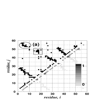
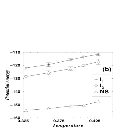
The SH3 domain is a -sheet protein (upper triangle of Fig. 1a). Our previous thermodynamic studies[6] of the c–Crk SH3 domain revealed only two stable states at equilibrium conditions: folded and unfolded. Both states coexist with equal probability at the folding transition temperature, , at which the temperature dependence of the potential energy has a sharp change, and the specific heat has a maximum (experimentally[18], this temperature corresponds to C). Thus, our model reproduces the experimentally-determined thermodynamics of the SH3 domain[30, 31, 18].
2.1 Initial Unfolded Ensemble
Our initially unfolded ensemble consists of 1100 protein conformations that we sample from a long equilibrium simulation at a high temperature at equal time intervals of time units (t.u). This time separation is long enough to ensure that the sampled conformations have low structural similarity among themselves. We calculate the frequency map — the plot of the probability of any two amino acids forming a contact — of this unfolded ensemble (lower triangle of Fig. 1a). At , only nearest and next nearest contacts have high frequency, and the frequency decreases rapidly with the sequence separation between the amino acids.
When we quench the system from to a target temperature, (see Methods), the system relaxes in approximately t.u. Due to the finite size of our heat bath, the heat released by the protein upon folding increases the final temperature of the system by units above . After relaxation, the protein stays for a certain time in the unfolded state, then undergoes a folding transition. During this time interval, the protein explores unfolded conformations, and we calculate the frequency map of the unfolded state for different target temperatures.
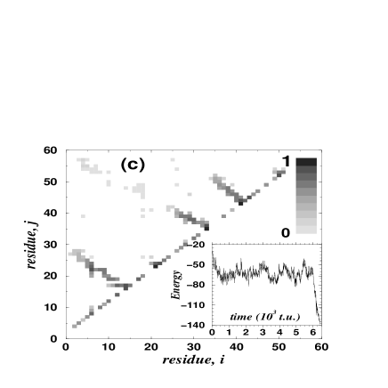
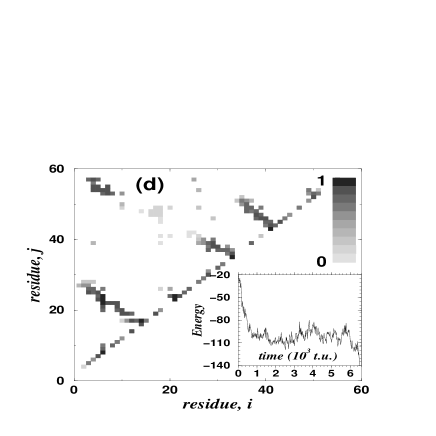
At , the secondary structure is unstable (Fig. 1b), with average frequency (see Methods). Successful folding requires the cooperative formation of contacts throughout the protein in a nucleation process[6, 7]. At , the secondary structure is more stable (Fig. 1c, ). Thus, the conformational search for the native state (NS) is optimized by limiting the search to the formation of a sufficient number of long range contacts. At , the lowest temperature studied, secondary structure elements form during the rapid collapse of the model protein in the first t.u. (Fig. 1d, ). During collapse, some tertiary contacts — contacts between secondary elements — may also form. The formation of these contacts prior to the proper arrangement of secondary structure elements may lead the protein model to a kinetic trap. Finally, folding proceeds at this temperature through a thermally activated search for the NS.
2.2 Kinetic Partition Temperature
In order to determine the temperature below which we can distinguish fast and slow folding pathways, we compute the distribution of folding times (Fig. 2a–e), as well as the average (Fig. 2f) and standard deviation . The ratio measures the average folding time in units of the standard deviation . This quantity is particularly useful when the value of the standard deviation correlates with the value of the average as we change . For instance, single-exponential distributions have .
We expect for , because at these high temperatures the folding transitions become rare events and are single-exponential distributed. As we decrease , we expect just below , because the folded state becomes more stable than the unfolded state, and the folding transitions are favored. Distributions with indicate a narrow distribution centered in , so that most of the simulations undergo a folding transition for times of the order of the average folding time. However, if we continue decreasing , we expect some folding transitions to be kinetically trapped, and the folding time distribution will spread over several orders of magnitudes. Such distributions have . Thus, there is a temperature below where the maximum of occurs, and which signals the onset of slow folding pathways. We use the maximum of to calculate .
Fig. 2g suggests that , which corresponds to a maximally compact distribution of folding times***Assuming a linear relation between experimental and simulated temperatures and taking into account[18] that C, we estimate C (Fig. 2d). We find that the ratio approaches one as we increase the temperature above , and the distribution of folding times approximates a single-exponential distribution. In particular, the distribution of folding times fits the single-exponential distribution for , the closest temperature to that we study. The ratio decreases monotonically below , indicating that the distribution of folding times spreads over several orders of magnitude. This is the consequence of an increasing fraction of folding simulations kinetically trapped (Fig. 2a-b). The average folding time is minimal not at , but at a lower temperature (Fig. 2f). At this temperature, we find that the protein becomes temporarily trapped in approximately of the folding transitions. On the other hand, the remaining simulations undergo a folding transition much faster, thus minimizing . Interestingly, , even though the distribution of folding times at this temperature is non-exponential.
2.3 Folding Pathways
Below , an increasing fraction of the simulations undergo folding transitions that take a time up to three orders of magnitude above the minimal . In addition, increases dramatically (Fig. 2f). At the lowest temperatures studied, we distinguish between the majority of simulations that undergo a fast folding transition (the fast pathway) and the rest of the simulations that undergo folding transitions with folding times spanning three orders of magnitude (the slow pathways). At the low temperature , the potential energy of the fast pathway has on average the same time evolution of all the simulations at , indicating that there are no kinetic traps in the fast pathway.
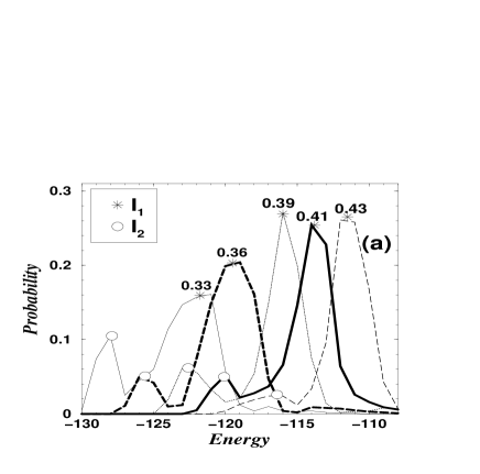
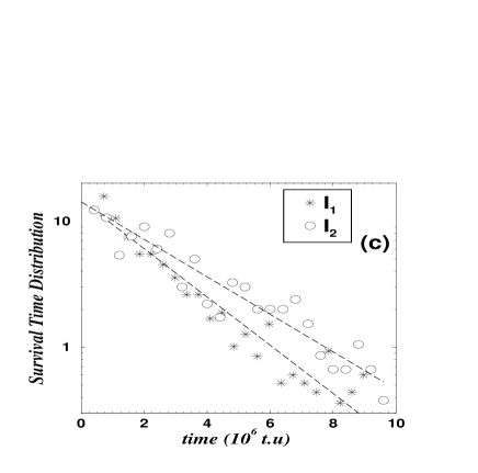
For each folding simulation that belongs to the slow pathways, we sample the potential energy at equal time intervals of t.u. until folding is finished (see Methods). Then, we collect all potential energy values and construct a distribution of potential energies. We find that below , the distribution is markedly bimodal (Fig. 3a). The positions of the two peaks along the energy coordinate do not correspond to the equilibrium potential energy value of the folded state (Fig. 3b). Therefore we hypothesize the existence of two intermediates in the slow pathways. We denote the two putative intermediates as and for the high energy and low energy peaks, respectively. As temperature decreases, the peaks shift to lower energies, but the energy difference between the two peaks, approximately six energy units, remains constant (Fig. 3b). A constant energy difference implies that the two putative intermediates differ by a specific set of native contacts. As temperature decreases, other contacts not belonging to this set become more stable and are responsible for the overall energy decrease. At , we record the distribution of survival times for both intermediates and find that they fit a single-exponential distribution, supporting the hypothesis that each intermediate is a local free energy minima and has a major free energy barrier (Fig. 3c).
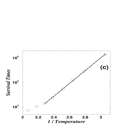
To further test the single free energy barrier hypothesis, we select a typical conformation representing intermediate and perform 200 folding simulations, each with a different set of initial velocities for a set of temperatures in the range . For each simulation, we record the time that the protein stays in the intermediate and find that the average survival time fits the Arrhenius law for temperatures below (Fig. 3d). This upper bound temperature roughly coincides with the temperature below which becomes noticeable in the histogram of potential energies (Fig. 3a). This result indicates that the free energy barrier to overcome intermediate becomes independent of temperature for low temperatures, or analogously, that the same set of native contacts must form (or break) to overcome the intermediate.
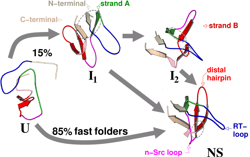
Next, we determine the structure of the two intermediates. For each intermediate, we randomly select three conformations and find that they are structurally similar. Conformations belonging to intermediate have a set of long-range contacts (C1) with a high occupancy and a set of long-range contacts (C2) with no occupancy at all (Fig. 3e). Contacts in C1 represent a -sheet made by three strands: the two termini and the strand following the RT-loop, which we name strand “A” (see in Fig. 4). Contacts in C2 represent the base of the n-Src loop and the contacts between the RT-loop and the distal hairpin (see in Fig. 4). In addition, has a set of medium-range contacts (C3) with high occupancy (Fig. 3e) representing the distal hairpin and a part of the n-Src loop. For a slow folding transition, the -sheet (C1 contacts) forms in the early events and strand “A” can no longer move freely. This constrained motion prevents strand “A” from forming contacts with one of the strands of the distal hairpin, which we name as strand “B” (see in Fig. 4). Similarly, strand “B” cannot move freely because it is a part of C3. The missing contacts between strand “A” and strand “B” are the contacts that form the base of the n-Src loop.
Conformational changes leading the protein away from intermediate involve either dissociation of the -sheet, thus breaking some contacts of C1, or dissociation of the distal hairpin, thus breaking some contacts of C3. We find that the latter dissociation may lead the protein conformation to intermediate . Intermediate has contacts of C1, but lacks the set of contacts (C4) that form the base of the the distal hairpin (see NS in Fig. 4).
Once we identify the structure of the intermediates, we investigate whether intermediate is present at larger temperatures when no distinction can be made concerning fast and slow folding pathways. To test this hypothesis, we sample the protein conformation during the folding transition at equal time intervals of t.u. for each of the simulations, and compare these conformations to intermediate with a similarity score function (see Methods). For each folding transition, we record only the highest value of the similarity score, thus obtaining highest score values. At , the histogram of the highest scores is bimodal, with of the folding simulations passing through intermediate (Fig. 3f). We find that at , simulations that undergo the folding transition through show kinetics of folding no different than those of the rest of simulations.
3 DISCUSSION
It was shown[6] that the simplified protein model and
interaction potentials that we use here reproduced in a certain range of
temperatures the
experimentally-determined two-state thermodynamics
of the SH3 domain[18]. The qualitative predictive power of the
model encouraged us to study the folding kinetics in a wider range of
temperatures. From our relaxation studies of the initial unfolded ensemble,
we observe that the structure of the unfolded state is highly sensitive to
. The role of the unfolded state in determining the folding
kinetics has already been pointed out in recent experimental and theoretical
studies[39, 40, 41, 42]. We observe
nucleation[6], folding with minimal kinetic barriers, and
thermally activated mechanisms for the different observed unfolded states.
In previous studies, various methods have been developed to determine the temperature that signals the onset of slow folding pathways. Socci et al.[43, 44] determined a glass transition temperature, , at which the average folding time is half way between and , where is the minimun average folding time and is the total simulation time. This method is sensitive to the a priori selected . The authors varied in the range , and they found a error in the calculation of . Also, Gutin et al.[45] estimated a critical temperature, , at which the temperature dependence of the equilibrium potential energy leveled off. From their results, one can evaluate a error in their calculation of . Both and are temperatures that authors use to characterize the onset of multiple folding pathways. In our study we use , which signals the breaking of time translational invariance of equilibrium measurements for temperatures below this value[46]. We estimate a error in our calculation of from uncertainties in the location of in Fig. 2g.
At , secondary structure elements are partially stable, and the search for the NS reduces to the formation of tertiary contacts. Furthermore, is a relatively high temperature that prevents the stabilization of improper arrangements of the protein conformation, thus minimizing the occurrence of kinetic traps. Below , the model protein exhibits two intermediates with well-defined structural characteristics. This modest number of intermediates is a direct consequence of the prevention of non-native contacts. This prevention reduces dramatically the number of protein conformations. Furthermore, since a low energy value implies that most of the native interactions have formed, there are few conformations having both low energy and structural differences with the NS[11].
It is found experimentally [47, 48, 49, 2, 50, 1, 3, 51, 52, 53, 54] that proteins exhibit only a discrete set of intermediates. Even though in real proteins amino acids that do not form a native contact may still attract each other, experimental and theoretical studies confirm that native contacts have a leading role in the folding transition. Protein engineering experiments[55, 33, 56, 57, 58] show that transition states in two-state globular proteins are mostly stabilized by native interactions. To quantitatively determine the importance of native interactions in the folding transition, Paci et al.[59] studied the transition states of three two-state proteins with a full-atom model. They found that on average, native interactions accounted for approximately of the total energy of the transition states. Of relevance to our studies of the SH3 domain are the full-atom study[60] and the protein engineering experiments[33, 36] showing that the transition state of the src-SH3 domain protein is determined by the NS. On the other hand, evidence exists that in some proteins, non-native contacts are responsible for the presence of intermediates. In their study of the homologous Im7 and Im9 proteins, Capaldi et al.[61] identified a set of non-native interactions responsible for a intermediate state in the folding transition of Im7 protein. Mirny et al.[62] performed Monte Carlo simulations of two different sequences with the same NS in the lattice. One sequence presented a series of pathways with misfolded states due to non-native interactions.
We investigate the kinetics of formation of the two intermediates in a wide range of temperatures. At low temperatures, simulations that undergo folding through intermediate reveal that contacts between the two termini form earlier than the contacts belonging to the folding nucleus[6, 7]. This result coincides with an off-lattice study[63] of a 36-monomer protein by Abkevich et al. In this study, the authors found an intermediate in the folding transition of their model protein. Inspection of the intermediate revealed no nucleus contacts, but a different set of long-range contacts already formed. In addition, we learned of the work by Viguera et al.[64] after completion of our study. They reported that a mutant of the -spectrin SH3 domain undergoes a folding transition through one intermediate. The authors observed that the newly-introduced long-range contacts had already been formed in the denaturated state, preceding the formation of the transition state of the protein. Thus, environmental conditions that favor stabilization of long-range contacts other than the nucleus contacts may induce intermediates in the folding transition.
Alternatively, short-range contacts in key positions of the protein structure may also be responsible for slow folding pathways. After completion of our study, Karanicolas et al.[65, 66] reported their studies on the Gō model of the forming binding protein WW domain. The authors found a slow folding pathway in the model protein, and a cluster of four short-range native contacts that are responsible for this pathway. However, the authors observed that it was the absence, not the presence, of these native contacts in the unfolded state that generated bi-phasic folding kinetics. Thus, environmental conditions that favor destabilization of short range contacts may promote the formation of intermediate states in the folding transition.
We also investigate the survival time of intermediate , and find that the free energy barrier separating from the NS is independent of temperature. Thus, the average survival time follows Arrhenius kinetics. The value of the free energy barrier is approximately energy units, indicating that about six native contacts break when the protein conformation reaches the transition state that separates from the NS. At the low temperatures where intermediates and are noticeable, thermal fluctuations are still large enough so that the observed survival times of should be much smaller, if only any six native contacts were to break. Thus we hypothesize that it is allways the same set of native contact that must break in the transition . Our observations of the transition support this hypothesis. In this transition, we find that the set of contacts C4 allways breaks.
At , we do not detect the intermediates from kinetics measurements of the average folding time, or analogously, from the folding rate. Thus we analyze the folding transition with the similarity score function that tests the presence of intermediate . Then we find that this intermediate is populated in of the folding transitions. In a recent study[67], Gorski et al. reported the existence of an intermediate in the folding transition of protein Im9 under acidic conditions (). This finding led authors to formulate the hypothesis that Im9 has an intermediate at normal conditions (), but it is too unstable to be detected with current kinetic experimental techniques. Interestingly, the homologous protein Im7 ( sequence identity) undergoes folding transition through an intermediate in all tested experimental conditions[68, 67, 61], supporting the authors’ hypothesis. Thus, changes in both the environmental conditions and the amino acid sequence may uncover hidden intermediates in the folding transition of a two-state protein. In addition, a detailed study at may reveal the intermediates. This is particularly useful for computer simulations, because simulations at low temperatures when intermediates are easily identifiable may require several orders of magnitude longer than simulations at .
3.1 Conclusion
We perform molecular dynamics analysis of the folding transition of the Gō model of the c-Crk SH3 domain in a broad range of temperatures. At the folding transition temperature, we observe that only the folded and unfolded states are populated. However, as we decrease the temperature, parameters monitoring the folding process such as potential energy and root mean square distance with respect to the native state, , suggest the presence of intermediates. We determine the kinetic partition temperature below which we observe two folding intermediates, and , and above which we do not observe accumulation of intermediates. Below , intermediate forms when the two termini and the strand following the RT-loop form a -sheet, prior to the formation of the folding nucleus. This intermediate effectively splits the folding transition into fast and slow folding pathways. Dissociation of part of the -sheet leads the protein to the native state. We also find that stabilization of this -sheet and subsequent dissociation of the distal hairpin may lead to intermediate .
The key structural characteristics of intermediate allow us to define a similarity score function that probes the presence of the intermediate in a folding transition. We find that is populated even at . This result suggests that one can obtain information regarding the existence of putative intermediates by studying the folding trajectories at . However, at this temperature, no intermediates are noticeable if one limits the analysis only to the distribution of folding times.
We observe that the folding pathways of the model SH3 domain are highly sensitive to temperature, suggesting the important role of the environmental conditions in determining the folding mechanism. Our findings suggest that the SH3 domain, a two-state folder, may exhibit stable intermediates under extreme experimental conditions, such as very low temperatures.
4 MATERIALS AND METHODS
4.1 Model Protein and Interactions
We adopt a coarse-grained description of the protein by which each amino acid is reduced to its Cβ atom (Cα in case of Gly). Details of the model, the surrounding heat bath, and the selection of structural parameters are discussed in detail in a previous study[6]. The selection of the set of interaction parameters among amino acids is of crucial importance for the resulting folding kinetics of the model protein [11, 12, 14]. Experimental and theoretical studies of globular proteins [6, 7, 69, 70, 71, 72, 73, 74, 36, 75, 76, 57] suggest that native topology is the principal determinant of the folding mechanism. Thus, we employ a variant of the Gō model of interactions[77, 12, 78, 79, 80, 81] — a model based solely on the native topology — in which we prevent formation of non-native interactions, since we are solely interested in the role that native topology and native interactions may have in the formation of intermediates. We perform simulations and monitor the time evolution of the protein and the heat bath with the discrete molecular dynamics algorithm [82, 83, 84, 85, 86, 87, 88]. The higher performance of this algorithm over conventional molecular dynamics allows one to increase the computational speed up to three orders of magnitude.
4.2 Frequencies and Folding Simulations
To calculate the frequency map at , we probe the presence of the native contacts in each of the initially unfolded conformations. Then, we compute the probability of each native contact to be present. To calculate the frequency map at , we select one particular folding transition and we probe the presence of the native contacts during the time interval that spans after the initial relaxation and before the simulation reaches the folding time . To compute , we stop the folding simulation when of the native contacts form. Then, we trace back the folding trajectory and record when the root mean square distance with respect to the native state, , becomes smaller than Å. We consider all protein conformations occurring for as belonging to the folded state and of no relevance to the folding transition.
4.3 Similarity Score Function
We introduce the similarity score function, , where is the number of native contacts belonging to the set of contacts C1, and is the number of native contacts belonging to set C2 (Fig. 3e). C1 has contacts and C2 has contacts. If the protein is unfolded, then , thus . Similarly, if the protein is folded, then and , thus again. Finally, if the protein adopts the intermediate structure, then and , thus .
We acknowledge E. I. Shakhnovich for insightful discussions, R. Badzey for careful reading of the manuscript, and the Petroleum Research Fund for support.
References
- 1. Kiefhaber, T. (1995). Kinetics traps in Lysozyme folding. Proc. Natl. Acad. Sci. USA, 92, 9029–9033.
- 2. Choe, S. E., Matsudaira, P. T., Osterhout, J., Wagner, G. & Shakhnovich, E. I. (1998). Folding kinetics of villin 14T, a protein domain with a central beta-sheet and two hydrophobic cores. Biochemistry, 37, 14508–14518.
- 3. Silverman, S. K., Deras, M. L., Woodson, S. A., Scaringe, S. A. & Cech, T. R. (2000). Multiple folding pathways for the p4-p6 RNA domain. Biochemistry, 39, 12465–12475.
- 4. Kitahara, R. & Akasaka, K. (2003). Close identity of a pressure-stabilized intermediate with a kinetic intermediate in protein folding. Proc. Natl. Acad. Sci. USA, 100, 3167–3172.
- 5. Briggs, M. S. & Roder, H. (1992). Early hydrogen-bonding events in the folding reaction of ubiquitin. Proc. Natl. Acad. Sci. USA, 89, 2017–2021.
- 6. Borreguero, J. M., Dokholyan, N. V., Buldyrev, S. V., Shakhnovich, E. I. & Stanley, H. E. (2002). Thermodynamics and folding kinetics analysis of the SH3 domain from discrete molecular dynamics. J. Mol. Biol. 318, 863–876.
- 7. Ding, F., Dokholyan, N. V., Buldyrev, S. V., Stanley, H. E. & Shakhnovich, E. I. (2002). Direct molecular dynamics observation of protein folding transition state ensemble. Biophys. J. 83, 3525–3532.
- 8. Fersht, A. R. & Daggett, V. (2002). Protein folding and unfolding at atomic resolution. Cell, 108, 573–582.
- 9. Karplus, M. & McCammon, J. A. (2002). Molecular dynamics simulations of biomolecules. Nature Struct. Biol. 9, 646–652.
- 10. Ozkan, S. B., Dill, K. A. & Bahar, I. (2002). Fast–folding protein kinetics, hidden intermediates, and the sequential stabilization model. Prot. Sci. 11, 1958–1970.
- 11. Plotkin, S. S. & Onuchic, J. N. (2002). Structural and energetic heterogeneity in protein folding. I. theory. J. Chem. Phys. 116, 5263–5283.
- 12. Thirumalai, D., Klimov, D. K. & Dima, R. I. (2002). Insights into specific problems in protein folding using simple concepts. Adv. Chem. Phys, 120, 35–76.
- 13. Tiana, G. & Broglia, R. A. (2001). Statistical analysis of native contact formation in the folding of designed model proteins. J. Chem. Phys. 114, 2503–2510.
- 14. Pande, V. S., Grosberg, A. Y. & Tanaka, T. (2000). Heteropolymer freezing and design: towards physical models of protein folding. Rev. Mod. Phys. 72, 259–314.
- 15. Eaton, W. A., Mun̄oz, V., Hagen, J. S. J., Jas, G. S., Lapidus, L. J., Henry, E. R. & Hofrichter, J. (2000). Fast kinetics and mechanisms in protein folding. Annu. Rev. Biophys. Biomol. Struct. 29, 327.
- 16. Bryngelson, J. D. & Wolynes, P. G. (1989). Intermediates and barrier crossing in a random energy model (with applications to protein folding). J. Phys. Chem. 93, 6902–6915.
- 17. Jackson, S. E. (1998). how do small single-domain proteins fold? Folding & Design, 3, R81–R91.
- 18. Filimonov, V. V., Azuaga, A. I., Viguera, A. R., Serrano, L. & Mateo, P. L. (1999). A thermodynamic analysis of a family of small globular proteins: SH3 domains. Biophys. Chem. 77, 195–208.
- 19. Chiti, F., Taddei, N., White, P. M., Bucciantini, M., Magherini, F., Stefani, M. & Dobson, C. M. (1999). Mutational analysis of acylphosphatase suggests the importance of topology and contact order in protein folding. Nature Struct. Biol. 6, 1005–1009.
- 20. Houliston, R. S., Liu, C. S., Singh, L. M. R. & Meiering, E. M. (2002). pH and urea dependence of amide hydrogen-deuterium exchange rates in the beta-trefoil protein hisactophilin. Biochemistry, 41, 1182–1194.
- 21. Khorasanizadeh, S., Petrs, I. D. & Roder, H. (1996). Evidence for a three-state model of protein folding form kinetics analysis of ubiquitin variants with altered core residues. Nature Struct. Biol. 3, 193 205.
- 22. Bachmann, A. & Kiefhaber, T. (2001). Apparent two-state tendamistat folding is a sequential process along a defined route. J. Mol. Biol. 306, 375–386.
- 23. Sanchez, I. E. & Kiefhaber, T. (2003). Evidence for sequential barriers and obligatory intermediates in apparent two-state protein folding. J. Mol. Biol. 325, 367–376.
- 24. Ikai, A. & Tandford, C. (1973). Kinetics of unfolding and refolding of proteins. I Mathematical analysis. J. Mol. Biol. 73, 145–163.
- 25. Matouschek, A., Jr, J. T. K., Serrano, L., Bycroft, M. & Fersht, A. R. (1990). Transient folding intermediates characterized by protein engineering. , 346, 440 445.
- 26. Fersht, A. R. (2000). A kinetically significant intermediate in the folding of barnase. Proc. Natl. Acad. Sci. USA, 97, 14121–14126.
- 27. Wu, X. D., Knudsen, B., Feller, S. M., Sali, J., Cowburn, D. & Hanafusa, H. (1995). Structural basis for the specific interaction of lysine-containing proline-rich peptides with the n-terminal SH3 domain of c-Crk. Structure, 2, 215–226.
- 28. Branden, C. & Tooze, J. (1999). Introduction to Protein Structure. Garland Publishing Inc, New York.
- 29. Berman, H. M., Westbrook, J., Feng, Z., Gilliland, G., Bhat, T. N., Weissig, H., Shindyalov, I. N. & Bourne, P. E. (2000). The protein data bank. Nucleic Acids Research, 28, 235–242.
- 30. Viguera, A. R., Martinez, J. C., Filimonov, V. V., Mateo, P. L. & Serrano, L. (1994). Thermodynamic and kinetic-analysis of the SH3 domain of Spectrin shows a 2-state folding transition. Biochemistry, 33, 10925–10933.
- 31. Villegas, V., Azuaga, A., Catasus, L., Reverter, D., Mateo, P. L., Aviles, F. X. & Serrano, L. (1995). Evidence for a 2-state transition in the folding process of the activation domain of human procarboxypeptidase-a2. Biochemistry, 46, 15105–15110.
- 32. Grantcharova, V. P. & Baker, D. (1997). Folding dynamics of the Src SH3 domain. Biochemistry, 36, 15685–15692.
- 33. Grantcharova, V. P., Riddle, D. S., Santiago, J. N. & Baker, D. (1998). Important role of hydrogen bonds in the structurally polarized transition state for folding of the src SH3 domain. Nature Struct. Biol. 8, 714–720.
- 34. Knapp, S., Mattson, P. T., Christova, P., Berndt, K. D., Karshikoff, A., Vihinen, M., Smith, C. I. & Ladenstein, R. (1998). Thermal unfolding of small proteins with SH3 domain folding pattern. Proteins: Struct. Func. & Genet. 23, 309–319.
- 35. Martinez, J. C., Viguera, A. R., Berisio, R., Wilmanns, M., Mateo, P. L., Filimonov, V. V. & Serrano, L. (1999). Thermodynamic analysis of alpha-spectrin SH3 and two of its circular permutants with different loop lengths: Discerning the reasons for rapid folding in proteins. Biochemistry, 38, 549–559.
- 36. Riddle, D. S., Grantcharova, V. P., Santiago, J. V., Ruczinski, A. E. & Baker, D. (1999). Experiment and theory highlight role of native state topology in SH3 folding. Nature Struct. Biol. 6, 1016–1024.
- 37. Guerois, R. & Serrano, L. (2000). The SH3-fold family: experimental evidence and prediction of variations in the folding pathways. J. Mol. Biol. 304, 967–982.
- 38. Klimov, D. K. & Thirumalai, D. (1996). Factors governing the foldability of proteins. Proteins, 26, 411–441.
- 39. Zagrovic, B., Snow, C. D., Khaliq, S., Shirts, M. R. & Pande, V. S. (2002). Native-like mean structure in the unfolded ensemble of small proteins. J. Mol. Biol. 323, 153–164.
- 40. Millet, I. S., Doniach, S. & Plaxco, K. W. (2002). Toward a taxonomy of the denatured state: Small angle scattering studies of unfolded proteins. Adv. Prot. Chem. 62, 241–262.
- 41. Plaxco, K. W. & Gross, M. (2001). Unfolded, yes, but random? Never! Nature Struct. Biol. 8, 659–660.
- 42. Garcia, P., Serrano, L., Durand, D., Rico, M. & Bruix, M. (2001). NMR and SAXS characterization of the denatured state of the chemotactic protein CheY: Implications for protein folding initiation. Prot. Sci. 10, 1100–1112.
- 43. Socci, N. D. & Onuchic, J. N. (1994). Folding kinetics of proteinlike heteropolymers. J. Chem. Phys. 101, 1519–1528.
- 44. Socci, N. D., Onuchic, J. N. & Wolynes, P. G. (1996). Diffusive dynamics of the reaction coordinate for protein folding funnels. J. Chem. Phys. 15, 5860–5868.
- 45. Gutin, A., Sali, A., Abkevich, V., Karplus, M. & Shakhnovich, E. I. (1998). Temperature dependence of the folding rate in a simple protein model: search for a “glass” transition. J. Chem. Phys. 108, 6466–6483.
- 46. Dokholyan, N. V., Pitard, E., Buldyrev, S. V. & Stanley, H. E. (2002). Glassy behavior of a homopolymer from molecular dynamics simulations. Phys. Rev. E, 65, 030801:1–4.
- 47. Bhutani, N. & Udgaonkar, J. B. (2001). GroEL channels the folding of thioredoxin along one kinetic route. J. Mol. Biol. 345, 1167–1179.
- 48. Tezcan, F. A., Findley, W. M., Crane, B. R., Ross, S. A., Lyubovitsky, J. G., Gray, H. B. & Winkler, J. R. (2002). Using deeply trapped intermediates to map the cytochrome c folding landscape. Proc. Natl. Acad. Sci. USA, 99, 8626–8630.
- 49. Juneja, J. & Udgaonkar, J. B. (2002). Characterization of the unfolding of ribonuclease A by a pulsed hydrogen exchange study: Evidence for competing pathways for unfolding. Biochemistry, 41, 2641–2654.
- 50. Park, S. H., ONeil, K. T. & Roder, H. (1997). An early intermediate in the folding reaction of the B1 domain of protein G contains a native-like core. Biochemistry, 36, 14277–14283.
- 51. Segel, D. J., Bachmann, A., Hofrichter, J., Hodgson, K. O., Doniach, S. & Kiefhaber, T. (1999). Characterization of transient intermediates in lysozyme folding with time-resolved small-angle X-ray scattering. J. Mol. Biol. 288, 489–499.
- 52. Heidary, D. K., O’Neill, J. C., Roy, M. & Jennings, P. A. (2000). An essential intermediate in the folding of dihydrofolate reductase. Proc. Natl. Acad. Sci. USA, 97, 5866–5870.
- 53. Rumbley, J., Hoang, L., Mayne, L. & Englander, S. W. (2001). An amino acid code for protein folding. Proc. Natl. Acad. Sci. USA, 98, 105–112.
- 54. Simmons, D. A. & Konermann, L. (2002). Characterization of transient protein folding intermediates during myoglobin reconstitution by time-resolved electrospray mass spectrometry with on-line isotopic pulse labeling. Biochemistry, 41, 1906–1914.
- 55. Fersht, A. R. (1995). Characterizing transition-states in protein folding — an essential step in the puzzle. Curr. Opin. Struc. Biol. 1, 79–84.
- 56. Ternstrom, T., Mayor, U., Akke, M. & Oliveberg, M. (1999). From snapshot to movie: phi analysis of protein folding transition states taken one step further. Proc. Natl. Acad. Sci. USA, 96, 14854–14859.
- 57. Clementi, C., Nymeyer, H. & Onuchic, J. N. (2000). Topological and energetic factors: what determines the structural details of the transition state ensemble and ”en-route” intermediates for protein folding? An investigation for small globular proteins. J. Mol. Biol. 5, 937–953.
- 58. Northey, J. G. B., Nardo, A. A. D. & Davidson, A. R. (2002). Hydrophobic core packing in the SH3 domain folding transition state. Nature Struct. Biol. 9, 126–130.
- 59. Paci, E., Vendruscolo, M. & Karplus, M. (2002). Native and non-native interactions along protein folding and unfolding pathways. Proteins: Struct. Func. & Genet. 47, 379–392.
- 60. Shea, J. E., Onuchic, J. N. & III, C. L. B. (2002). Probing the folding free energy landscape of the src-SH3 protein domain. Proc. Natl. Acad. Sci. USA, 99, 16064–16068.
- 61. Capaldi, A. P., Kleanthous, C. & Radford, S. E. (2002). Im7 folding mechanism: misfolding on a path to the native state. Nature Struct. Biol. 9, 209–216.
- 62. Mirny, L. A., Abkevich, V. & Shakhnovich, E. I. (1996). Universality and diversity of the protein folding scenarios: a comprehensive analysis with the aid of a lattice model. Folding & Design, 1, 103–116.
- 63. Abkevich, V. I., Gutin, A. M. & Shakhnovich, E. I. (1994). Specific nucleus as the transition state for protein folding: evidence from the lattice model. Biochemistry, 33, 10026–10036.
- 64. Viguera, A. R. & Serrano, L. (2003). Hydrogen-exchange stability analysis of Bergerac-Src homology 3 variants allows the characterization of a folding intermediate in equilibrium. Proc. Natl. Acad. Sci. USA, 100, 5730–5735.
- 65. Karanicolas, J. & III, C. L. B. (2003). The structural basis for biphasic kinetics in the folding of the ww domain from a formin-binding protein: Lessons for protein design? Proc. Natl. Acad. Sci. USA, 100, 3954–3959.
- 66. Nguyen, H., Jager, M., Moretto, A., Gruebele, M. & Kelly, J. W. (2003). Tuning the free-energy landscape of a WW domain by temperature, mutation, and truncation. Proc. Natl. Acad. Sci. USA, 100, 3948–3953.
- 67. Gorski, S. A., Capaldi, A. P., Kleanthous, C. & Radford, S. E. (2001). Acidic conditions stabilise intermediates populated during the folding of Im7 and Im9. J. Mol. Biol. 312, 849–863.
- 68. Ferguson, N., Capaldi, A. P., James, R., Kleanthous, C. & Radford, S. E. (1999). Rapid folding with and without populated intermediates in the homologous four-helix proteins im7 and im9. J. Mol. Biol. 286, 1597–1608.
- 69. Plaxco, K. W., Simons, K. T. & Baker, D. (1998). Contact order, transition state placement and the refolding rates of single domain proteins. J. Mol. Biol. 277, 985–994.
- 70. Mun̄oz, V. & Eaton, W. A. (1999). A simple model for calculating the kinetics of protein folding from three-dimensional structures. Proc. Natl. Acad. Sci. USA, 96, 11311–11316.
- 71. Fersht, A. R. (2000). Transition-state structure as a unifying basis in protein-folding mechanisms: contact order, chain topology, stability, and the extended nucleus mechanism. Proc. Natl. Acad. Sci. USA, 97, 1525–1529.
- 72. Alm, E. & Baker, D. (1999). Prediction of protein-folding mechanisms from free-energy landscapes derived from native structures. Proc. Natl. Acad. Sci. USA, 96, 11305–11310.
- 73. Galzitskaya, O. V. & Finkelstein, A. V. (1999). A theoretical search for folding/unfolding nuclei in three-dimensional protein structures. Proc. Natl. Acad. Sci. USA, 96, 11299–11304.
- 74. Shea, J. M., Onuchic, J. N. & Brooks, C. L. (2000). Energetic frustration and the nature of the transition state in protein folding. J. Chem. Phys. 113, 7663–7671.
- 75. Du, R., Pande, V. S., Grosberg, A. Y., Tanaka, T. & Shakhnovich, E. I. (1999). On the role of conformational geometry in protein folding. J. Chem. Phys. 111, 10375–10380.
- 76. Micheletti, C., Banavar, J. R., Maritan, A. & Seno, F. (1999). Protein structures and optimal folding from a geometrical variational principle. Phys. Rev. Lett. 82, 3372–3375.
- 77. Jang, H., Hall, C. K. & Zhou, Y. Q. (2002). Protein folding pathways and kinetics: molecular dynamics simulations of beta-strand motifs. Biophys. J. 83, 819–835.
- 78. Cieplak, M. & Hoang, T. X. (2001). Kinetic nonoptimality and vibrational stability of proteins. Proteins: Struct. Func. & Genet. 44, 200–25.
- 79. Shimada, J., Kussell, E. L. & Shakhnovich, E. I. (2001). The folding thermodynamics and kinetics of crambin using an all-atom Monte Carlo simulation. J. Mol. Biol. 308, 79–95.
- 80. Hoang, T. X. & Cieplak, M. (2000). Sequencing of folding events in Gō-type proteins. J. Chem. Phys. 113, 8319–8328.
- 81. Zhou, Y. Q. & Karplus, M. (1999). Folding of a model three-helix bundle protein: a thermodynamic and kinetic analysis. J. Mol. Biol. 293, 917–951.
- 82. Alder, B. J. & Wainwright, T. E. (1959). Studies in molecular dynamics. I. general method. J. Chem. Phys. 31, 459–466.
- 83. Zhou, Y. Q., Karplus, M., Wichert, J. M. & Hall, C. K. (1997). Equilibrium thermodynamics of homopolymers and clusters: molecular dynamics and Monte Carlo simulations of systems with square-well interactions. J. Chem. Phys. 24, 10691–10708.
- 84. Allen, M. P. & Tildesley, D. J. (1987). Computer simulation of liquids. Clarendon Press, Oxford.
- 85. Rapaport, D. C. (1997). The art of molecular dynamics simulation. Cambridge University Press, Cambridge.
- 86. Grosberg, A. Y. & Khokhlov, A. R. (1997). Giant molecules. Academic Press, Boston.
- 87. Dokholyan, N. V., Buldyrev, S. V., Stanley, H. E. & Shakhnovich, E. I. (1998). Molecular dynamics studies of folding of a protein-like model. Folding & Design, 3, 577–587.
- 88. Dokholyan, N. V., Buldyrev, S. V., Stanley, H. E. & Shakhnovich, E. I. (2000). Identifying the protein folding nucleus using molecular dynamics. J. Mol. Biol. 296, 1183–1188.
Figure 1: (a) Upper triangle: c–Crk SH3 domain contact map with native
contacts. The secondary structure elements are the clusters of contacts that
are organized perpendicularly to the map diagonal. Long-range contacts
between the two termini are enclosed in the circle, and long-range contacts
between the RT-loop and the distal hairpin are enclosed in the square. Lower
triangle: the frequency map for the initial set of 1100 unfolded
conformations at . (b) Frequency map of the unfolded state at
. We compute the frequencies for a particular folding
transition, whose potential energy trajectory we show in the inset (see
Methods). Same for (c) and (d) , the lowest temperature
studied.
Figure 2: (a–e) Histograms of folding times for selected temperatures. At
and , the two lowest temperatures studied, histograms have a
maximum for long folding times (), which suggests the
existence of putative intermediates. At , a maximum in the histogram
(), not present at , corresponds to short lived
kinetic traps. The distributions of folding times are unimodal at higher
temperatures. At , the histogram is compact, and has no tail of long
folding times. At , the histogram fits a single-exponential
distribution for times
larger than the relaxation time of t.u. (dashed line). We estimate
the errors of the histogram bars as the square root of each bar. (f) Average
folding time versus temperature. Each dot represents the folding time for a
particular folding transition. (g) Ratio of the average and the standard
deviation, , for the distribution of folding
times. The ratio approaches one above and zero below . The
ratio is maximal at , indicating a compact
distribution of folding times at this temperature.
Figure 3: (a) Distributions of the potential energies of the slow folding
pathways for temperatures below . The distributions are bimodal,
suggesting two putative intermediates and . (b) The potential
energy of the distribution peaks (✳ and ) increases with
temperature, but the energy difference between peaks remains constant. The
energy of the peaks is significantly larger than the equilibrium energy of
the folded state (). (c) Distributions of survival times at
for the high energy intermediate (✳), and , and the low energy
intermediate (), and
, fit to single-exponential distributions. (d)
Arrhenius-fit of the average survival time of intermediate below
. This upper bound temperature coincides with the temperature below
which the distribution of the potential energies (Fig. 3a) of
the slow folding pathways becomes bimodal. (e) Upper triangle: Absent
contacts (filled squares) and present contacts (crosses, “C4”) in
intermediate . Upon the transition , these
contacts reverse their presence (so that the filled squares are the present
contacts and the crosses are the absent contacts). There are five more
squares than crosses, which roughly accounts for the difference of six energy
units between the two intermediates. Lower triangle: long-range contacts
“C1” are present in intermediate , and long-range contacts
“C2” are absent. There are contacts in “C1” and contacts
in “C2”. (f) Probability that a folding transition at
contains a protein conformation with
similarity to intermediate (see Methods).
Figure 4: Schematic diagram of fast and slow folding pathways. At , approximately in of the simulations, the model protein undergoes a folding transition through the slow folding pathways. We show the protein structure in and using the secondary structural elements of the native state, although some of these elements are not formed. In intermediate , both termini and the strand A form a -sheet (in the ellipse). The corresponding set of native contacts is C1. Dissociation of the -sheet leads to rearrangements of the protein conformation and successful folding to the native state (NS). However, dissociation of the distal hairpin (in red) leads to more localized rearrangements that may lead the protein to intermediate . Upon transition, contacts of C2 (the two ellipses in ) form, but contacts of C4 (the ellipse in NS) break.