X-ray multi-energy radiography with scintillator-photodiode detectors
Abstract
For reconstruction of the spatial structure of
many-component objects, it is proposed to
use multi-radiography with detection of X-ray
by combined detector arrays using detectors of
scintillator-photodiode type. A theoretical model has been
developed of multi-energy radiography for thickness measurements
of multi-layered systems and systems with defects. Experimental
studies of the sensitivity, output signal of various inspection systems based
on scintillators and , and object image reconstruction (with organics
and non-ogranics materials singled out) have been carried out.
Key-words:
multi-radiography, non-destructive testing
PACS numbers: 07.85.-m ; 81.70.Jb ; 87.59.Bh ; 95.75.Rs
1. The radiographic method (with signal conversion to digital form) is one of the main directions of modern non-destructive testing (NDT) [1, 2]. As radiation sources, X-ray tubes are generally used, with characteristic radiation energy from tens to hundreds . When this radiation is absorbed in the studied objects, the processes that dominate are photo effect and Compton scattering. Linear attenuation coefficients at medium radiation energies are well known for most substances and materials. In introscopy of the objects of large thickness, radioactive sources with higher energies of penetrating X-ray and gamma-radiation can also be used.
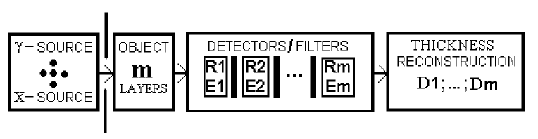
Inspection and technical diagnostics (TD) are based on scanning (linear or planar) and subsequent topography of the three-dimensional structure of the object. It is often needed to carry out quantitative analysis of the internal structure of materials. When the geometry is complex, as well as for systems of variable thickness, multi-layered, multiply connected or multi-component structure, conventional NDT methods (”one-energy”, but non-monochromatic) could be insufficient. The use of more informative and more complex tomographic methods is not always possible due to technical or economical reasons. Important progress can be achieved here in relationship with a multi-energy approach. Radiographic monitoring with separate detection of radiation (in different ranges of the energy spectrum) at several selected energies can give additional information on the internal structure of the studied object. A block diagram of such method is presented in Fig.1.
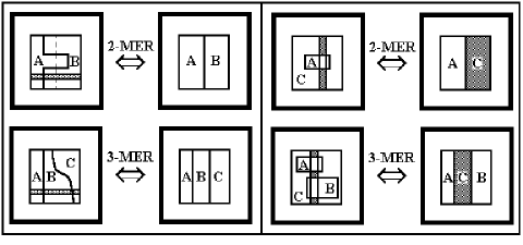
2. In developing of the said aspect of multi-energy radiography (MER), especially efficient are simple schemes of two- and three-energy monitoring. Fig.2 shows characteristic cases of mutual position of simple objects , and as parts of a “complex” object or overlapping in the projection the defects A and B on the main background . In this case, carrying out of structuroscopy (determination of thickness or defectoscopy) is equivalent to solution of the inverse problems for 2- and 3-MER, respectively. It follows from the theory that in the general case the number of reconstructed thicknesses is the same as the multiplicity of radiography, i.e., the number of separately detected radiation ranges). Consequent local scanning of the object allows us to reconstruct the profile of its internal three-dimensional structure also in the case of variable cross-section of the components that form it. To determine thickness of separate components or size of inclusions, one has to assume their chemical composition to be approximately known. This refers to the two parameters that are principal for radiography – effective atomic number and density of each specific material. Or, linear attenuation coefficients should be specified for corresponding substances. For independent determination of these and , it is also possible to use means of MER [3].
3. Theoretical model for thickness reconstruction by means of MER uses the universal character of exponential attenuation of the quantized radiation in monitoring objects and detectors. Passing over to logarithmic (arbitrary) units of the detected signal normalized to the background value (when the object is absent), radiography equations can be presented in a simple form
| (1) |
| (2) |
where are reflexes (registration data) at corresponding maximum absorption energies within each monitoring range. Unknown are thicknesses . Matrix (of linear attenuation coefficients) will be specified, with energy dependencies on photo-effect , Compton scattering and pair generation effect . In the medium energy range up to , the latter scattering channel can be neglected. Solving the linear system is the inverse problem of MER. To obtain its univalent solution and to determine the thicknesses, the number of layers should correspond to the order of multi-energeticity, . The general solution has the form
| (3) |
where is the inverse matrix. In the case of 2-MER, it is convenient to write down explicit formulas
| (4) |
We do not present here the somewhat clumsy expressions for 3-MER case. In the general case, for determination of it is necessary and sufficient that determinant . This implies a physical condition for MER feasibility:
| (5) |
where is the total noise level in the system expressed in energy units. For one-energy radiography, separation of the reconstructed “images” of the composed object by scanning at one camera angle is not possible. This experimentally and theoretically proven fact corresponds to the uncertainty of expressions (4) when their denominator becomes zero at .
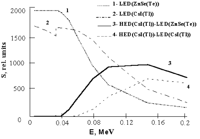
4. For practical developments of MER, an important factor is detector sensitivity of the inspecting system. In the Concern “Institute for Single Crystals”, combined detectors of “scintillator-photodiode” type have been developed, which are characterized by improved sensitivity (contrast and detecting). The two-energy system was realized on the basis of a “sandwich” structure comprising two detectors of “scintillator-photodiode” type. It includes a low-energy detector (LED) based on ZnSe(Te) and a high-energy detector (HED) based on CsI(Tl). Both theoretical calculations and experiments show that such combination is the most efficient for multi-radiographic inspection. In experiments on determination of the detector sensitivity (Fig.3) and output signal (Fig.4), the X-ray source used had anode voltage and current (a tube with a -shaped anode). Sensitivity and output signal was determined in arbitrary units.
In choosing detectors, the following features were accounted for. Scintillator ZnSe(Te) has relatively small atomic number , but its density is high enough to ensure efficient absorption of the ionizing radiation in the low energy region. Light output of ZnSe(Te) can reach 100-130% with respect to CsI(Tl) at absorbing thickness of . As a result, all this ensures substantial advantages of zinc selenide for radiation detection in the range as compared with other scintillators and good filtration of this part of the X-ray radiation spectrum. Our calculations have also shown that optimum thickness values of scintillators for the two-energy radiograph with are: for LED ZnSe(Te) – ; for HED CsI(Tl) – .
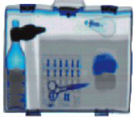
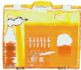
Fig.3 and Fig.4 show results of our measurements of the relative detecting sensitivity and the output signal (reflex) for combined scintielectronic detector arrays of different types. The data obtained confirm advantages of the chosen type and design of the 2-energy inspection system. This physical configuration has been realized in the Polyscan-4 two-energy introscope [4]. Images of a multi-component object obtained using this inspection system are shown in Fig.5. This system also allows distinction between images corresponding to materials with high and low atomic number, e.g., to detect organic materials against the background of inorganics.

5. The developed scheme of multi-radiography can be directly used for different control evaluations, especially in topography of several surimposed “layers”(or defects) or when analysis under different camera angles is impossible. Quantitative determination of thicknesses in a many-component structure makes it possible to physically discern between physically surimposed parts of one and the same piece or object. This substantially increases contrast sensitivity of MER as compared with conventional methods, which is important not only for technology, but also for medical applications (separate diagnostics of soft and bone tissues). Therefore, the proposed radiographic method of multi-energy reconstruction of geometrical structure of the objects can be useful for many applications in the field of NDT and TD. This conclusion is also supported by the already achieved positive results in industrial production of 2- and 3-energy detectors of different types and modifications, e.g., [5, 6].
[1] 15th Word Conference on NDT, Rome (Italy), 15-21 Oct., 2000, Abstracts Book, 800 p.
[2] R.M. Harrison, Nucl. Instr. and Meth. A310, pp. 24-34 (1991).
[3] S.V. Naydenov, V.D. Ryzhikov, Technical Physics Letters, vol. 28, # 5, pp. 357-360 (2002).
[4] The X-Ray Introscopy System of Luggage Customs Control ”Poliscan-4”, Prospects, developed by STC RI & SCB ”Polisvit” PO ”Kommunar”; e-mail: stcri@isc.kharkov.com .
[5] Rapiscan Prospects. USA. - 2002, http://www.rapiscan.com .
[6] Heimann Prospects. Germany. - 2002, http://www.heimannsystems.com .
a)
![[Uncaptioned image]](/html/physics/0206014/assets/x5.png)
b)
 c)
c)