Insulating behavior of -DNA on the micron scale
Abstract
We have investigated the electrical conductivity of -DNA using DNA covalently bonded to Au electrodes. Thiol-modified dTTP was incorporated into the ‘sticky’ ends of bacteriophage -DNA using DNA polymerase. Two-probe measurements on such molecules provide a hard lower bound for the resistivity at bias potentials up to 20 volts, in conflict with recent claims of moderate to high conductivity. By direct imaging, we show that the molecules are present after the measurements. We stress the importance of eliminating salt residues in these measurements.
The question whether DNA is electrically conducting has generated broad interest. The initial spurt of interest arose in photoexcitation experiments which were interpreted in terms of long-range electron transfer barton . In the past few years, there have been upwards of 20 papers reporting the results of more direct electrical measurements ranging from contactless meaurements at microwave frequencies to DC measurements. A distressingly wide range of conductivity values – from to – has been reported fink ; braun ; dekker ; spanish ; DekkerAPL . Proximity-induced superconductivity in DNA has also been claimed superconducting . Recently, local polarization measurements by ‘electrostatic force microscopy’ have been used to show that -DNA is insulating spanish2 ; Sohn . We note, however, that the force-microscopy experiments probe conductivity at relatively weak bias potentials.
In many of the DC measurements, contact with the metal electrodes (usually Au) was achieved by laying down the molecules directly on the electrodes. Although expedient, this approach raises several concerns. It is very difficult to prove that the DNA molecule is in direct physical contact with the electrodes. Even if contact is attained, the weak physical adhesion between DNA and Au may produce an insulating contact and possibly account for the wide variation in reported resistivities sciencecomment . A recent experiment on octanedithiol science has shown that deliberate chemical bonding between organic molecules and metal electrodes is a pre-requisite for achieving reproducible conductivity results. Thus a better approach would be to achieve direct chemical binding between the open ends of -DNA and Au. The bonds should be strong enough to withstand shear forces in a flow, and should survive the measurement process. A second concern is the shunting effect of buffer residue. Because of its finite conductance, the buffer salts which coat the electrodes and substrate produce a spurious conductance signal. Hence adequate salt removal is important. We report the results of experiments performed along these lines. Our results show that -DNA is a good insulator up to bias potentials of 20 volts.
Chemical binding between organic molecules and Au is usually achieved by the Au-thiol (SH) chemical bond mirkin . Commercially available oligonucleotides modified to incorporate the thiol group usually have carbon-chain spacers (C3 or C6) between the thiol group and DNA braun ; DekkerAPL ; glen , which may present barriers to electron transfer. To avoid the spacer problem, we adopted an approach in which the DNA base-pair is bound directly to gold electrodes by a Au-thiol bond. This approach should provide the most direct conductance channel between the gold electrode and the putative electronic “-way” proposed for the DNA helix history .
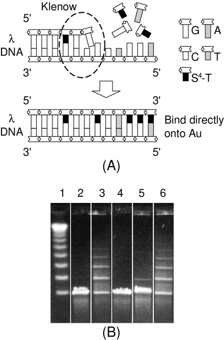
-DNA is a double-stranded DNA helix comprised of 48,502 base pairs (length m). At the extremities, there are single-stranded 12-base 5’ overhangs (‘sticky ends’), with the complementary sequences
where A,C,G,T are the nucleotides adenine, cytosine, guanine and thymine, respectively. Our technique relies on the incorporation of T’s modified to include the desired thiol group modT ; LeziusRao . The ‘sticky’ ends are filled in by a standard reaction chemistry using the Klenow fragment of DNA polymerase and the three deoxynucleoside triphosphates dATP, dGTP, and S4-dTTP (see Fig. 1-A). Because of the preponderance of modified dTTPs (and absence of dCTP) in solution, we can incorporate a significant number of modified T’s at both ends of each DNA molecule mismatch . To prevent the Klenow fragment from excising T’s that are not Watson-Crick matched to the template, we use a mutated form of the Klenow fragment which lacks the 3’5’ proof-reading activity Klenow .
We tested the incorporation of the nucleotides into the DNA ends by a ligation assay ligation . Unmodified -DNA is readily ligated by T4 DNA ligase to form multimers. In the modified -DNA, however, the sticky ends – now filled in by the incorporated bases – are blunt, and multimer formation is strongly suppressed. The reaction products were analyzed by pulsed-field gel electrophoresis. As shown in Fig. 1-B, unmodified -DNA (“natural-”) was efficiently ligated (lane 3). To control for the possibility that unincorporated S4-dTTP inhibited ligation, -DNA monomers were ligated in the presence of all 3 dNTP’s minus the polymerase (“control-”), and the ligation was also efficient (lane 6). The majority of the thiol-modified -DNA (“HS-”), however, remained as monomers (lane 5). This provides strong evidence that the protocol is effective in incorporating bases into the ends of -DNA.
Using standard photolithography, we constructed Au electrodes on a quartz substrate in parallel strips, 4 m wide and 5 mm long, and separated by 4 or 8 m. The Au surfaces were rigorously cleaned cleangold before depositing the modified DNA. At several stages during these experiments, it was important to observe the molecules in an optical microscope. To image the thiol-modified DNA molecules, we stained them with the fluorescent intercalating dye TOTO1 (Molecular Probes), and then loaded them on the chip. After a 20-min. incubation period, many of the molecules were observed to be attached to the electrodes at one end. The unattached molecules were carefully rinsed in TE TE . The chip was then covered with a clean coverslip, and a flow of the buffer solution was applied perpendicular to the electrodes. We observed that DNA molecules anchored at one end were stretched by the buffer flow to bridge the space between the electrodes. Many of these molecules subsequently attached to the second electrode by their free end. After this occurs, the flow may be repeatedly reversed to demonstrate that the anchored DNA molecules bow out with the flow while their ends remain anchored (Panels A and B of Fig. 2. See video in Ref. video ). This is direct evidence that chemical binding between the ends to Au is much stronger than physical adhesion of the rest of the molecule to either quartz or Au. (For the specific DNA samples used in the resistivity measurements, we carried out the dye-staining step after the measurements to avoid inadvertent damage from dye intercalation.)
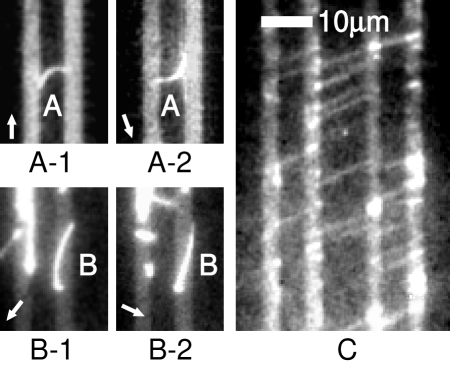
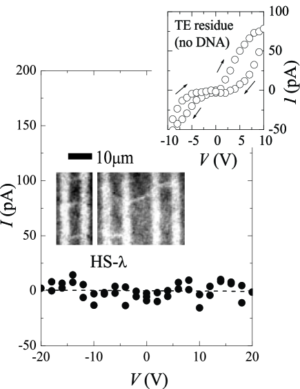
As discussed above, a crucial step in the experiment was the removal of buffer. In preliminary experiments, we repeatedly observed a finite, semiconductor-like, history-dependent conductance after loading DNA solution and removing the buffer solution. The inset of Fig. 3 is a representative - curve of the buffer salt residue, 1TE. Such spurious signals are of particular concern when DNA is laid down on electrodes in a thick bundle, because the salts trapped between the DNA molecules can form conduction paths. The spurious background vanished after we adopted the following procedure. Chips containing bridging DNA molecules were carefully rinsed in 5 mM ammonium acetate (NH4Ac, pH 6.6), a volatile buffer that can be completely removed in high vacuum. A drawback of this rinsing is that a large fraction of the anchored DNA molecules are cut after rinsing and drying. However, rinsing in 10 mM MgSO4/40 mM Tris-HCl (pH 8) before the NH4Ac rinsing introduces Mg2+ ions which coat the quartz surface with weak positive charges DekkerAPL . As the negatively charged DNA molecules stick to the substrate by electrostatic interaction, damage due to NH4Ac rinsing is minimized. Panel C of Fig. 2 shows a typical image of anchored DNA after Mg2+ and NH4Ac rinsing. [Rinsing with the MgSO4 solution also led to binding of unmodified -DNA. However, the yield of anchored molecules was much smaller.]
To perform the electrical measurements, unstained thiol-modified -DNA was attached to the Au electrodes as described above. After the final NH4Ac rinsing, the chip was dried in the dark to avoid possible photon-induced damage. Two-probe - measurements were performed in moderately high vacuum ().
A typical room-temperature - curve, measured on -DNA spanning electrodes 4 m apart, is shown in the main panel of Fig.3. The voltage was swept between 20 V. A linear fit to the data in Fig. 3 yields . Using a cross-section of 3 nm2 per molecule, and the estimated number of bridging molecules (1000), we obtain the bound on the resistivity of in electric fields up to . Measurements performed on several chips yielded consistent results. No current was detected within the noise level of our measurement(10 pA), despite sustained and deliberate efforts to improve electrical contacts between the base pair stack of -DNA and Au.
Immediately after the measurements, a buffer solution with an appropriate amount of TOTO1 dye dissolved in 1TE was loaded on the chip. By direct optical microscopy inspection of the post-stained DNA, we confirmed that there were 1000 DNA molecules bridging the electrodes, and thus the measurements did not destroy the DNA.
As a final check that the observed images are those of -DNA, we introduce DNase to digest the molecules DNase . Complete deletion of all fluorescent DNA molecules was observed. These tests leave very little room for doubt that a large number of intact DNA molecules were chemically bound to the electrodes during the electrical measurements denature .
The bound cm in our experiment and the large bias potential applied (20 V) is at odds with many recent reports of moderately high conductivity. In some DC experiments, the DNA molecules formed bundles or networks between the microfabricated electrodes ZnDNA ; DNAnetwork ; dyeDNA . As noted above, high conductance may arise from residual salts trapped between the DNA strands. Contamination from other sources (C or Re) may be a problem as well in the experiment on proximity-induced superconductivity in -DNA superconducting . Microwave absorption experiments have been used to infer that at 295 K in -DNA gruner . The high microwave conductivity, 106 times larger than our bound, is very difficult to reconcile with our data. If -DNA had such a high uniform conductivity, all the applied potential should fall across the contacts (2-3 nm) to produce an -field V/cm, high enough to produce a large tunneling current, if not breakdown of the contact barrier altogether. This is not observed. Possibly, the microwave is detecting very short dissipative regions embedded in the insulating molecule.
Two groups recently used electrostatic force microscopy to probe the electrostatic polarization of DNA spanish2 ; Sohn . Our results are consistent with their conclusion that DNA is insulating. However, the electrostatic force microscopy technique probes conductivity in the limit of weak bias potentials, so it does not rule out a transition to moderately large conductivity above a bias threshold of several volts (as reported in some experiments dekker ; Watanabe ). The present experiments show that insulating behavior extends to bias potentials as high as 20 volts.
We are grateful to P. M. Chaikin for invaluable suggestions, and acknowledge fruitful discussions with Shirley S. Chan, J. Tegenfeldt, P. Silberzan, Christelle Prinz, S. Park and R. Huang. This research is supported by a U.S. National Science Foundation MRSEC grant (DMR 98-09483) and by the National Inst. Health (NIH HG01506).
References
- (1) C. J. Murphy et al., Science 262, 1025 (1993); S. O. Kelley and J. K. Barton, Science 283, 375 (1999).
- (2) H.-W. Fink and C. Schonenberger, Nature 398, 407 (1999).
- (3) E. Braun et al., Nature 391, 775 (1998).
- (4) D. Porath, et al., Nature 403, 635 (2000).
- (5) P. J. de Pablo et al., Phys. Rev. Lett. 85, 4992 (2000).
- (6) A. J. Storm et al., Appl. Phys. Lett. 79, 3881 (2001).
- (7) A. Yu. Kasumov et al., Science 291, 280 (2001).
- (8) C. Gómez-Navarro et al., Proc. Natl. Acad. Sci. USA 99, 8484 (2002).
- (9) M. Bockrath et al., Nano Lett. 2, 187 (2002).
- (10) K. W. Hipps, Science 294, 536 (2001).
- (11) X. D. Cui et al., Science 294, 571 (2001).
- (12) C. A. Mirkin et al., Nature 382, 607 (1996).
- (13) User Guide to DNA Modification, Glen Research, 1999. See the refs. therein.
- (14) D. D. Eley and D. I. Spivey, Trans. Faraday Soc. 58, 411 (1962).
- (15) In the modified nucleotide 4-thiothymidine-5’-triphosphate (S4-dTTP, Trilink Biotechnologies), a sulfur atom is covalently bound to the C4 site of the thymine molecule. Its close proximity to the hydrogen ion at the N3 site results in a resonant state that forms the desired thiol group.
- (16) A. G. Lezius and K. H. Scheit, European J. Biochem. 3, 85 (1967); T. V. S. Rao et al., Bioorg. Med. Chem. Lett. 10, 907 (2000).
- (17) The incorporation reaction was 100 g/ml -DNA, 0.1 mM dATP, 0.1 mM dGTP and 0.2 mM S4-dTTP in 1 EcoPol buffer(10 mM Tris-HCl, 5 mM MgCl2 and 7.5 mM dithiothreitol (DTT); pH7.5 @ 25∘C; New England Biolabs), for a total volume of 100 l. Five units of Klenow fragment (3’5’ exo-) were added. The mixture was incubated at 37 ∘C for 30 min. To stop the reaction, 1l of 0.5 M EDTA was added, and the mixture heated at 75∘C for 20 min. To exchange the buffer, the product was dialyzed against 30 ml 0.1TE at room temperature for 45 min across a 50 nm Millipore membrane.
- (18) One 5’ overhang of the -DNA ends with an A, which is Watson-Crick matched to S4-T. The other overhang ends with G’s, which are matched to C’s. While sulfur-modified dCTP is available, the sulphur is remote from H atoms and the resonant thiol group can not form. To maximize the probability of modified T’s at both ends, we excluded dCTP altogether from the reaction, so that S4-T:G pairing could occur in place of C:G. The mutated Klenow fragment used here is incapable of detecting the mis-matches.
- (19) The Klenow fragment is a large fragment of DNA polymerase I lacking the error-correcting 5’3’ exonuclease activity (this function damages the -DNA ‘sticky’ ends). Here, we employed a mutated form of the Klenow fragment (3’5’ exo-, New England Biolabs) in which the proof-reading 3’5’ exonuclease activity is deleted. S4-dTTP binds more weakly to A than unmodified dTTP LeziusRao , and hence the proof-reading activity could possibly remove incorporated S4-dTTP.
- (20) The reaction was carried out in 1 ligation buffer (50 mM Tris-HCl, 10 mM MgCl2, 10 mM DTT, 1 mM ATP and 25 g/ml BSA; pH 7.5@25 ∘C) at a -DNA concentration of 50 g/ml. 400 units of T4 DNA ligase (New England Biolabs) was added to the reaction volume of 20 l, and incubated at room temperature for 5 h.
- (21) 1% agarose gel in 0.5TBE buffer; =6 V/cm; 14 ∘C; 16 hours; switch time ramped from 0.1 sec to 40 sec.
- (22) To clean the Au surface, freshly prepared chips were soaked in fuming HNO3 for 30 min, and further cleaned in a mixture of 18 M deionized water, ammonium hydroxide and hydrogen peroxide (5:1:1 by volume, 70 ∘C) for 20 min. The chips were thoroughly rinsed in deionized and distilled water (DD H2O), and kept in Ar-flushed DD H2O for no more than 30 min before loading DNA samples. The Au surface was never allowed to dry during the process.
- (23) 1TE buffer contains 10 mM Tris (tris(hydroxymethyl)-aminomethane) and 1 mM EDTA (ethylene diamine tetra acetic acid), pH 8.0 .
- (24) See videos at http://suiling.princeton.edu/research/ DNAconductance/DNAconduction.html.
- (25) The strong fluorescence observed after the resistivity measurements strongly argues against denaturation of DNA into single strands when affixed to quartz in vacuum. TOTO1 displays strong fluorescence only when bound to double-strand DNA. Moreover, AFM images of B-DNA in vacuum have recently been obtained by Takayuki Uchihashi et al., Langmuir 16, 1349 (2000); ibid Jpn. J. Appl. Phys. 39, L887 (2000).
- (26) A. Rakitin et al., Phys. Rev. Lett. 86, 3670 (2001).
- (27) L. Cai, H. Tabata, and T. Kawai, Appl. Phys. Lett. 77, 3105 (2000).
- (28) J. Gu et al., Appl. Phys. Lett. 80, 688 (2001).
- (29) To digest the DNA, we used 10 units of DNase I (RNase-free, Roche Molecular Biochemicals) dissolved in 10 l reaction buffer (40 mM Tris-HCl, 10 mM MgSO4, and 1mM CaCl2).
- (30) P. Tran, B. Alavi, and G. Gruner, Phys. Rev. Lett. 85, 1564 (2000).
- (31) H. Watanabe et al., Appl. Phys. Lett. 79, 2462 (2001).