DNA Dynamics in A Water Drop
Abstract
Due to its polyionic character the DNA double helix is stable and biologically active only in salty aqueous media where its charge is compensated by solvent counterions. Monovalent metal ions are ubiquitous in DNA environment and they are usually considered as the possible driving force of sequence-dependent modulations of DNA structure that make it recognizable by proteins. In an effort to directly examine this hypothesis, MD simulations of DNA in a water drop surrounded by vacuum were carried out, which relieves the requirement of charge neutrality. Surprisingly, with zero concentration of counterions a dodecamer DNA duplex appears metastable and its structure remains similar to that observed in experiments.
pacs:
87.14.Gg; 87.15.Aa; 87.15.HeIt has been long recognized that, because of the polyionic nature of DNA, solvent counterions are required for its stability, and that gross structural changes in DNA can be provoked by changing the concentration, the charge or the type of counterions Saenger (1984). More recently it has been proposed that free ions might also act as biological regulators because, by binding to DNA, they can provoke conformational deformations recognized by specific proteins Jayaram and Beveridge (1996); Young et al. (1997a, b); Hud and Feigon (1997); Rouzina and Bloomfield (1998); Williams and Maher (2000). The most controversial is the role of the common monovalent cations Na+ and K+. They are ubiquitous in the DNA environment and can be readily available for any purpose. Until recently, they remained invisible in experimental DNA structures because it is difficult to detect them in water, and it has been suggested that they are perhaps responsible for the most widespread deformations of the double helix, namely, narrowing of the minor groove and bending. It is assumed that counterions are sequestered in the minor groove of some sequences, which brakes the symmetry of the repulsive electrostatic forces and provokes deformations. This model is general and it easily explains other puzzling effects in DNA structure.
The above hypothesis is supported by many recent studies. Penetration of monovalent cations into the minor DNA groove has been confirmed by X-ray diffraction Rosenberg et al. (1973); Bartenev et al. (1983); Tereshko et al. (1999); Sines et al. (2000), NMR spectroscopy Hud et al. (1999); Denisov and Halle (2000), and MD simulations Young et al. (1997a); Feig and Pettitt (1999); Strahs and Schlick (2000); Štefl and Koča (2000); Hamelberg et al. (2001). However, it appears difficult to find a discriminating set-up for testing the cause and consequence relationship between the ions and the DNA structure. All available evidences have more than one interpretation making this problem highly controversial McFail-Isom et al. (1999); Chiu et al. (1999); McConnell and Beverdige (2000). For example, correlations between ion positions and the local groove width observed in MD cannot answer whether the ions perturb DNA or they just bind “opportunistically” in the sites of low potential near already deformed double helix Hamelberg et al. (2001). To clarify the issue of cause and effect one would have to remove solvent ions and check if the supposed counterion effects disappear with them. Unfortunately, a counterion-free DNA does not exist in nature whereas the most reliable computational procedures presently employed require that the simulation cell that holds DNA carries zero net charge. Therefore, in both simulations and experiments the counterion effects cannot be completely eliminated.
In an effort to directly address this question I have adapted the Particle Mesh Ewald algorithm Darden et al. (1993); Essmann et al. (1995) for modeling dynamics of DNA in a water drop surrounded by vacuum. Particle-mesh calculations with vacuum boundary conditions are long known in physics Hockney and Eastwood (1981), but, to my knowledge, they were never applied to chemical or biological systems. Free vacuum boundaries are intuitively most simple and they allow one to relieve the problems of charge neutrality and possible artifacts from interactions between periodical images. I describe here the first such “naive” all atom simulations of DNA in water, with unperturbed Coulomb electrostatics. It appears that, with zero concentration of counterions, a dodecamer DNA duplex is metastable and its structure remains similar to that observed in experiments.
The Dickerson-Drew dodecamer (CGCGAATTCGCG, Wing et al. (1980)) in a canonical B-DNA conformation Arnott and Hukins (1972) is surrounded by a spherical drop of 4000 TIP3P water molecules Jorgensen et al. (1983). Initially, the drop had around 50 Å in diameter and in dynamics it remained roughly spherical. A rectangular unit cell is constructed around the drop with the minimal separation of 25 Å between the water molecules and the cell sides. The cell is replicated periodically, which gives an infinite lattice of water drops with at least 50 Å spacing between the closest neighbors. A shifted Coulomb’s law is used, with for and for longer distances, where Å, which eliminates any interactions between periodical images. Because this shifting does not affect the forces, the system behaves in dynamics as if surrounded by infinite vacuum. Within the drop the electrostatics are effectively evaluated with a cut-off of 50 Å, which is larger than the DNA size and applies only to a small fraction water molecules at opposite poles of the drop. Larger distances were also tested, but showed no noticeable difference. The Van-der-Waals interactions are computed with a conventional cut-off of 9 Å.
| Xdisp | Inclin | Rise | Twist | RMSD-A111Heavy atom root mean square deviation from the corresponding canonical DNA form. | RMSD-B111Heavy atom root mean square deviation from the corresponding canonical DNA form. | |
| A-DNA | -5.4 | +19.1 | 2.6 | 32.7 | 0.0 | 6.2 |
| B-DNA | -0.7 | -6.0 | 3.4 | 36.0 | 6.2 | 0.0 |
| Tj1222Water drop calculation without counterions. | -2.2 | +6.9 | 3.3 | 33.8 | 4.52 | 2.36 |
| Tj2333Water drop neutralized by 22 ions. | -2.0 | +6.5 | 3.2 | 34.1 | 4.57 | 2.18 |
| Tj3444Conventional PME calculation with counterions and periodical boundaries. | -2.1 | +6.9 | 3.3 | 33.6 | 4.46 | 2.41 |
The shifted Coulomb interactions are evaluated by using the SPME method Essmann et al. (1995). The shifting is taken into account in the reciprocal sum by replacing the Fourier transform of with that of the shifted law. The resulting series is absolutely convergent regardless of the system charge. The values of energies and forces thus obtained have been verified by direct computation without cut-offs. Modifications in the direct sum are not necessary if we are only interested in the forces and not in the absolute energy values. It can be shown that this simple procedure is essentially equivalent to mathematically more complex earlier formulations of the Ewald method for cluster calculations Hockney and Eastwood (1981); Martyna and Tuckerman (1999). In dynamics, the shape of the drop fluctuates and the size of the unit cell is adjusted accordingly at every time step. Evaporated water molecules are detected and excluded from calculations to prevent explosion in the cell size. After every 50 ps the calculation is stopped and water molecules that have left the drop are re-introduced with zero velocities by scattering them randomly near the surface of the drop. The rate of evaporation was around 84 mol/ns, that is on average four molecules had to be re-introduced at each stop.
All calculations were carried out with AMBER98 parameters Cornell et al. (1995); Cheatham et al. (1999) by using the ICMD method Mazur (1997, 1999a, 2001a) with the time step of 0.01 ps. Two control simulations of the same dodecamer have been carried out for comparison. In the first, 22 ions were included in the same water drop. The second represents a conventional calculation in a rectangular unit cell including DNA, 3901 water molecules, and 22 ions by the original SPME method with periodical boundary conditions. The trajectories were continued to 5 ns.
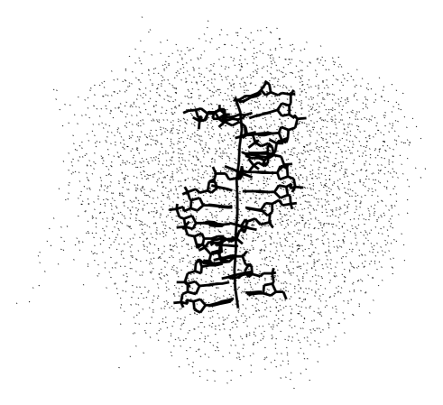
Figure 1 shows a snapshot of the charged drop from the last nanosecond of the trajectory. The system appears metastable in the nanosecond time range. The DNA molecule shows no signs of denaturation. The water media remains continuous without internal bubbles. The surface of the drop is covered by spiky “protuberances” which reduce with increased drop size and disappear if DNA is neutralized by counterions. The DNA structures averaged over the last 2.5 ns of the three trajectories are characterized in Table 1. These data indicate that with zero counterion concentration this dodecamer DNA molecule remains in B-form. It is understood that with increased DNA length it should have exploded because otherwise the electrostatic energy would go to infinity. However, no such trend is seen in Table 1, which indicates that critical lengths of catastrophic deformations are much larger whereas the dodecamer B-form is metastable in water even if it is charged.
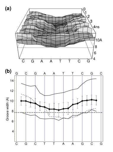
(b) The minor groove profile averaged over the last nanosecond of Tj1. The central trace represents the average groove width with rms fluctuations shown as error bars. The upper and lower solid traces show the maximal and minimal values, respectively. The dotted trace exhibits the profile observed in the experimental X-ray structure Wing et al. (1980). The canonical B-DNA groove width is marked by the horizontal dashed line. Despite the narrowing, the groove width remains larger than the canonical value Mazur (1999b), which corresponds to the lower average twist.
The scales of differences between the DNA conformations in the three trajectories are well characterized by the data in Table 1 and they were always far from statistically significant. The corresponding experimental data are available only for the twist, namely, its value drops by 0.1∘ when the NaCl concentration is reduced from 0.3M to 0.05M Anderson and Bauer (1978). A shift by 0.1∘ in the average twist is too small to be detected in a 5 ns simulation because, for this relatively small molecule, its fluctuations between consecutive 1 ns averaged structures can reach 1.0∘. The absolute twist value is lower than in experiment, which is a known general feature earlier discussed in the literature Cheatham et al. (1999).
The most famous feature of this DNA molecule is the middle AATT fragment. It is long known from experiments that the minor DNA groove always narrows in this and some similar sequences, called A-tracts, and widens outside of them. Figure 2 exhibits dynamics of the minor groove in Tj1 and its average profile over the last nanosecond. It has a characteristic waving shape with a narrowing in the middle. The amplitude of this modulation is similar to that in the experimental X-ray structure. The minimal width is 1.5 Å larger than experimental value, which is probably linked mechanically to the lower average twist. Similar results were obtained for the other two trajectories and they are close to earlier reported simulation studies carried out with non-zero counterion concentrations Young et al. (1997b).
The sequence-dependent groove-width modulations in DNA are well established experimentally and, in the recent years, they have been proposed to result from interactions with bound monovalent metal ions commonly undetectable in X-ray crystal maps Jayaram and Beveridge (1996); Williams and Maher (2000); Hamelberg et al. (2001). The present results evidence that it is not the case, supporting recent conclusions of different groups Denisov and Halle (2000); McConnell and Beverdige (2000); Chiu et al. (1999). They explain also why groove modulations and intrinsic DNA bending could be reproduced in MD simulations with simplified treatment of electrostatic interactions that ignored specific counterion effects Mazur (1998, 2000, 2001b).
According to the counterion condensation theory, DNA in aqueous environment should be always covered by a shell of counterions and its charge should be compensated by around 75% regardless of the bulk ion concentrations Manning (1978). The results presented here do not contradict this theory but they are somewhat at odds with an implicit assumption that the counterion cloud is critical for the native DNA structure. This was surprising, at least for the author, and suggests that we are still far from complete understanding of interactions that control the DNA structure. The correlations observed in earlier MD simulationsHamelberg et al. (2001) apparently are due to binding of counterions in sites of low potential near an already narrowed minor groove, therefore, these interactions are structure-specific rather than sequence-specific, and they cannot be the driving force of the corresponding DNA deformations.
References
- Saenger (1984) W. Saenger, Principles of Nucleic Acid Structure (Springer Verlag, New York, 1984).
- Jayaram and Beveridge (1996) B. Jayaram and D. L. Beveridge, Annu. Rev. Biophys. Biomol. Struct. 25, 367 (1996).
- Young et al. (1997a) M. A. Young, B. Jayaram, and D. L. Beveridge, J. Am. Chem. Soc. 119, 59 (1997a).
- Williams and Maher (2000) L. D. Williams and L. J. Maher, III, Annu. Rev. Biophys. Biomol. Struct. 29, 497 (2000).
- Young et al. (1997b) M. A. Young, G. Ravishanker, and D. L. Beveridge, Biophys. J. 73, 2313 (1997b).
- Hud and Feigon (1997) N. V. Hud and J. Feigon, J. Am. Chem. Soc. 119, 5756 (1997).
- Rouzina and Bloomfield (1998) I. Rouzina and V. A. Bloomfield, Biophys. J. 74, 3152 (1998).
- Rosenberg et al. (1973) J. M. Rosenberg, N. C. Seeman, J. J. P. Kim, F. L. Suddath, H. B. Nicholas, and A. Rich, Nature 243, 150 (1973).
- Bartenev et al. (1983) V. N. Bartenev, E. I. Golovanov, K. A. Kapitonova, M. A. Mokulskii, L. I. Volkova, and I. Y. Skuratovskii, J. Mol. Biol. 169, 217 (1983).
- Tereshko et al. (1999) V. Tereshko, G. Minasov, and M. Egli, J. Am. Chem. Soc. 121, 3590 (1999).
- Sines et al. (2000) C. C. Sines, L. McFail-Isom, S. B. Howerton, D. VanDerveer, and L. D. Williams, J. Am. Chem. Soc. 122, 11048 (2000).
- Hud et al. (1999) N. V. Hud, V. Sklenár̆, and J. Feigon, J. Mol. Biol. 286, 651 (1999).
- Denisov and Halle (2000) V. P. Denisov and B. Halle, Proc. Natl. Acad. Sci. USA 97, 629 (2000).
- Feig and Pettitt (1999) M. Feig and B. M. Pettitt, Biophys. J. 77, 1769 (1999).
- Strahs and Schlick (2000) D. Strahs and T. Schlick, J. Mol. Biol. 301, 643 (2000).
- Štefl and Koča (2000) R. Štefl and J. Koča, J. Am. Chem. Soc. 122, 5025 (2000).
- Hamelberg et al. (2001) D. Hamelberg, L. D. Williams, and W. D. Wilson, J. Am. Chem. Soc. 32, 7745 (2001).
- McFail-Isom et al. (1999) L. McFail-Isom, C. C. Sines, and L. D. Williams, Curr. Opin. Struct. Biol. 9, 298 (1999).
- Chiu et al. (1999) T. K. Chiu, M. Zaczor-Grzeskowiak, and R. E. Dickerson, J. Mol. Biol. 292, 589 (1999).
- McConnell and Beverdige (2000) K. J. McConnell and D. L. Beverdige, J. Mol. Biol. 304, 803 (2000).
- Darden et al. (1993) T. Darden, D. York, and L. Pedersen, J. Chem. Phys. 98, 10089 (1993).
- Essmann et al. (1995) U. Essmann, L. Perera, M. L. Berkowitz, T. Darden, H. Lee, and L. G. Pedersen, J. Chem. Phys. 103, 8577 (1995).
- Hockney and Eastwood (1981) R. W. Hockney and J. W. Eastwood, Computer Simulation Using Particles (McGraw-Hill, New-York, 1981).
- Wing et al. (1980) R. Wing, H. Drew, T. Takano, C. Broka, S. Tanaka, K. Itakura, and R. E. Dickerson, Nature 287, 755 (1980).
- Arnott and Hukins (1972) S. Arnott and D. W. L. Hukins, Biochem. Biophys. Res. Communs. 47, 1504 (1972).
- Jorgensen et al. (1983) W. L. Jorgensen, J. Chandreskhar, J. D. Madura, R. W. Impey, and M. L. Klein, J. Chem. Phys 79, 926 (1983).
- Lavery and Sklenar (1988) R. Lavery and H. Sklenar, J. Biomol. Struct. Dyn. 6, 63 (1988).
- Martyna and Tuckerman (1999) G. J. Martyna and M. E. Tuckerman, J. Chem. Phys. 110, 2810 (1999).
- Cheatham et al. (1999) T. E. Cheatham, III, P. Cieplak, and P. A. Kollman, J. Biomol. Struct. Dyn. 16, 845 (1999).
- Cornell et al. (1995) W. D. Cornell, P. Cieplak, C. I. Bayly, I. R. Gould, K. M. Merz, D. M. Ferguson, D. C. Spellmeyer, T. Fox, J. W. Caldwell, and P. A. Kollman, J. Am. Chem. Soc. 117, 5179 (1995).
- Mazur (1997) A. K. Mazur, J. Comput. Chem. 18, 1354 (1997).
- Mazur (1999a) A. K. Mazur, J. Chem. Phys. 111, 1407 (1999a).
- Mazur (2001a) A. K. Mazur, in Computational Biochemistry and Biophysics, edited by O. M. Becker, A. D. MacKerell, Jr, B. Roux, and M. Watanabe (Marcel Dekker, New York, 2001a), pp. 115–131.
- Mazur (1999b) A. K. Mazur, J. Mol. Biol. 290, 373 (1999b).
- Anderson and Bauer (1978) P. Anderson and W. Bauer, Biochemistry 17, 594 (1978).
- Mazur (1998) A. K. Mazur, J. Am. Chem. Soc. 120, 10928 (1998).
- Mazur (2000) A. K. Mazur, J. Am. Chem. Soc. 122, 12778 (2000).
- Mazur (2001b) A. K. Mazur, J. Comput. Chem. 22, 457 (2001b).
- Manning (1978) G. S. Manning, Q. Rev. Biophys. 2, 179 (1978).