Evolutionary conservation of the folding nucleus
Abstract
In this Communication we present statistical analysis of conservation profiles in families of homologous sequences for nine proteins whose folding nucleus was determined by protein engineering methods. We show that in all but one protein (AcP) folding nucleus residues are significantly more conserved than the rest of the protein. Two aspects of our study are especially important: 1) grouping of amino acids into classes according to their physical-chemical properties and 2) proper normalization of amino acid probabilities that reflects the fact that evolutionary pressure to conserve some amino acid types may itself affect concentration of various amino acid types in protein families. Neglect of any of those two factors may make physical and biological “signals” from conservation profiles disappear.
Running title: Conservation of folding nucleus
Submitted to Journal of Molecular Biology
Harvard University, Department of
Chemistry and Chemical Biology
12 Oxford Street, Cambridge MA 02138
E-mail: leonid@origami.harvard.edu, eugene@belok.harvard.edu
http://paradox.harvard.edu/~leonid
Introduction
It is now widely accepted that folding of small single-domain proteins follows “nucleation-condensation” mechanism [Abkevich et al., 1994, Itzhaki et al., 1995, Fersht, 1997, Shakhnovich, 1997, Guo & Thirumalai, 1995, Pande et al., 1998] whereby relatively small fragment of protein structure is formed in the transition state between unfolded and folded states. Residues belonging to this fragment constitute specific folding nucleus (SFN). Considerable experimental [Itzhaki et al., 1995, Main et al., 1999, Martinez et al., 1998, Chiti et al., 1999] and theoretical [Abkevich et al., 1994, Klimov & Thirumalai, 1998, Li et al., 2000, Dokholyan et al., 2000] effort has been devoted to identification of folding nuclei in real proteins and various models as well as factors that determine its location in structure and in sequence.
One of the most intriguing aspect of nucleation-condensation mechanism of protein folding is its relation to protein evolution. Indeed residues constituting folding nucleus can be metaphorically considered “accelerator pedals” of folding [Mirny et al., 1998a] since mutations in those positions affect folding rate to a much greater extent than elsewhere in a protein. One can conclude that if there is evolutionary control of folding rate it should have resulted in additional pressure applied on folding nucleus residues, and such pressure can be manifested in noticeable additional conservation of nucleus residues.
This idea was first proposed in [Shakhnovich et al., 1996] where it was applied to prediction of nucleus residues from protein structure. Many sequences were designed to fit the structure of Chymotripsin Inhibitor 2 (CI2) with low energy. Positions conserved among the designed sequences were identified as a putative nucleus. This way blind predictions of folding nucleus in CI2 were made that were verified in independent experiments [Itzhaki et al., 1995].
In related studies papers Ptitsyn studied conservatism in distant yet related by sequence homology members of Cytochrome C [Ptitsyn, 1998] and myoglobin [Ptitsyn & Ting, 1999] families. In both cases he found conserved clusters of residues without an obvious functional role which he suggested to belong to folding nucleus of those proteins. Michnick and Shakhnovich [Michnick & Shakhnovich, 1998] carried out an analysis of conservation in natural and designed sequences for families of three structurally related proteins - ubiquitin, raf and ferredoxin and predicted possible folding nucleus for those proteins.
Neverteheless the notion of folding nucleus conservation has drawn some controvercy in the lietrature. While earlier papers [Shakhnovich et al., 1996, Michnick & Shakhnovich, 1998, Ptitsyn, 1998, Ptitsyn & Ting, 1999] suggested conservation of folding nucleus in some proteins, a more recent paper by Plaxco and coauthors [Plaxco et al., 2000] argued to the opposite. These authors looked at conservatism profile in several protein families for which protein engineering analysis of folding transition states has been carried out, and did not observe correlation between conservation and experimentally measured -values. This made them conclude that there is no evolutionary pressure to control the folding rates.
In this work we study evolutionary conservation of the folding nucleus for several homologous proteins. Conservation of the folding nucleus is systematically compared with the conservation in the rest of the protein sequence. In contrast to previous studies, we perform rigorous statistical test to assess significance of higher conservation in the folding nucleus. The main result of this study is that for all studied proteins, except AcP, folding nucleus is significantly more conserved than the rest of the protein. We explain the difference between our thorough statistical analysis and that of Plaxco et al [Plaxco et al., 2000] by pointing out to some technical shortcomings in the earlier work [Plaxco et al., 2000].
Results and Discussion
To study evolutionary conservation of the folding nucleus we turn to nine proteins for which nucleus has been experimentally identified from protein engineering analysis: CI2, FKBP12, ACBP, CheY, Tenascin, CD2.d1, U1A, AcP and ADA2h. For each of them we obtain a multiple sequence alignment from HSSP database [Dodge et al., 1998] (or PFAM [Bateman et al., 2000] database if HSSP contains too few sequences). We compute variability at position of the alignment as
| (1) |
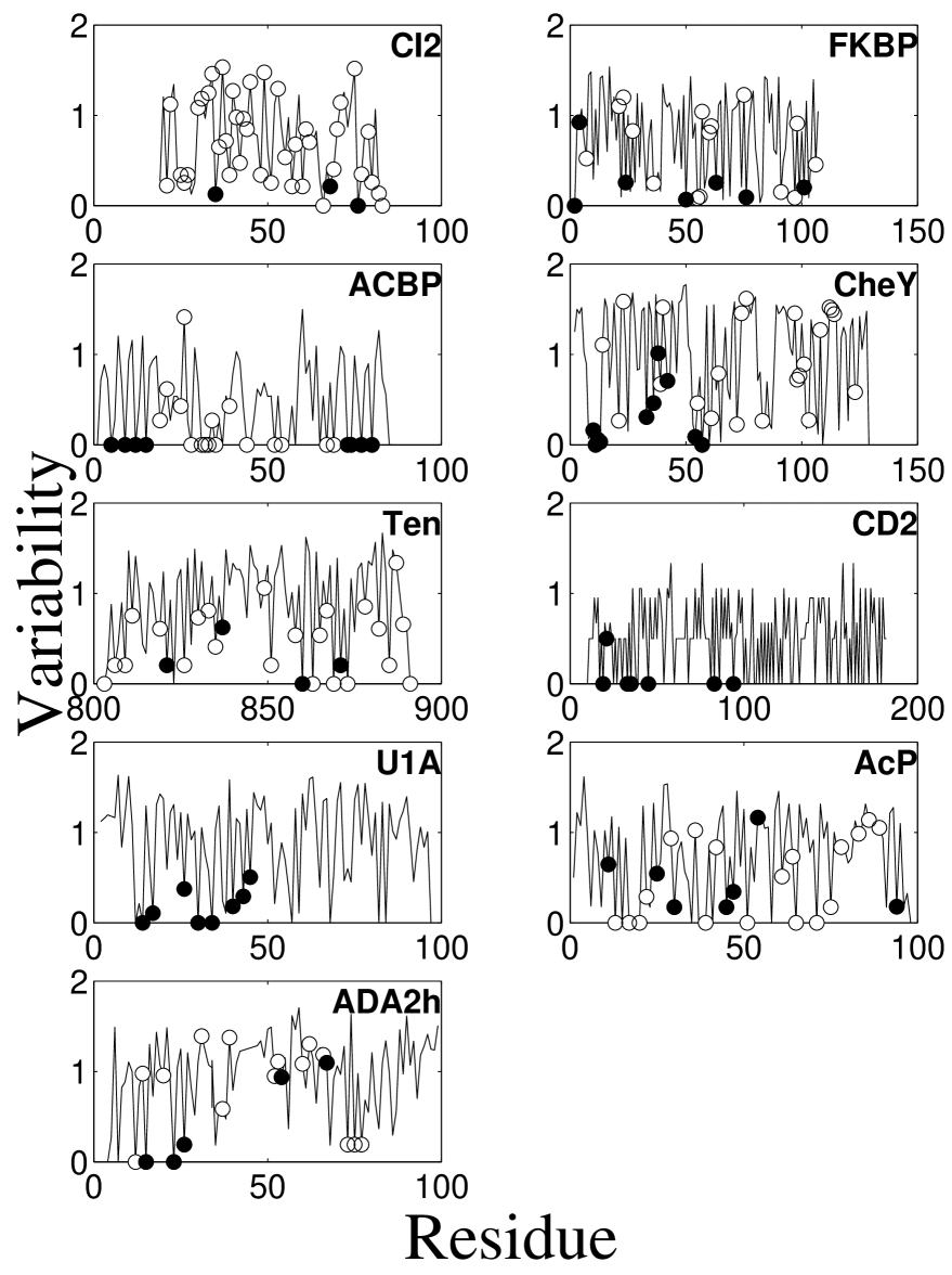
where is the frequency of residues from class in position . We use six classes of residues to reflect physical-chemical properties of amino acids and their natural pattern of substitutions: aliphatic [A V L I M C], aromatic [F W Y H], polar [S T N Q], basic [K R], acidic [D E], and special (reflecting their special conformational properties) [G P]. As a result of this classification mutations within a class are ignored (e.g. ), while mutations that change the class are taken into account. Figure 1 presents variability profile for studied proteins with nucleation positions marked by filled circles. ††margin: Fig.1 Importantly, we defined the folding nucleus as it was identified by the original experimental groups (Table 1).
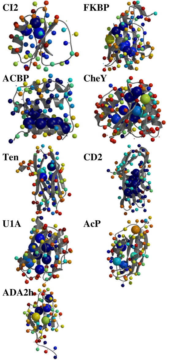
Figure 2 ††margin: Fig.2 clearly shows that nucleus residues are almost always among the most conserved ones for all studied proteins. It also shows that nucleus residues are not the only conserved ones: many other residues (predominantly in the cores of the proteins) are also conserved.
In order to evaluate statistical significance of nucleus conservation we compare evolutionary conservation of the folding nucleus with the conservation of all residues in the protein using the following statistical test. We start from the null hypothesis H0 that nucleus residues are no more conserved than the whole protein sequence. To test this hypothesis we compute median variability of the nucleus residues () and compare it with the distribution of medians variability of the same number of residues randomly chosen in the same protein (). The distribution is obtained by choosing random sets of residues ( is the number of residues in the nucleus). Then the fraction of instances with gives the probability of accepting H0. In other words, is the probability that observed lower variability of the folding nucleus is obtained by chance. Hence, indicates statistically significant strong evolutionary conservation of the folding nucleus. Below we use confidence level .
Table 2 presents computed values. The main result of this work is that in all proteins, except AcP, residues in the folding nucleus are significantly more conserved than the rest of the protein.
Next we study how obtained results depend on the way amino acids are grouped into classes (see Table 2). When classification scheme from [Branden & Tooze, 1998] (BT) is used, still all proteins except AcP exhibit significant conservation of the folding nucleus. This clearly demonstrates that observed conservation of the folding nucleus is not a consequence of a particular choice of the classification scheme.
However, when amino acids are not grouped into classes, nucleus exhibits significant conservation only in four out of nine proteins. Taken together these results indicate that substitutions in the folding nucleus may occur, but they are limited to residues that belong to the same class (i.e. have similar physical-chemical properties [Thompson & Goldstein, 1996]).
To study what physical-chemical properties are conserved in the folding nucleus we used various classification schemes. Starting from all 20 amino acids, we grouped some of them into classes and repeated the analysis, including the statistical tests (see Table 2). The goal is to find a minimal classification (i.e. grouping the minimal number of amino acids together) that provides statistically significant conservation of the folding nucleus. Our results show that classification where only I, L, and V are grouped in one class while all other amino acids each represent their own class satisfies this requirement (see Table 2).This classification provides significant conservation of the nucleus for all proteins except AcP with , and for all proteins except AcP and FKBP12 with . This result demonstrates that are the most common substitutions in the nucleus (and in the protein core in general [Henikoff & Henikoff, 1992, Benner et al., 1994]). These substitutions are tolerated in the nucleus as they do not change much neither stability of the native fold nor the folding rate. Analysis of available experimental data (L.Li unpublished) shows that changes in stability upon mutations are in average kCal mol-1 for the native state and kCal mol-1 for the transition state.
Note that grouping of residues into classes to assess conservation is similar to the use of substitution matrices in sequence alignment techniques. The underlying idea for both methods is to take into account natural physical-chemical similarity between amino acids and their substitution patterns. Plaxco et all used all 20 types of amino acids and failed to identify strong conservation of the folding nucleus [Plaxco et al., 2000]. Similarly, a method that relies on simple sequence identity cannot detect distant homology. However distant homology between sequences can be detected using proper substitution matrices [Abagyan & Batalov, 1997, Brenner et al., 1998]. The use of substitution matrices is physically meaningful since they weight, e.g., match higher then , while a method that relays on percentage of sequence identity weights and equally. Likewise, our amino acid classification scheme does not count as a mutation, while it certainly considers substitutions like as mutations to be counted.
Although, on average, nucleus is more conserved, than the rest of the protein, not all nucleating residues are strongly conserved. For example, in CheY two out of ten nucleation residues are not conserved. In ADA2h two out of five and in tenascin one out of four residues are not conserved. Some nucleus residues may be less conserved because they belong to “extended nucleus” [Mirny & Shakhnovich, 1999] or because of limitation of our residues classification scheme that puts aromatic and aliphatic residues into two different groups, while aromatic-aliphatic substitutions may occur in the core of some proteins (i.e. tenascin, ADA2h) usually as a result of correlated mutations that are not treated properly in this approach (but are taken into account in the conservation-of-conservation approach [Mirny & Shakhnovich, 1999]). Another interesting observation is that the only protein that exhibits no preferential conservation of the folding nucleus is AcP, which is the slowest folding protein among all studied two-state folding proteins (). Perhaps, this protein did not undergo evolutionary selection for faster folding and hence its folding nucleus is under no additional pressure to be conserved.
Note that, as expected, several other residues in studied proteins are as conserved as the nucleating ones. (see Fig.2) Those are the residues of the active site, core hydrophobic residues responsible for stabilization of the native structure and others. This suggests that although folding nucleus is conserved it can not be uniquely identified just by analysis of a single protein family as a pattern of conservation is dominated by residues conserved for protein stability and function (see [Mirny & EI,]). Thus a consistent analysis should discriminate between residues that are conserved for functional reasons, for stability reasons and for kinetic reasons (folding nucleus), like it was done in a more detailed conservation-of-conservation analysis in [Mirny & Shakhnovich, 1999].
Why do results of our analysis differ from those of Plaxco et al [Plaxco et al., 2000]? First, we took into account physical-chemical properties of amino acids and their natural substitution patterns to group amino acids into classes. As we showed, substitutions of large aliphatic residues (I,L,V) are frequent in folding nuclei and this confused previous analysis that did not apply any amino acid classification scheme. While Plaxco et al claimed in their paper [Plaxco et al., 2000] (without providing a supporting evidence) that grouping of amino acids into classes did not change their conclusions, our analysis shows that proper classification of amino acids is crucial for detecting conservation in the folding nucleus.
Second, Plaxco et al used a different method to compute sequence variability:
| (2) |
This equation differs from eq.(1), used in this study, in normalization by - the “background” frequency of residue type in all proteins. Although the difference may seem technical, equations (1) and (2) are based on two different models of evolution. We argue that while equation 2 may be adequate for DNA sequence analysis [Stormo, 1998] it is not appropriate for analysis of protein evolution.
Equation 2 implicitly assumes that amino acid composition is fixed a priori in each protein. Hence equation (2) tends to underestimate conservation of “frequent” amino acids (L,A,S etc), while overestimating conservation of less frequent amino acids (W,C,H etc). In contrast, equation (1) assumes that conservation requirement itself affects the composition, i.e. higher conservation of an amino acid leads to its higher frequency in proteins.
To illustrate this point consider a toy protein that consists of two types of residues: hydrophobic H and polar P. Assume that of amino acids in this proteins are in the core and are in the loops. Also assume that in the toy world selection for stability requires a 100% conservation of H amino acids in the core, while loops are under no evolutionary pressure and H and P are equally probable in the loops. Then and . At conserved core positions , while in the loops . Hence, the use of equation (2 leads to a counterintuitive and apparently wrong result , i.e. that loops are more conserved than 100% conserved core! Clearly this result shows inadequacy of equation (2) as applied to protein evolution with unconstrained composition. Similarly, application of equation 2 to real proteins leads to unreasonably low conservation of the hydrophobic core as compared to exposed loops (data not shown).
A possible way to compensate for variations in amino acid composition of proteins is to define the sequence entropy as in [Schneider, 1999]:
| (3) |
where the second term gives the “background” variability due to amino acid composition. This term however does not depend on and hence does not change the relative variability.
Interestingly, the use of equation (2) by Plaxco et al [Plaxco et al., 2000] gave rise to a surprising result that active sites in proteins are generally no more conserved than the rest of the protein (see Fig.2 of [Plaxco et al., 2000]). Conservation of known active sites was used as a control in [Plaxco et al., 2000] for their method of analysis based on equation 2 which it apparently failed.
Finally, Plaxco et al did not study conservation of the folding nucleus. Instead, they focused on the residues that featured high -values in protein engineering experiments and compared them with low -value residues. As we explained above residues in the folding nucleus do not necessarily exhibit high -values, and many low -value residues are conserved in evolution as they contribute to stabilization of the native structure. Comparison with low -value residues instead of comparison with the whole protein also confused previous analysis since most of -values have been measured for amino acids located in the the core of a protein and hence these amino acids are on average more conserved. Here, in contrast, we used the folding nucleus as it was identified for each protein by the original experimental group and compared its conservation with the conservation of all amino acids in the protein.
In summary, we showed that folding nucleus is indeed conserved in most of the proteins whose folding transition states are known from protein engineering analysis. That does not mean that folding nucleus residues are the the only conserved ones in any family of homologous proteins. That also may not mean that folding nucleus is more conserved than other residues in the protein core, as nucleus is equally important for protein stability and for fast folding. Our result show that the folding nucleus is more conserved than the rest of the protein. As stated earlier it is difficult to uniquely identify folding nucleus by looking at a conservation profile in just one family of homologous sequences. Nevertheless conservation of folding nucleus found in this paper and in other works [Mirny & Shakhnovich, 1999, Li et al., 2000] points out to an exciting possibility that folding rates may be of biological significance. Biological significance of this fact needs to be assessed in future studies.
References
- [Abagyan & Batalov, 1997] Abagyan, R. & Batalov, S. (1997). Do aligned sequences share the same fold? J Mol Biol, 273:355–68.
- [Abkevich et al., 1994] Abkevich, V., Gutin, A., & Shakhnovich, E. (1994). Specific nucleus as the transition state for protein folding: Evidence from the lattice model. Biochemistry, 33:10026–10036.
- [Bateman et al., 2000] Bateman, A., Birney, E., Durbin, R., Eddy, S., Howe, K., & Sonnhammer, E. (2000). The pfam protein families database. Nucleic Acids Res, 28:263–6.
- [Benner et al., 1994] Benner, S., Cohen, M., & Gonnet, G. (1994). Amino acid substitution during functionally constrained divergent evolution of protein sequences. Protein Eng, 7:1323–32.
- [Branden & Tooze, 1998] Branden, C. & Tooze, J. (1998). Introduction to Protein Structure. Garland Publishing, Inc., New York.
- [Brenner et al., 1998] Brenner, S., Chothia, C., & Hubbard, T. (1998). Assessing sequence comparison methods with reliable structurally identified distant evolutionary relationships. Proc Natl Acad Sci U S A, 95:6073–8.
- [Chiti et al., 1999] Chiti, F., Taddei, N., White, P., Bucciantini, M., Magherini, F., Stefani, M., & Dobson, C. (1999). Mutational analysis of acylphosphatase suggests the importance of topology and contact order in protein folding. Nature Structural Biology, in press, 6:1005–1009.
- [Dodge et al., 1998] Dodge, C., Schneider, R., & Sander, C. (1998). The hssp database of protein structure-sequence alignments and family profiles. Nucleic Acids Res, 26:313–5.
- [Dokholyan et al., 2000] Dokholyan, N., Buldyrev, S., Stanley, H., & Shakhnovich, E. (2000). Identifying the protein folding nucleus using molecular dynamics. Journ. Mol. Biol., 296:1183–1188.
- [Fersht, 1997] Fersht, A. (1997). Nucleation mechanism of protein folding. Curr. Opin. Struct. Biol., 7:10–14.
- [Guo & Thirumalai, 1995] Guo, Z. & Thirumalai, D. (1995). Nucleation mechanism for protein folding and theoretical predictions for hydrogen-exchange labelling experiments. Biopolymers, 35:137–139.
- [Hamill et al., 2000] Hamill, S., Steward, A., & Clarke, J. (2000). The folding of an immunoglobulin-like greek key protein is defined by a common-core nucleus and regions constrained by topology. J Mol Biol, 297:165–168.
- [Henikoff & Henikoff, 1992] Henikoff, S. & Henikoff, J. (1992). Amino acid substitution matrices from protein blocks. Proc Natl Acad Sci U S A, 89:10915–9.
- [Itzhaki et al., 1995] Itzhaki, L., Otzen, D., & Fersht, A. (1995). The structure of the transition state for folding of chymotrypsin inhibitor 2 analyzed by protein engineering methods: Evidence for a nucleation-condensation mechanism for protein folding. J.Mol.Biol., 254:260–288.
- [Klimov & Thirumalai, 1998] Klimov, D. & Thirumalai, D. (1998). Lattice models for proteins reveal multiple folding nuclei for nucleation-collapse mechanism. J.Mol.Biol., 282:471–492.
- [Kragelund et al., 1999] Kragelund, B., Osmark, P., Neergaard, T., Schiodt, J., Kristiansen, K., Knudsen, J., & Poulsen, F. (1999). The formation of a native-like structure containing eight conserved hydrophobic residues is rate limiting in two-state protein folding of acbp. Nature Struct Biol, 6:594–601.
- [Li et al., 2000] Li, L., Mirny, L., & Shakhnovich, E. (2000). Kinetics, thermodynamics and evolution of non-native interactions in protein folding nucleus. Nature. Struct. Biol, 7:336–341.
- [Lopez-Hernandez & Serrano, 1996] Lopez-Hernandez, E. & Serrano, L. (1996). Structure of the transition state for folding of the 129 aa protein chey resembles that of a smaller protein, ci2. Folding & Design, 1:43–55.
- [Lorch et al., 1999] Lorch, M., Mason, J., Clarke, A., & Parker, M. (1999). Effects of core mutations on the folding of a beta-sheet protein: implications for backbone organization in the i-state. Biochemistry, 38:1377–85.
- [Main et al., 1999] Main, E., Fulton, K., & Jackson, S. (1999). Folding pathway of fkbp12 and characterisation of the transition state. J. Mol.Biol., 291:429–444.
- [Martinez et al., 1998] Martinez, J., Pissabarro, T., & Serrano, L. (1998). Obligatory steps in protein folding and the conformational diversity of the transition state. Nature Structural Biology, 5:721–729.
- [Michnick & Shakhnovich, 1998] Michnick, S. & Shakhnovich, E. (1998). A strategy for detecting the conservation of folding-nucleus residues in protein superfamilies. Folding & Design, 3:239–251.
- [Mirny et al., 1998a] Mirny, L., Abkevich, V., & Shakhnovich, E. (1998a). How evolution makes proteins fold quickly. Proc Natl. Acad. Sci. USA, 95:4976–4981.
- [Mirny et al., 1998b] Mirny, L., Abkevich, V., & Shakhnovich, E. (1998b). How evolution makes proteins fold quickly. Proc Natl Acad Sci U S A, 95:4976–81.
- [Mirny & EI, ] Mirny, L. & EI, S. Protein folding theory: from lattice to all-atom models. Annual Review in Biophysics and Biophysical Chemistry, 30:in press.
- [Mirny & Shakhnovich, 1999] Mirny, L. & Shakhnovich, E. (1999). Universally conserved residues in protein folds. reading evolutionary signals about protein function, stability and folding kinetics. J.Mol.Biol., 291:177–196.
- [Pande et al., 1998] Pande, V., Grosberg, A., Rokshar, D., & Tanaka, T. (1998). Pathways for protein folding: is a “new view” needed? Curr Opin Struct Biology, 8:68–79.
- [Plaxco et al., 2000] Plaxco, K., Larson, S., Ruczinski, I., Riddle, D., Buchwitz, B., Davidson, A., & Baker, D. (2000). Evolutionary conservation in protein folding kinetics. J. Mol.Biol., 298:303–312.
- [Ptitsyn, 1998] Ptitsyn, O. (1998). Protein folding and protein evolution: Common folding nucleus in different subfamilies of c-type cytochromes? J.Mol.Biol, 278:655–666.
- [Ptitsyn & Ting, 1999] Ptitsyn, O. & Ting, K. (1999). Non-functional conserved residues in globins and their possible role as a folding nucleus. J.Mol.Biol, 291:671–682.
- [Schneider, 1999] Schneider, T. (1999). Measuring molecular information [letter]. J Theor Biol, 201:87–92.
- [Shakhnovich, 1997] Shakhnovich, E. (1997). Theoretical studies of protein-folding thermodynamics and kinetics. Curr. Opin. Struct. Biol., 7:29–40.
- [Shakhnovich et al., 1996] Shakhnovich, E., Abkevich, V., & Ptitsyn, O. (1996). Conserved residues and the mechanism of protein folding. Nature, 379:96–98.
- [Stormo, 1998] Stormo, G. (1998). Information content and free energy in dna–protein interactions. J Theor Biol, 195:135–7.
- [Ternstrom et al., 1999] Ternstrom, T., Mayor, U., Akke, M., & Oliveberg, M. (1999). From snap-shot to movie: phi-value analysis of protein folding transition states taken one step further. Proc Natl Acad Sci USA, 96:14854–14859.
- [Thompson & Goldstein, 1996] Thompson, M. & Goldstein, R. (1996). Constructing amino acid residue substitution classes maximally indicative. Proteins, 25:28–37.
- [Vilegas et al., 1998] Vilegas, V., Martinez, J., Avilez, F., & Serrano, L. (1998). Structure of the transition state in the folding process of human procarboxypeptidase a2 activation domain. J Mol Biol., 283:1027–1036.
Figure Captions
Fig.1 Variability profiles (sequence entropy) for nine different proteins computed using MS residue classes. Circles indicate positions at which -values have been experimentally measured. Residues forming the folding nucleus are shown by filled circles.
Fig.2 Nine studied proteins with Cβ atoms colored according to the degree of their conservation (evaluated in Fig.1): from blue (high conservation) to light-blue, green, yellow and red (no conservation). Folding nucleus residues are shown by twice as large spheres. Notice conserved (blue) cores of the proteins and non-conserved (yellow and red) surfaces. Also notice several conserved non-nucleus residues in the protein core.
| Protein | PDB | Folding Nucleus | Reference |
|---|---|---|---|
| CI2 | 2ci2I | A35 L68 I76 | [Itzhaki et al., 1995] |
| Tenascin | 1ten | I821 Y837 I860 V871 | [Hamill et al., 2000] |
| CD2.d1 | 1hnf | L19 I21 I33 A45 V83 L94 W35 | [Lorch et al., 1999] |
| CheY | 3chy | D12 D13 D57 V10 V11 V33 A36 D38 A42 V54 | [Lopez-Hernandez & Serrano, 1996] |
| ADA2h | 1aye | I15 L26 F67 V54 I23 | [Vilegas et al., 1998] |
| AcP | 1aps, 2acy | Y11 P54 F94 | [Chiti et al., 1999] |
| U1A | 1urn | I43 V45 L30 F34 I40 I14 L17 L26 | [Ternstrom et al., 1999] |
| ACBP | 1aca | F5 A9 V12 L15 Y73 I74 V77 L80 | [Kragelund et al., 1999] |
| FKBP12 | 1fkj | V2 V4 V24 V63 I76 I101 | [Main et al., 1999] |
| MS | BT | no grouping | [I,L,V], [W,F,Y] | [I,L,V] | [I,L,V] | [I,L,V] | |
| [R,K] [D,E] | [W,F,Y] | [W,F] | |||||
| 6 | 5 | 20 | 14 | 16 | 17 | 18 | |
| CI2 | 0.0041 | ||||||
| FKBP12 | 0.0187 | ||||||
| ACBP | |||||||
| CheY | |||||||
| Ten | |||||||
| CD2.d1 | |||||||
| U1A | |||||||
| AcP |