Molecular Dynamics Studies of Sequence-directed Curvature in Bending Locus of Trypanosome Kinetoplast DNA
Abstract
The macroscopic curvature induced in the double helical B-DNA by regularly repeated adenine tracts (A-tracts) plays an exceptional role in structural studies of DNA because this effect presents the most well-documented example of sequence specific conformational modulations. Recently, a new hypothesis of its physical origin has been put forward, based upon the results of molecular dynamics simulations of a 25-mer fragment with three A-tracts phased with the helical screw. Its sequence, however, had never been encountered in experimental studies, but was constructed empirically so as to maximize the magnitude of bending in specific computational conditions. Here we report the results of a similar investigation of another 25-mer B-DNA fragment now with a natural base pair sequence found in a bent locus of a minicircle DNA. It is shown that the static curvature of a considerable magnitude and stable direction towards the minor grooves of A-tracts emerges spontaneously in conditions excluding any initial bias except the base pair sequence. Comparison of the bending dynamics of these two DNA fragments reveals both qualitative similarities and interesting differences. The results suggest that the A-tract induced bending obtained in simulations reproduces the natural phenomenon and validates the earlier conclusions concerning its possible mechanism.
Introduction
It is now generally accepted that the double helical DNA can somehow translate its base pair sequence in tertiary structural forms. The simplest such form is a bend. Large bends in natural DNA were discovered nearly twenty years ago for sequences containing regular repeats of , called A-tracts [1, 2]. Since then this intriguing phenomenon has been thoroughly studied, with several profound reviews of the results published in different years [3, 4, 5, 6, 7, 8]. Every A-tract slightly deviates the helical axis towards its minor groove, which results in significant macroscopic curvature when they are repeated in phase with the helical screw. However, in spite of considerable efforts spent in attempts to clarify the the physical origin of this phenomenon it still remains a matter of debate because all theoretical models proposed until now contradict some of the experimental results. This problem is of general importance because the accumulated large volume of apparently paradoxical observations on the subject points to our lack of understanding of the fundamental principles that govern the DNA structure.
A variety of theoretical methods based upon computer molecular modeling have been used in order to get insight in the mechanism of DNA bending [9, 10, 11, 12, 13]. The most valuable are unbiased simulations where sequence dependent effects result only from lower level interactions and are not affected by a priori empirical knowledge of relevant structural features [14]. Such calculations can reveal the essential physical factors and eventually shed light upon the true mechanism of DNA bending. We have recently reported about the first example of stable static curvature emerging spontaneously in free molecular dynamics simulations a B-DNA fragment with A-tracts phased with the helical screw, and proposed a new mechanism of bending that could explain our results as well as experimental data [15, 16]. However, the sequence used in these computational experiments was artificial in the sense that it was designed empirically so as to accelerate the development and maximize the amplitude of bending. It was never studied experimentally, therefore, even though it was similar to canonical DNA benders, one could not exclude that the static bending observed in calculations was of a different origin than that found in experiments. Here we report the results of a similar study of a complementary 25-mer DNA duplex . This fragment is found in the center of the first bent DNA locus investigated experimentally in vitro [1, 2]. It belongs to a minicircle DNA from the kinetoplast body of Leishmania tarentolae and, together with several additional A-tracts, provides planar curvature that apparently serves in vivo to facilitate the loop closure. We have only replaced the 3’-terminal tract in the original fragment [2] by because our preliminary empirical observations suggested that 3’-terminal tracts usually need larger time to converge to characteristic conformations with a narrow minor groove.
We show that two independent long MD trajectories starting from straight conformations corresponding to canonical A and B-DNA forms both converge to statically bent structures with similar bending direction and magnitude, thus giving the first example of a natural DNA fragment where this phenomenon is reproduced in simulations. The results are qualitatively similar to our first report [16] as regards the kinetics of convergence and comparison with different theoretical models of bending. At the same time, along with convergence of the overall macroscopic DNA from, we find here a remarkably larger than earlier degree of convergence in local backbone and base pair conformations. These results confirm that A-tract induced DNA bending found in calculations corresponds to the experimental phenomenon. They provide additional information concerning its mechanism and the intriguing relationship between the base pair sequence and the DNA structure.
Results
Two long MD trajectories have been computed for the complementary duplex . The first trajectory referred to below as TJB started from the fiber canonical B-DNA structure and continued to 10 ns. The second trajectory (TJA) started form the fiber canonical A-DNA conformation and continued to 20 ns. The longer duration of TJA was necessary to ensure structural convergence. The computational protocols are detailed in Methods.
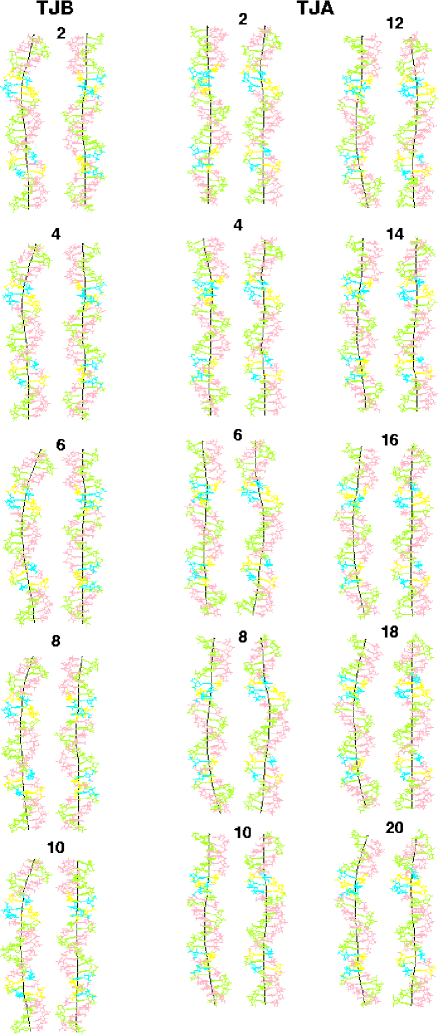
Figure 1 shows two series of representative structures from the two trajectories. Each structure is an average over a nanosecond interval, with these intervals equally spaced in time. In TJB the molecule was curved always in the same direction, but both the shape of the helical axis and the magnitude of bending varied. The first two structures shown in Fig. 1a are locally bent between the upper two A-tracts while their lower parts are nearly straight. In contrast, in the last three structures, the planar curvature is smoothly distributed over the entire molecule. In TJA, distinguishable stable bending emerged only after a few nanoseconds, but after the fourth nanosecond all average conformations were smoothly bent. In contrast to TJB, however, the bending direction was not stable, and by comparing the two time series of perpendicular projections one may see that during the first 15 nanoseconds the bending plane slowly rotated. During the final five nanoseconds the overall bending direction was stable. In the last conformation, an S-shaped profile of the helical axis is found in the perpendicular projection, which indicates that there are two slightly misaligned bends located between the three A-tracts. The orientations of the helices in this figure was chosen separately for the two trajectories, and one can notice that the left projection in plate (a) is close to the right one in plate (b), in other words, the final bend directions differed by approximately 90∘. In TJA, the intersection of the minor groove with the bending plane occurs close to the center of the middle A-tract while in TJB this point is shifted towards its 3’ end. In both cases, however, the narrowed minor grooves of the A-tracts appear at the inside edge of the curved axis.
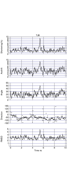
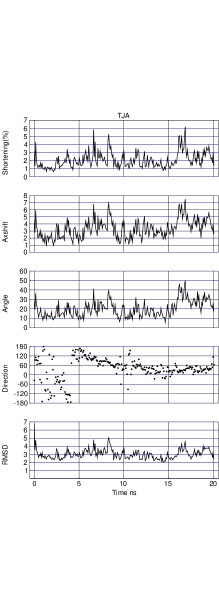
Figure 2 displays fluctuations of various parameters that characterize the overall bending of the double helix. In both trajectories the rmsd from the canonical B-DNA usually fluctuates between 2 and 4 Å and correlates with the three parameters shown in Fig. 2 that measure the bending magnitude. Note that in TJA there was a short period of strong initial bending which have not been detected in Fig. 1. The most significant difference between the two plates in Fig. 2 is in the dynamics of the bending direction. In TJB, the final orientation of the bend was found early and remained quite stable, although the molecule sometimes straightened giving, simultaneously, a low bending magnitude and large temporary fluctuations of direction. In contrast, during the first 15 nanoseconds of TJA, the bending plane made more than a half turn with respect to the coordinate system bound to the molecule, that is a transition occurred between oppositely bent conformations by passing through a continuous series of bent states. Temporary transitions to the straight state were short-living and always reversed, with the bending resumed in the previous direction. Owing to this turn the bend orientations in TJA and TJB converged considerably although not exactly. The zero direction in Fig. 2 corresponds to bending towards the minor groove at the fifth AT base pair of the middle A-tract. We see that, at the end of TJA, it is shifted from zero by an angle corresponding to rotation in one base pair step, which gives a bend towards the minor groove close to the center of the middle A-tract. In TJB, the direction deviates from zero in the opposite sense by an angle corresponding to roughly two base pair steps, resulting in a residual divergence of approximately 90∘ between the two trajectories. The slow kinetics of convergence exhibited in Fig. 2 indicates, however, that still better accuracy, if ever possible, would require much longer time.
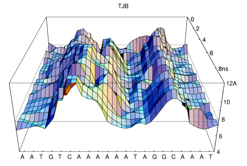
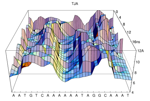
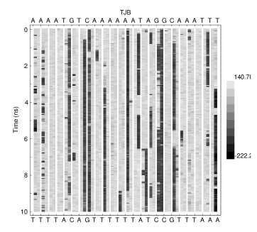
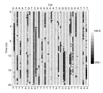
Figure 3 displays the time evolution of the profile of the minor grooves in TJB and TJA. At the end of both trajectories the minor groove width exhibits modulations phased with the helical screw. It is significantly widened between the A-tracts and narrowed within them by approximately 1 Å with respect to the canonical level. This magnitude of narrowing corresponds well to the values observed in experimental structures of A-tract containing B-DNA oligomers [20, 21, 22, 23, 24, 19]. In TJB, the overall waving profile established early and remained more or less constant. Interestingly, during two rather long periods, a secondary minimum occurred close to the 5’ end of the middle A-tract, and at the same time the main central minimum sifted towards the 3’ end of this A-tract. These motions involve almost an entire helical turn and, apparently, are concerted, which demonstrates the possibility of medium range structural correlations along the double helix. Comparison of Figs. 1a and 3a suggests that there is no simple one-to-one relationship between bending and minor groove modulations. Notably, the right smaller and narrower widening corresponds to a stable and strong bending point of the helical axis, while the left one, which is evidently larger, gives less or no bending. In TJA, the final configuration of the minor groove established only during the last few nanoseconds, but the final profile has the same number and similar positions of local maxima and minima as that in TJB. The overall minor groove dynamics in TJA looks rather complicated and its relationship with the quasi-regular rotation of the bending plane demonstrated in Figs. 1b and 2b is not readily seen.
Figure 4 displays dynamics of backbone transitions in the two trajectories. A few common features can be noticed that have been encountered previously [15, 16]. For instance, in A-tracts, the BII conformers are commonly found in ApA steps and almost never in TpT steps. They tend to alternate with BI in consecutive ApA steps. transitions often occur concertedly along the same strand as well as in opposite strands. The dynamics comprises all time scales presented in these calculations and clearly involves slower motions as well. Note, for instance, a particularly stable BII conformer in the only GpA step available in the two strands. On the other hand, there is some similarity in the distributions of the BII conformers in the two trajectories, which is a new feature compared to our previous report [15, 16]. It is seen in Fig. 4 that in total ten BII conformers were found at the end of TJB and eight in TJA. Among them six and five, respectively, occurred in non A-tract sequences. In three base pair steps the BII conformers are found in both final conformations, with all of them in non A-tract sequences. A careful examination the two plates in Fig. 4 shows that, although in A-tracts the preferred sites of BII conformations differ, in the intervening sequences their dynamics is rather similar in TJB and TJA. This trend is demonstrated in Fig. 5 where inter-trajectory correlations are examined for the specific base pair step propensities to BI and BII conformers. We have not included here the TpT steps in A-tracts because they strongly prefer the BI conformation and, therefore, are trivially correlated. It is evident that, except for ApA steps in A-tracts, there was certain correlation in the average populations of BII conformers for each specific base pair step in the two trajectories. The ApA steps apparently can adopt both conformations with little effect of the sequence context and the overall structure.
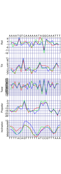
Figure 6 shows variation of several helicoidal parameters along the duplex in three representative one nanosecond averaged structures. Two of them were taken from TJA, namely, the 16th and 18th nanosecond averages which we refer to as TJA16 and TJA18. They illustrate the scale and the character of fluctuations of these parameters in the course of the same dynamics. The third conformation is the last average of TJB (TJB10) and it illustrates convergence of helical parameters in independent trajectories. We have chosen TJA16 and TJA18 because, as seen in Fig. 1, the corresponding two structures are particularly similar. They are both smoothly bent in a virtually identical direction and their rmsd is only 0.95 Å. All parameters shown in the figure, except the inclination, exhibit jumping alternations between consecutive base pair steps. Although they look chaotic, there is a considerable similarity between TJA16 and TJA18 and less significant, but still evident similarity of the two with TJB10. Notably, a remarkable correspondence of alterations of the twist is observed in the right-hand half of the sequence. At the same time, even the TJA16 and TJA18 plots sometimes diverge. Note, for instance, that the alteration in their roll traces are phased in the central A-tract, but dephased in the other two, with a particularly significant difference around the TpA step. These results show that, in a statically curved double helix, the base pair helicoidal parameters fluctuate around certain specific values that are stable in the nanosecond time scale. There is, however, more than one combination of these parameters for the same overall bend. At the same time, the evident convergence of the corresponding distributions in TJA and TJB suggests that, at least for this particular base pair sequence, the number of such redundant combinations should not be very large.
Discussion
The computational experiments described here give the first example of a natural DNA fragment with phased A-tracts which in free unbiased MD simulations adopts spontaneously a statically bent shape. In the analogous earlier calculations the static curvature has been demonstrated for a different A-tract containing sequence constructed artifically and never tested in experiments [15, 16]. The qualitative similarity between these two simulations is evident. Trajectories starting from canonical A and B-DNA forms both travel within the B-DNA family and, in straight, states yield rmsd from B-DNA of around 2 Å. TJA enters the B-DNA family with a significant temporary bending during the first 500 ps. Later it becomes bent in an arbitrary direction and next changes the bend direction by slowly rotating the bending plane. This rotation slows down after 10 ns, with the final orientation much closer to that in TJB than the initial one. In both cases the residual discrepancy was in the range of 60∘ – 90∘ [16]. The final minor groove profiles are not identical, although similar for TJA and TJB, as well as the distributions of the and backbone conformers and base pair helicoidal parameters. The present results, therefore, suggest that the A-tract induced DNA bending observed in calculations here and before corresponds to the experimental phenomenon.
At the same time, there are several clear differences. Notably, the preferred bending direction here is closer the centers of the minor grooves of the A-tracts, whereas the magnitude of bending is somewhat less than in the previous calculations. The bending angle in the average structures shown in Fig. 1 fluctuates between 12∘ and 25∘ in TJB and between 7∘ and 28∘ in TJA, with the maximal values reached at the end in both cases. The previous artifical sequence was constructed to maximize the bending and it showed the corresponding values beyond 35∘ [15, 16]. According to the experimental estimates made for “good benders” in an optimal sequence context, the magnitude of bending is around 18∘ per A-tract [8], which in our case gives 36∘ for the overall bend because the principal bending elements are the two intervening zones between the A-tracts. The bends observed here are somewhat below this estimate. However, in experiments, the bending magnitude differs significantly between different A-tract sequences and depends upon many environmental parameters that are not controlled in simulations. One can expect to observe in calculations sequence variations of the bending magnitude that may not exactly follow those in experiments. Therefore, whatever the possible reasons of the apparent discrepancy, the overall correspondence of the computed bending magnitudes to the experimental estimates should be considered as surprisingly good. Yet another difference is a larger than earlier degree of similarity in the profiles of the minor groove, in the distributions of conformers, and helicoidal parameters in trajectories starting from A and B-DNA forms. It was the most surprising observation of our previous report that reasonably good convergence in terms of the macroscopic bent shape of the double helix was not accompanied by the parallel convergence of microscopic conformational parameters. Here the two trajectories manifest clear signs of convergence for base pair step parameters as well as for the backbone conformations. Additional studies are necessary in order to tell if this difference is a sequence specific property or just an occasional effect.
In spite of these differences the results presented here support our previous conclusion concerning the qualitative disagreement of the computed structural dynamics of the double helix and the most popular earlier theories of bending [16]. In Figs. 3, 4, and 6, multiple examples are found of strong variability of local helical parameters in bent substates, which argues against the local interactions and the preferred base pair orientations as the cause of bending. All three A-tracts are characterized by a narrowed minor groove and local minima in the traces of the propeller twist and the inclination of base pairs. Nevertheless, their internal structures are not homogeneous and vary from one base pair step to another. Moreover, the structures of the three are not the same and present another example of an ensemble of conformational substates with a common overall shape. This pattern is qualitatively different from that implied by the junction models of bending [26, 27]. At the same time, the present results are well interpreted in the framework of our model that sees the principal cause of the bending in the backbone stiffness and the postulated excess of its specific length over that in the idealized regular B-DNA double helix [16]. Several non trivial observations support this view.
The first observation is the microheterogeneity of the ensemble of conformations that provide the same bent form of the double helix during the last nanoseconds of both trajectories. Once the backbone have found its preferred waving shape on the surface of the B-DNA cylinder it fixes the bending direction. The thermal motion of bases is allowed, but they are forced to respect this mechanical constraint, giving rise to an ensemble of conformations with different base orientations, but the same bent form of the double helix
The second observation is an always waving minor groove profile which does not change during temporary short-living straightening. The waving profile is the direct consequence of the postulated excess of the specific backbone length over that in the regular B-DNA with even grooves. The main immediate cause of bending is the necessity to compress the backbone in the minor groove widenings if the parallel base pair stacking is to be preserved [16]. The backbone stiffness tends to cause destacking from the minor groove side, which results in bending towards the major groove. Symmetrical destacking is also possible, however, and transitions between various types of stacking perturbations makes possible time variations of the magnitude of bending with a constant backbone profile.
Finally, our model explains well the persistent bends in incorrect directions and the rotation of the bending plane observed in TJA. According to this view the excessive backbone length and its stiffness force the backbone to wander on the surface of the B-DNA cylinder whatever the base pair sequence is. In the dynamics starting from the A-DNA structure the duplex enters the B-DNA family in strongly non-equilibrium conditions, with rapidly changing different energy components. The backbone quickly extends to its preferred specific length taking some waving profile and causing bending in an arbitrary direction. During the subsequent slow evolution it remains always waving, and that is why there is always a preferred bend direction which is not lost during occasional straightening.
Methods
As in the previous report [16], the molecular dynamics simulations were carried out with the internal coordinate method (ICMD) [28, 29]. The minimal model of B-DNA was used, with fixed all bond length, rigid bases, fixed valence angles except those centered at sugar atoms and increased effective inertia of hydrogen-only rigid bodies as well as planar sugar angles [30, 16]. The time step was 10 fsec. AMBER94 [31, 32] force field and atom parameters were used with TIP3P water [33] and no cut off schemes. The heating and equilibration protocols were same as before [30, 34]. The Berendsen algorithm [35] was applied during the production run, with a relaxation time of 10 ps, to keep the temperature close to 300 K. The coordinates were saved once in 2.5 ps.
The initial conformation for TJB was prepared by vacuum energy minimization starting from the fiber B-DNA model constructed from the published atom coordinates [17]. 375 water molecules were next added by the hydration protocol designed to fill up the minor groove [30]. The initial conformation for TJA was prepared by hydrating the minor groove of the corresponding A-DNA model [17] without preliminary energy minimization. The necessary number of water molecules was added after equilibration to make it equal in TJA and TJB.
During the runs, after every 200 ps, water positions were checked in order to identify those penetrating into the major groove and those completely separated. These molecules, if found, were removed and next re-introduced in simulations by putting them with zero velocities at random positions around the hydrated duplex, so that they could readily re-join the core system. This time interval was chosen so as to ensure a small enough average number of repositioned molecules which was ca 1.5.
REFERENCES
- [1] J. C. Marini, S. D. Levene, D. M. Crothers, and P. T. Englund, Bent helical structure in kinetoplast DNA, Proc. Natl. Acad. Sci. USA 79, 7664 (1982).
- [2] H.-M. Wu and D. M. Crothers, The locus of sequence-directed and protein-induced DNA bending, Nature 308, 509 (1984).
- [3] S. Diekmann, in Nucleic Acids and Molecular Biology, Vol. 1, edited by F. Eckstein and D. M. J. Lilley (Springer-Veralg, Berlin Heidelberg, 1987), pp. 138–156.
- [4] P. J. Hagerman, Sequence-directed curvature of DNA, Annu. Rev. Biochem. 59, 755 (1990).
- [5] D. M. Crothers, T. E. Haran, and J. G. Nadeau, Intrinsically bent DNA, J. Biol. Chem. 265, 7093 (1990).
- [6] D. M. Crothers and J. Drak, Global features of DNA structure by comparative gel electrophoresis, Meth. Ensymol. 212, 46 (1992).
- [7] W. K. Olson and V. B. Zhurkin, in Structure and Dynamics. Vol. 2: Proceedings of the Ninth Conversation, State University of New York, Albany, NY 1995, edited by R. H. Sarma and M. H. Sarma (Adenine Press, New York, 1996), pp. 341–370.
- [8] D. M. Crothers and Z. Shakked, in Oxford Handbook of Nucleic Acid Structure, edited by S. Neidle (Oxford University Press, New York, 1999), pp. 455–470.
- [9] V. B. Zhurkin, Y. P. Lysov, and V. I. Ivanov, Anisotropic flexibility of DNA and the nucleosomal structure, Nucl. Acids Res. 6, 1081 (1979).
- [10] E. von Kitzing and S. Diekmann, Molecular mechanics calculations of and of the curved molecule , Eur. Biophys. J. 14, 13 (1987).
- [11] V. P. Chuprina and R. A. Abagyan, Structural basis of stable bending in DNA containing tracts. Different types of bending, J. Biomol. Struct. Dyn. 1, 121 (1988).
- [12] V. B. Zhurkin, N. B. Ulyanov, A. A. Gorin, and R. L. Jernigan, Static and statistical bending of DNA evaluated by Monte Carlo simulations, Proc. Natl. Acad. Sci. USA 88, 7046 (1991).
- [13] S. R. Sanghani, K. Zakrzewska, S. C. Harvey, and R. Lavery, Molecular modelling of : Sequence elements responsible for curvature, Nucl. Acids Res. 24, 1632 (1996).
- [14] D. Sprous, M. A. Young, and D. L. Beveridge, Molecular dynamics studies of axis bending in : Effects of sequence polarity on DNA curvature, J. Mol. Biol. 285, 1623 (1999).
- [15] A. K. Mazur, A-tract induced DNA bending is a local non-electrostatic effect, Preprint http: // xxx.lanl.gov/abs/ physics/0002010, (2000).
- [16] A. K. Mazur, The physical origin of intrinsic bends in double helical DNA, Preprint http: // xxx.lanl.gov/abs/ physics/0004040, (2000).
- [17] S. Arnott and D. W. L. Hukins, Optimised parameters for A-DNA and B-DNA, Biochem. Biophys. Res. Communs. 47, 1504 (1972).
- [18] R. E. Dickerson, M. Bansal, C. R. Calladine, S. Diekmann, W. N. Hunter, O. Kennard, R. Lavery, H. C. M. Nelson, W. K. Olson, W. Saenger, Z. Shakked, H. Sklenar, D. M. Soumpasis, C.-S. Tung, E. von Kitzing, A. H.-J. Wang, and V. B. Zhurkin, Definitions and nomenclature of nucleic acid structure parameters, J. Mol. Biol. 205, 787 (1989).
- [19] A. K. Mazur, Internal correlations in minor groove profiles of experimental and computed B-DNA conformations, J. Mol. Biol. 290, 373 (1999).
- [20] R. E. Dickerson and H. R. Drew, Structure of a B-DNA dodecamer. II. Influence of base sequence on helix structure, J. Mol. Biol. 149, 761 (1981).
- [21] H. C. M. Nelson, J. T. Finch, B. F. Luisi, and A. Klug, The structure of an oligo(dA):oligo(dT) tract and its biological implications, Nature 330, 221 (1987).
- [22] A. D. DiGabriele, M. R. Sanderson, and T. A. Steitz, Crystal lattice packing is important in determining the bend of a DNA dodecamer containing an adenine tract, Proc. Natl. Acad. Sci. USA 86, 1816 (1989).
- [23] K. J. Edwards, D. G. Brown, N. Spink, J. V. Skelly, and S. Neidle, Molecular structure of the B-DNA dodecamer d(CGCAAATTTGCG)2. An examination of propeller twist and minor-groove water structure at 2.2A resolution, J. Mol. Biol. 226, 1161 (1992).
- [24] A. D. DiGabriele and T. A. Steitz, A DNA dodecamer containing an adenine tract crystallizes in a unique lattice and exhibits a new bend, J. Mol. Biol. 231, 1024 (1993).
- [25] R. Lavery and H. Sklenar, The definition of generalized helicoidal parameters and of axis curvature for irregular nucleic acids, J. Biomol. Struct. Dyn. 6, 63 (1988).
- [26] S. D. Levene and D. M. Crothers, A computer graphics study of sequence-directed bending of DNA, J. Biomol. Struct. Dyn. 1, 429 (1983).
- [27] J. G. Nadeau and D. M. Crothers, Structural basis for DNA bending, Proc. Natl. Acad. Sci. USA 86, 2622 (1989).
- [28] A. K. Mazur, Quasi-Hamiltoian equations of motion for internal coordinate molecular dynamics of polymers, J. Comput. Chem. 18, 1354 (1997).
- [29] A. K. Mazur, Symplectic integration of closed chain rigid body dynamics with internal coordinate equations of motion, J. Chem. Phys. 111, 1407 (1999).
- [30] A. K. Mazur, Accurate DNA dynamics without accurate long range electrostatics, J. Am. Chem. Soc. 120, 10928 (1998).
- [31] W. D. Cornell, P. Cieplak, C. I. Bayly, I. R. Gould, K. M. Merz, D. M. Ferguson, D. C. Spellmeyer, T. Fox, J. W. Caldwell, and P. A. Kollman, A second generation force field for the simulation of proteins, nucleic acids and organic molecules, J. Am. Chem. Soc. 117, 5179 (1995).
- [32] T. E. Cheatham, III, P. Cieplak, and P. A. Kollman, A modified version of the Cornell et al. force field with improved sugar pucker phases and helical repeat, J. Biomol. Struct. Dyn. 16, 845 (1999).
- [33] W. L. Jorgensen, Transferable intermolecular potential functions for water, alcohols and ethers. application to liquid water., J. Am. Chem. Soc. 103, 335 (1981).
- [34] A. K. Mazur, A minimal model of B-DNA, Preprint http: // xxx.lanl.gov/abs/ physics/9907028, (1999).
- [35] H. J. C. Berendsen, J. P. M. Postma, W. F. van Gunsteren, A. DiNola, and J. R. Haak, Molecular dynamics with coupling to an external bath, J. Chem. Phys. 81, 3684 (1984).
Appendix
This section contains comments from anonymous referees of a peer-review journal where this paper was been considered for publication, but rejected (see also [16]).
A Journal of Molecular Biology
1 First referee
These companion manuscripts describe a series of molecular dynamics trajectories obtained for DNA sequences containing arrangements of oligo dA - oligo dT motifs implicated in intrinsic DNA bending. Unlike previous MD studies of intrinsically bent DNA sequences, these calculations omit explicit consideration of the role of counterions. Because recent crystallographic studies of A-tract-like DNA sequences have attributed intrinsic bending to the localization of counterions in the minor groove, a detailed understanding of the underlying basis of A-tract-dependent bending and its relationship to DNA-counterion interactions would be an important contribution.
Although the MD calculations seem to have been carried out with close attention to detail, both manuscripts suffer from some troubling problems, specifically:
The sequence investigated here is a 25-bp segment of the well-characterized L. tarentolae kinetoplast-DNA bending locus. Two trajectories, TJA and TJB, were computed starting from canonical A-form and B-form structures, respectively. Although the author argues that greater structural convergence between TJA and TJB has taken place in these simulations, there is still a significant disparity concerning the observed bending directions in these two structures. Moreover, the extent of bending in this simulated helix is significantly less than that observed in the previous study, which is unexpected because of out-of-phase placement of the third A tract in the previous sequence. This behavior is not explained and seems difficult to rationalize.