Tumour Therapy with Particle Beams
Abstract
Photons are exponentially attenuated in matter producing high doses close to the surface. Therefore they are not well suited for the treatment of deep seated tumours. Charged particles, in contrast, exhibit a sharp increase of ionisation density close to the end of their range, the so-called Bragg-peak. The depth of the Bragg-peak can be adjusted by varying the particle’s energy. In parallel with the large energy deposit the increase in biological effectiveness for cell killing at the end of the range provides an ideal scalpel for the surgeon effectively without touching the surface tissue. Consequently proton therapy has gained a lot of ground for treating well localized tumours. Even superior still are heavy ions, where the ionisation pattern is increased by the square of their charge .
Introduction
It has been known for a long time that tissue, in particular tumour tissue, is sensitive to ionising radiation. Therefore it is only natural that tumours have been treated with various types of radiation, like -rays and electrons. -rays are easily available from radioactive sources, like 60Co, and electrons can be accelerated to -energies by relatively inexpensive linear accelerators. The disadvantage of -rays and electrons is that they deposit most of their energy close to the surface. To reduce the surface dose in tumour treatment requires rotating the source or the patient so that the surface dose is distributed over a larger volume. In contrast, protons and heavy ions deposit most of their energy close to the end of their range (Bragg-peak). The increase in energy loss at the Bragg-peak amounts to a factor of about 5 compared to the surface dose, depending somewhat on the particle’s energy. Heavy ions offer, in addition, the possibility to monitor the destructive power of the beam by observing annihilation radiation by standard positron-emission tomography techniques (PET). The annihilation radiation is emitted by -active nuclear fragments produced by the incident heavy ion beam itself.
Energy loss of particles in tissue [1]
A photon beam is attenuated in matter according to
| (1) |
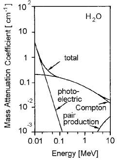
where is the initial intensity and the beam intensity at the depth . is the linear mass attenuation coefficient which depends on the photon energy and the target charge . is shown in figure 1 for a target composed of water, which is essentially equivalent to tissue. The main interaction mechanisms which contribute to are the photoelectric effect , Compton scattering and pair-production . For energies typical for radioactive sources Compton scattering dominates. The absorption profile of photons in matter exhibits a peak close to the surface followed by an exponential decay.
Charged particles suffer energy loss by ionisation. This energy loss is described by the Bethe-Bloch formula:
| (2) |
where
| (3) |
| – | charge of the beam particle | |
| – | charge of the absorber material | |
| – | mass number of the absorber material | |
| – | electron mass | |
| – | velocity of light | |
| – | Avogadro’s number | |
| – | classical electron radius | |
| – | velocity of the particle divided by | |
| – | maximum transferable energy | |
| to an atomic electron | ||
| – | mean excitation energy of the target material | |
| – | density parameter |
For protons interacting in water (or tissue) equation (2) can be approximated by
| (4) |
where
| (5) |
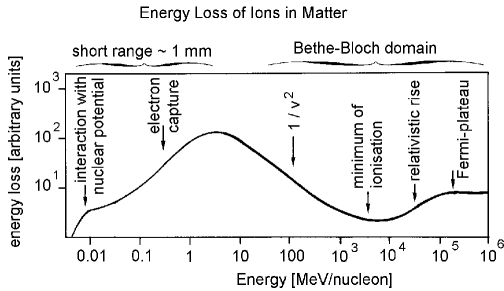
which gives an energy loss of 4.2 for 200 protons at the surface and close to the end of their range. For heavy ions the energy loss is essentially scaled by . When charged particles reach the end of their range the energy loss first rises like but when they are very slow they capture electrons from the target material and their effective charge decreases and hence their energy loss rapidly falls to zero.
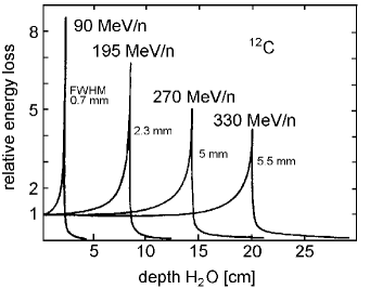
A typical energy loss curve for ions as a function of their energy is sketched in figure 2 Kraft1 . The energy loss of 12C ions as a function of the depth in water is shown in figure 3 Kraft1 ; Kraft2 . The tail of the energy loss beyond the Bragg-peak originates from fragmentation products of 12C ions, which are faster than the 12C ions and have a somewhat longer range.
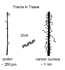
In the ionisation process a generally small fraction of the particle’s energy is transferred to the atomic electrons. In rare cases these electrons can get a larger amount of energy. The -electrons deviate from the main ionisation trail and produce a fuzzy-like track (figure 4, Kraft1 ).
In addition to ionisation light particles, like electrons, can also undergo bremsstrahlung . Since the probability for this process is inversely proportional to the square of the mass of the beam particle, bremsstrahlung can be neglected for particles heavier than the electron for energies relevant to tumour therapy Grupen1 .
The above mentioned fragmentation of heavy ions leads to the production of positron emitters. For the 12C case, lighter isotopes like 11C and 10C are produced. Both isotopes decay with short half-lives ; to boron according to
| (6) | |||||
The positrons have a very short range, typically below . After coming to rest they annihilate with electrons of the tissue giving off two monochromatic photons of which are emitted back-to-back
| (7) |
These photons can be detected by positron-emission tomography techniques and can be used to monitor the destructive effect of heavy ions on the tumour tissue.
Production of particle beams
The treatment of deep seated tumours requires charged particles of typically 100 to per nucleon, i.e. 100 to protons or 1.2 to C ions. These particles are accelerated in either a linear accelerator or in a synchrotron. As an example figure 5 shows a typical set-up for the production of heavy ions. 12C atoms are evaporated from an ion source and pre-accelerated. Thin foils are used to strip off all electrons from the ions. The 12C nuclei are then injected into a synchrotron, where they are accelerated by radiofrequency cavities to the desired energy. The ions are kept on track by dipole bending magnets and they are focussed by quadrupoles. After having reached the final energy they are ejected by a kicker magnet, which directs the particles to the treatment room. Their path is monitored by tracking

chambers (multi-wire proportional counteres, ion chambers or drift-chambers). If beam losses occur veto-counters (mostly scintillation counters) ensure that only a pencil beam is steered to the treatment room.
Nowadays, mainly protons and heavy ions are used for tumour therapy. Other possibilities consist of the use of negative pions Dyson ; Curtis ; Goodman , which are produced by high energy protons in a beam dump according to
| (8) |
where the are momentum selected and collimated. After losing their energy by ionisation the negative pions are captured in the tumour tissue by nuclei at the end of their range and produce so-called ‘stars’ in which neutrons are created. The Bragg-peak of the negative pions along with the local production of neutrons which have a high biological effectiveness leads to an efficient cell killing in the tumour at the end of the pion’s range.
Neutrons are also possible candidates for tumour treatment Lennox . For this purpose the tumour is sensitized by a boron compound before neutron treatment. The boron compound must be selected in such a way that it is preferentially deposited in the tumour region. Neutrons are then captured by the boron according to:
| (9) |
The produced -particles (He-nuclei) have a very short range ( several ) and high biological effectiveness. Best results are obtained with epithermal neutrons () produced by protons on light targets (e.g. Be).
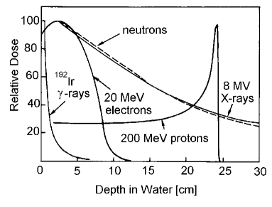
Direct irradiation with neutrons – without sensitizing the tumour – has the disadvantage that neutrons show a similar dose depth curve like 60Co -rays thus producing a high amount of biologically very effective damage in the healthy tissue around the tumour (see figure 6 NAC ).
Applications in Tumour Therapy
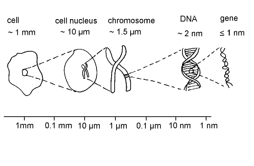
The target for cell killing is the DNA in the cell nucleus (see figure 7 (after Kraft1 )). The size of the DNA-molecule compares favorably well with the width of the ionisation track of a heavy ion. The DNA contains two strands containing identical information. A damage of one strand by ionising radiation can easily be repaired by copying the information from the unaffected strand to the damaged one. Therefore the high ionisation density at the end of a particle’s range matches well with the requirement to produce double strand breaks in the DNA, which the cell will not survive. Heavy ions like 12C seem to be optimal for this purpose. Ions heavier than carbon would even be more powerful in destroying tumour tissue, however, their energy loss in the surrounding tissue and in the entrance region already reaches a level where the fraction of irreparable damage is too high, while for lighter ions (like 12C) mostly repairable damage is produced in the healthy tissue outside the targeted tumour. The cell killing rate in the tumour region thus benefits from two properties of protons or ions like carbon:
-
•
the increased energy loss of protons and ions at the end of their range and
-
•
the increased biological effectiveness of double strand breaks at high ionisation density.
The cell killing rate is eventually related to the equivalent dose H in the tumour region, which can be expressed by
| (10) |
where is the tumour mass and RBE the increased relative biological effectiveness. The integral extends over the tumour region.
As mentioned above the rate and location of cell killing can be monitored by observing the annihilation photons which result from the -decay of fragments formed by the beam.
These physical and biological principles are employed in an efficient way by the raster scan technique Kraft2 ; Kraft3 ; Kraft4 . A pencil beam of heavy ions (diameter ) is aimed at the tumour. The beam location and spread is monitored by tracking chambers with high spatial resolution. In the treatment planning the tumour is subdivided into three-dimensional pixels (“voxels”). Then the dose required to destroy the tumour, which is proportional to the beam intensity, is calculated for every voxel. For a fixed depth in tissue an areal scan is performed by magnetic deflection sweeping the beam across the area in a similar way as a TV image is produced (see figure 8, Kraft3 ; Kraft4 ). The tumour volume is filled from the back by energy variation ( range variation) of the beam. Typically 50 energy steps are used starting at the rear plane. For a depth profile from to one has to cover energies from /nucleon to /nucleon. When the beam energy is reduced the required dose for the plane under irradiation is calculated using the damage that the more energetic beam had already produced in its entrance region. This ensures that the lateral (caused by magnetic deflection) and longitudinal scanning
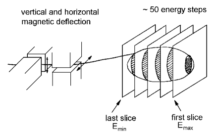
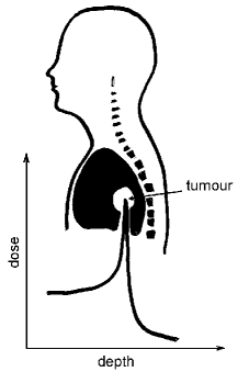
(by energy variation) covers the tumour completely. In figure 9 (after Kraft1 ) the dose distribution for individual energy settings and the resulting total dose is sketched and compared with the damage that X-rays from a 60Co-source would produce. An artist impression of the dose distribution for a lung and a brain tumour is given in figure 10.
Treatment facilities
Berkeley was the birthplace of therapy with hadrons. Since 1954 protons and later Helium-nuclei were used for treatment. Throughout the world treatment with protons is standard (Sweden, USA, Russia, Japan, Switzerland, England, Belgium, France, South Africa). In some places negative pions have been used in the past (USA, Canada, Switzerland). The most promising results have been obtained with heavy ions


(Berkeley, USA; Chiba, Japan; and Darmstadt, Germany). In total 25000 patients have been treated from 1954 to 1999.
Summary and Outlook
The inverse ionisation dose profile of charged particles has been known for a long time, from nuclear and particle physics. The instrumentation originally developed for elementary particle physics experiments has made it possible to design and monitor particle beams with great precision which can then be used for tumour therapy. Heavy ions seem to be ideal projectiles for tumour treatment. They are suitable for well localized tumours. The availability of treatment facilities is increasing. Naturally such a facility requires an expensive and complex accelerator for the charged particles. For beam steering and control sophisticated particle detectors and interlock systems are necessary to ensure the safety of patients.
Acknowledgements
The author has benefitted a great deal from information provided by G. Kraft from GSI-Darmstadt and from discussions with him. I acknowledge also the help of Mrs. L. Hoppe and C. Haucke for the drawing of the figures, Mrs. A. Wied for typing the text, Mr. Ngac An Bang for giving the paper the final LaTeX-touch, and Mr. D. Robinson for a careful reading of the manuscript.
References
- (1) C. Grupen ‘Particle Detectors’ Cambridge University Press 1996
- (2) G. Kraft ‘Radiobiology of Heavy Charged Particles’ GSI-Preprint 96-60, Nov. 1996
- (3) G. Kraft ‘Tumour Therapy with Ion Beams’ invited paper at the SAMBA-Symposium at the University of Siegen 1999, to be printed in Nucl.Instr.Meth. 2000
- (4) Medical Radiation Group, National Accelerator Centre, South Africa, Internet paper 1999
-
(5)
G. Kraft ‘The Impact of Nuclear Science on Medicine’
Nucl. Phys. A 654 (1999) 1058c - 1067c -
(6)
G. Kraft ‘Radiotherapy with Heavy Charged Particles’
http://www.gsi.de -
(7)
N.A. Dyson ‘Nuclear Physics with Application in Medicine and
Biology’ John Wiley & Sons Inc. N.Y. (1981) and
‘Radiation Physics with Application in Medicine and Biology’, Ellis Horwood, N.Y. (1993) - (8) S.B. Curtis, M.R. Raju ‘A Calculation of the Physical Characteristics of Negative Pion Beams Energy Loss Distribution and Bragg-Curves’ Rad. Research 34 (1968) 239
- (9) G.B. Goodman ‘Pion Therapy for Cancer – What are the Prospects’ TRIUMF-Preprint TRI-PP-92-134 (1992)
- (10) A.J. Lennox ‘Hospital-Based Proton Linear Accelerator for Particle Therapy and Radioisotope Production’ Fermilab-Pub. 90/217 (1990)