Further study of CdWO4 crystal scintillators as detectors for high sensitivity 2 experiments: scintillation properties and pulse-shape discrimination
L. Bardellia, M. Binia, P.G. Bizzetia, L. Carraresia, F.A. Danevichb, T.F. Fazzinia, B.V. Grinyovc, N.V. Ivannikovad, V.V. Kobychevb, B.N. Kropivyanskyb, P.R. Maurenziga, L.L. Nagornayac, S.S. Nagornyb, A.S. Nikolaikob, A.A. Pavlyukd, D.V. Podab, I.M. Solskye, M.V. Sopinskyyf, Yu.G. Stenind, F. Taccettia, V.I. Tretyakb, Ya.V. Vasilievd, S.S. Yurchenkob
a Dipartimento di Fisica, Universit di Firenze and INFN, 50019 Firenze, Italy
b Institute for Nuclear Research, MSP 03680 Kiev, Ukraine
c Institute for Scintillation Materials, 61001 Kharkov, Ukraine
d Nikolaev Institute of Inorganic Chemistry, 630090 Novosibirsk, Russia
e Institute for Materials, 79031 Lviv, Ukraine
f Lashkaryov Institute of Semiconductor Physics, 03028 Kiev, Ukraine
Abstract
Energy resolution, light yield, non-proportionality in the scintillation response, ratio, pulse shape for rays and particles were studied with CdWO4 crystal scintillators. Some indication for a difference in the emission spectra for rays and particles was observed. No dependence of CdWO4 pulse shape on emission spectrum wavelengths under laser, particles and ray excitation was observed. Dependence of scintillation pulse shape for quanta and particles and pulse-shape discrimination ability on temperature was measured in the range of C.
1 INTRODUCTION
Observations of neutrino oscillations manifest the non-zero neutrino mass and provide important motivation for high sensitivity experiments on neutrinoless double beta () decay. However, this process still remains unobserved, and only half-life limits for mode were obtained (see, e.g., reviews [1])111An evidence for decay of 76Ge has been claimed in [2]. However, it was criticized in [3, 4, 5]. Later the Heidelberg group has presented new data with improved statistics and after a reanalysis. A half-life y has been reported [6], which corresponds to the effective Majorana neutrino mass eV. One of the most sensitive experiments has been performed in the Solotvina Underground Laboratory [7] by the Kiev-Firenze collaboration with the help of enriched cadmium tungstate (116CdWO4) crystal scintillators [8, 9]. The half-life limit on decay of 116Cd was set as yr at 90% C.L., which corresponds to an upper bound on the effective Majorana neutrino mass eV [9]. This result is among the strongest world-wide restrictions on (in addition to the bounds obtained in experiments with 76Ge, 82Se, 100Mo, 130Te, and 136Xe).
Two by-product results obtained in the course of the Solotvina experiment with CdWO4 scintillators should also be mentioned: (i) the half-life ( yr) and the spectrum shape of the fourth-forbidden decay of 113Cd were measured [10]; (ii) indication for the decay of 180W with the half-life yr has been observed for the first time [11].
The Solotvina experiment demonstrates that CdWO4 crystals possess several unique properties required for high sensitivity decay experiments: low level of intrinsic radioactivity, good scintillation characteristics, and pulse-shape discrimination ability, which allow one to reduce background effectively. To enhance sensitivity to the neutrino mass to the level of eV, one has to increase the measuring time and the mass of enriched 116CdWO4, improve the energy resolution and reduce background of the detector. As it was shown by Monte Carlo calculations, the required sensitivity could be achieved by using 116CdWO4 crystals placed into a large volume of a high purity liquid. For instance, in the CAMEO project [12] it was proposed to place 100 kg of 116CdWO4 crystals into the BOREXINO counting test facility. With 1000 kg of 116CdWO4 crystals the neutrino mass limit can be pushed down to eV. An alternative solution should also be mentioned for a sensitive decay experiment with 116CdWO4 by using lead tungstate crystal scintillators as a high efficiency active shield [13].
In addition, as it was demonstrated in [14], CdWO4 crystals show good potential to develop thermal bolometers with energy resolution keV in a wide energy interval. Furthermore, CdWO4 can be used as a scintillating bolometer with registration of both light and heat signals [15]. Scintillating cryogenic detectors are highly promising to search for rare processes like dark matter and double beta decay thanks to their excellent energy resolution and particle discrimination ability.
Precise measurements of CdWO4 properties are necessary for development of methods to simulate such detectors.
The purpose of our work was investigation of different CdWO4 scintillation properties important for high sensitivity experiment: energy resolution, light yield, non-proportionality in the scintillation response, ratio, emission spectra and transparency, pulse shape for rays and particles and their temperature dependence.
2 MEASUREMENTS AND RESULTS
The luminescence of CdWO4 crystals was discovered about sixty years ago [16]. The different properties of CdWO4 scintillators were investigated (see Refs. [17, 18, 19, 20, 21, 22, 23, 24, 25, 26, 27, 28, 29, 30, 31, 32] and references therein). In 1960 G.B. Beard and W.H. Kelly have used a small natural CdWO4 crystal to search for alpha activity of natural tungsten [33]. To our knowledge, it was the first low background experiment applying this detector.
The main characteristics of CdWO4 scintillators are presented in Table 1. The material is non-hygroscopic and chemically resistant. All crystals used for measurements are listed in Table 2. All of them were grown by Czochralski method. The crystal CWO–1 was grown by the low-thermal-gradient Czochralski technique [34].
| Density (g/cm3) | 7.9 |
| Melting point (∘C) | 1271 |
| Structural type | Wolframite |
| Cleavage plane | Marked (010) |
| Hardness (Mohs) | |
| Wavelength of emission maximum (nm) | 480 |
| Refractive index | |
| Effective average decay time∗ (s) | 13 |
| ∗For rays, at room temperature. |
| ID | Size (mm) | Manufacturer |
|---|---|---|
| CWO–1 | IIC Novosibirska | |
| CWO–2 | IM Lvivb | |
| CWO–3 | ISM Kharkovc | |
| CWO–4 | ISM Kharkovc | |
| CWO–5 | IM Lvivb | |
| CWO–6 | ISM Kharkovc | |
| CWO–7 | IFNU Lvivd | |
| a Nikolaev Institute of Inorganic Chemistry, Novosibirsk, Russia | ||
| b Institute for Materials, Lviv, Ukraine | ||
| c Institute for Scintillation Materials, Kharkov, Ukraine | ||
| d Ivan Franko National University, Lviv, Ukraine | ||
2.1 Scintillation properties
2.1.1 Energy resolution
In the present work, the energy resolution was measured with three CdWO4 crystals: mm (CWO–1), mm (CWO–2), and mm (CWO–3).
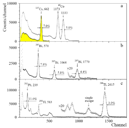
The CWO–1 crystal was ground at the side surface, the exit and top faces were polished. The crystal was wrapped by PTFE reflector tape and optically coupled to 3” photomultiplier (PMT) Philips XP2412. The measurements were carried out with 16 s shaping time to collect most of the charge from the anode of the PMT. The detector was irradiated by quanta of 60Co, 137Cs, 207Bi, and 232Th sources. The energy resolutions (full width at half maximum, FWHM) of 7.0% (137Cs, 662 keV), 5.8% (207Bi, 1064 keV), 5.0% (60Co, 1333 keV), and 3.9% (232Th, 2615 keV) were obtained (see Fig. 1).
The energy resolutions measured with the crystal CWO–2 in the same conditions are slightly worse. For instance, energy resolutions of 7.5%, 6.2%, and 4.6% were obtained with 662, 1064, and 2615 keV lines, respectively. In our opinion it is mainly due to the lower transparency of the crystal CWO–2 in comparison with the CWO–1 (see subsection 2.3 where the results of measurements of transmittance of the crystals are presented).
Energy resolutions of 6.8%, 5.6% and 3.4% for 662 keV (137Cs), 1064 keV (207Bi) and 2615 keV (232Th) lines, respectively, were measured with the small crystal CWO–3.
All these results are summarized in Fig. 2 where the fitting curves are also shown. The square root function with one free parameter was used for the fit: , where is the energy resolution and is energy of quanta in keV. The values , 4.12, and 3.07 were obtained for the CWO–1, CWO–2, and CWO–3 crystals, respectively.

2.1.2 Light yield
Light yield of CdWO4 was measured in [22] with the help of silicon photodiodes as photons/MeV (which is % of NaI(Tl)). In [26] a higher photon yield of a cadmium tungstate scintillator ( photons/MeV) was estimated on the basis of the measurements reported in [25]. This result was recently confirmed in [32], where values in the range photons/MeV were reported for CdWO4 crystal scintillators. In [31] the absolute light yield of CdWO4 scintillators photons per MeV was reported.
We try to estimate an absolute light yield of CdWO4 crystal scintillators by using data of energy resolution measurements. For an ideal scintillation detector the energy resolution for rays is given by [35]:
| (1) |
where is the mean number of photoelectrons produced in photocathode of PMT. The number of photoelectrons can be written as product:
| (2) |
where is mean number of photons created in a scintillator per 1 MeV of energy deposit, – the energy of quanta in MeV, – the fraction of scintillation photons arrived to the photocathode of PMT, – the quantum efficiency of the PMT photocathode to photons emitted by the scintillator.
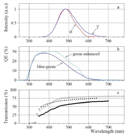
Value of can be calculated as the convolution of CdWO4 emission spectrum and spectral sensitivity of the PMT photocathode. We have obtained using the measured emission spectrum of CdWO4 (see subsection 2.2 and Fig. 6, a) and specification of the PMT (XP2412) with bialkali (blue-green sensitive) photocathode. For the high quality PMT with green-enhanced (RbCs) photocathode (EMI D724KFL, serial #13) produced by THORN EMI for the Solotvina experiment [9], we have obtained the value .
To estimate the value of , light propagation in the CdWO-2 detector was Monte Carlo simulated with the help of the GEANT4 package [36]. The emission spectrum and optical transmission curve of the CdWO4 crystal (see subsection 2.3), and the spectral sensitivity of the PMT photocathode were taken into account. An overall light collection of 27% was calculated. Such a modest value is mainly due to absorption of light and large refractive index of CdWO4 ( [25]).
Using formulas 1 and 2, an absolute light yield in the range photons/MeV was calculated for the CdWO4 scintillation crystal. At least the lower border of this estimation is in agreement with the results reported in [26, 32].
The absolute photon yield was also estimated with the help of the CWO–3 crystal scintillator. The energy resolution was measured in different conditions of light collection. However, we select a geometry (the polished CWO–3 crystal without reflector, covered by black cope, optically coupled to the PMT) which can be simulated with a comparatively high degree of accuracy. In this case we do not need to simulate diffused surfaces, light propagation from crystal with further reflection and return into scintillator, etc. The energy resolution of was measured for 662 keV quanta of 137Cs, while the value of the light collection for this detector was calculated as 23%. The photon yield was estimated to be of photons per 1 MeV of energy deposit, which is more than that reported in [26, 31, 32]. At the same time we realize that the main systematic error in the estimations of absolute light yield can be due to not quite correct calculations of the light collection. In our opinion further investigations are necessary to determine the absolute light yield of CdWO4 scintillators.
The relative photoelectron yield was measured with the CWO–1 crystal and NaI(Tl) mm of standard assembling. Both crystals were coupled to the same PMT XP2412 with the bialkali photocathode and were irradiated by quanta of 137Cs source. A transient digitizer based on the Analog Devices 12 bit ADC (AD9022) operated at 20 Mega Sample per second (20 MS/s) [29] was used to accumulate the pulse shapes from the detectors. To build the energy spectra of the CdWO4 and NaI(Tl) scintillators, the area of the pulses was calculated. In such a way we overcome the problem of different decay times of these scintillators. The relative photoelectron yield of the CWO–1 scintillator was measured as 26% of NaI(Tl).
2.1.3 Scintillation response at low energy
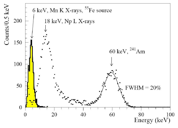
Fig. 3 shows the energy spectra of 241Am and 55Fe low energy gamma and X-ray lines measured with thin CdWO4 scintillator mm (CWO–4). Even the 6 keV peak of 55Fe is still resolved from the PMT noise. A low energy threshold of a CdWO4 detector is important to search for low energy processes, as for instance, the two neutrino double electron capture in 106Cd. Expected energy release in a crystal in this process is only keV.
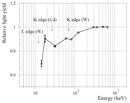
We have studied the non-proportionality in the scintillation response with the CWO–4 scintillator. The crystal was optically connected to EMI9256KB PMT operating at –1000 volts. The shaping time of the ORTEC (Model 572) amplifier was set to 10 s. The and X ray lines from the sources: 57Co (14.4 and 122.1 keV), 241Am (17.6 and 59.5 keV), 137Cs (32.1 and 661.7 keV), 133Ba (30.9, 81.0, 295.3 and 356.0 keV), 22Na (511 keV) were used for the measurements. Positions of the peaks were determined relatively to 661.7 keV line of 137Cs. The dependence of the relative photoelectron yield on the energy of X and lines is presented in Fig. 4. The behaviour of the scintillator response agrees with the results of other authors [37, 38]. This effect should be taken into account in experiments to search for low energy processes like, for instance, the neutrino accompanied double electron capture in 106Cd. The energy scale of a detector should be carefully measured in the region of interest.
2.1.4 ratio
Quenching factor for particles, in other words ratio222The ratio is defined as ratio of peak position in the energy scale measured with sources to the energy of particles., is important to interpret and suppress background caused by internal Thorium, Uranium and active Lanthanides contamination. In [11] the dependence of the ratio on the energy and direction of particles relatively to the main crystal axes was observed for CdWO4 crystals. To obtain particles with energies in the range MeV, a set of thin mylar films (with thickness of 0.65 mg/cm2) as absorbers were used. The average energies of particles after the absorbers were measured with the help of a surface-barrier detector. Disadvantage of such an approach is the substantial broadening of the particles energy after passing the absorbers.
In the present work the 3 MV Tandetron accelerator of the LABEC laboratory of the Sezione di Firenze of INFN [39] was used to obtain beams of alpha particles in the energy range MeV. By scattering of the beam on a thin gold foil energies of particles of 0.91, 1.86, 2.78, 4.18, and 6.99 MeV were obtained. The CWO–3 crystal was irradiated in the direction perpendicular to the (010) crystal plane. The obtained dependence of the ratio on the energy of particles is shown in Fig. 5. The energy spectra measured with 0.91, 2.78, and 6.99 MeV particles are shown in inset. In the energy interval MeV the ratio increases with increasing energy as , where is alpha particle energy in MeV. This result is in agreement with that reported in [11]. Such a behaviour of the ratio can be explained by the energy dependence of ionization density of particles [40]. It should be also noted, that ratio is not actually a property of a crystal, but more likely a certain characteristics of the detector depending on the shape and surface quality of a crystal, shaping time of electronics, etc.

2.2 Emission spectra
Emission spectra were measured under rays (60Co source) and particles (241AmPuCm source) excitation. The CdWO4 crystal, 42 mm in diameter and 25 mm height (CWO–5), was used for the measurements. The fluorescence light was analyzed in wavelength by the SPEX spectrometer. Intensities were integrated over 10 nm intervals. The results of the measurements are presented in Fig. 6, a. The emission spectra under irradiation are in a good agreement with result reported in [23]. A small difference in the emission spectra under particles and rays excitation was observed. However, this effect could be due to different absorption of the light emitted by the localized source ( particels) or diffused one ( quanta).
2.3 Light transmission and scattering
Transmittance of the CWO–2 crystal was measured in the spectral range 350-700 nm with the help of the spectrophotomether KSVU-23 equipped with reflection attachment. The transmission curve is shown in Fig. 6, c (filled circles). Taking into account the reflection losses, the value of cm for attenuation length of the CdWO4 crystal was obtained at the maximum of emission spectra (485 nm). Transmittance of the CWO–1 and CWO–3 crystals measured by the producers is also presented in Fig. 6, c. The crystals CWO–1 and CWO–3 show much better optical properties, namely the value of attenuation length is cm at the wavelength of the maximum of the emission spectrum.
Generally speaking, light attenuation in crystals is caused by absorption and scattering. The angular dependence of light intensity after passing the CWO–2 crystal was measured to estimate the light scattering in the crystal. Fig. 7 shows the layout of the measurement. A laser beam (expansion angle less than 1 mrad) of 632.8 nm wavelength and 0.5 mm diameter was used. The beam was directed normally to the face of the crystal. Intensity of the beam was measured by a Si-photodetector with diameter of 11 mm. The distance between the photodetector and the crystal was varied in the range 30–1350 mm.
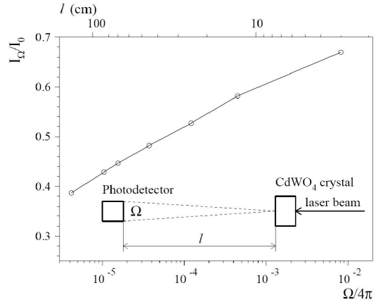
The measured dependence on the solid angle (Fig. 7) is well described by logarithmic function, and shows a considerable forward light scattering in the CdWO4 crystal. No dependence was observed in the measurements without crystal, as well as with the 30 mm-thick optical glass (K-8) installed instead of the crystal. The observed behaviour of light scattering can be explained by substantial amount of optical inhomogeneities whose sizes are comparable or exceed wavelength of the light [41]. Non-stoichiometric composition, presence of regions with distorted (or disturbed) structure, especially with partially amorphous structure, pores, voids, flaws, inclusions, can be causes of these inhomogeneities in CdWO4 crystals.
Processes of light scattering should be taken into account in simulation of light collection in CdWO4 scintillation detectors.
2.4 Scintillation decay
2.4.1 Pulse shape for rays and particles
Pulse shapes of CdWO4 scintillators were studied as described in [29, 11] with the help of a transient digitizer based on the 12 bit ADC (AD9022) operated at 20 MS/s. However, the integration time of the preamplifier in the present measurements was decreased (s in comparison with s in [29, 11]) to investigate possible fast components of scintillation decay. More recently, pulse shape for rays and particles was measured also with the help of the 12 bit 125 MS/s transient digitizer described in Ref. [42, 43]. Furthermore, single-electron counting method was applied to study the dependence of CdWO4 scintillation signal for particles and quanta on emission wavelength (see subsection 2.4.4).
To study pulse shape of scintillation decay for particles, the CdWO4 crystal mm (CWO–6) was irradiated by particles from collimated 241Am source in the direction perpendicular to the (010) crystal plane. The dimensions of the collimator were 0.75 2 mm. The energy of particles after passing of 2 mm air layer was calculated by GEANT3.4 program as 5.25 MeV [11]. 60Co was used as a source of quanta. Measurements were carried out at room temperature C.
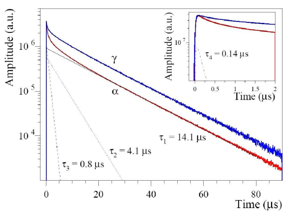
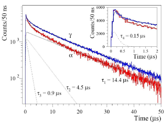
The shape of the light pulses produced by particles and rays in the CdWO4 scintillator measured by the 20 MS/s digitizer are shown in Fig. 8. To obtain the pulse shapes, large numbers of individual and events (with amplitudes corresponding to peak of 241Am) were summed. The first part of CdWO4 and pulses measured with the help of the 125 MS/s digitizer is presented in the inset of Fig. 8. A fit to the pulses was done by the function:
,
where are the relative intensities, – the decay constants for different light-emission components, and is integration constant of electronics (s). Four decay components were observed with s, s, s and s with different intensities for rays and particles (see Table 3). Similar results have been obtained with the crystal CWO–7 studied both with the 20 MS/s and 125 MS/s digitizers.
| Type of irradiation | Decay constants (s) and relative intensities | |||
|---|---|---|---|---|
| (A | (A | (A | (A | |
| particles | ||||
| rays | ||||
2.4.2 Pulse-shape discrimination between rays and particles
The difference of the pulse shapes allows to discriminate () events from those induced by particles. We applied for this purpose the optimal filter method proposed in [44] and already applied to CdWO4 scintilators in [29]. For each CdWO4 signal a numerical parameter (shape indicator, ) was calculated in the following way:
,
where the sum is over time channels starting from the origin of pulse and up to certain time (75 s for 20 MS/s digitizer and 64 s for 125 MS/s), is the digitized amplitude (at the time ) of the signal. The weight function was defined as: , where and are the reference pulse shapes for particles and quanta.
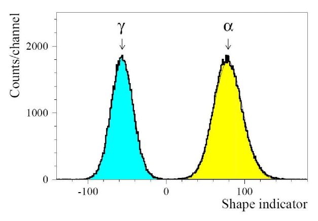
Clear discrimination between particles and rays was achieved using this approach, as one can see in Fig. 9 where the distributions measured by the 125 MS/s transient digitizer with the CWO–7 scintillation crystal for particles ( MeV) and quanta ( MeV) are shown. As a measure of discrimination ability (factor of merit, ), the following expression can be used:
,
where and are mean values for particles and quanta distributions (which are well described by Gaussian functions, see Fig. 9), and are the corresponding standard deviations. For the distributions presented in Fig. 9, the factor of merit is . This value is slightly better than that of obtained by using the 20 MS/s transient digitizer.
2.4.3 Pulse shape and fluorescence light wavelength under laser excitation
Measurements with pulses of ultraviolet light have been performed in order to investigate whether the fluorescence emission contains at least part of the components of different lifetime observed in particles and induced scintillation, and to search for the possible dependence of pulse shape on the wavelength of the emitted light. This part of the work has been performed at the European Laboratory for non-linear Spectroscopy (LENS, Florence).
The fluorescence of the CWO–7 crystal has been excited by fast ultraviolet pulses from a laser source ( nm), and the time dependence of the emitted light has been investigated in different intervals of wavelength, of 10 nm width, centered at 380, 440, 470, 500, 560, 600, and 650 nm [45]. In the experimental set-up the 1064 nm light from a YAG:Nd laser was used to excite a pair of non-linear crystals tuned to generate the fourth harmonics. The resulting 266 nm radiation was focused on the face of the CdWO4 crystal. The fluorescence light, analyzed in wavelength by the SPEX Spectrometer (22 cm focal length), was collected by an EMI9813 PMT, which was located close to the exit slits of the Spectrometer. The pulses from the anode of the PMT were integrated with a time constant of s, and sent to the input of a digital oscilloscope (HP TDS460). The digital output of the oscilloscope was transmitted to a computer and stored in memory for further analysis.
The pulse shapes (corresponding to the average of a large number of individual pulses) of the CdWO4 fluorescence light with the different wavelength wavelengths are shown in Fig. 10. Three components of the scintillation decay with decay times and intensities s (85%), s (11%), and s (4%) were observed. We were not able to measure the fast s decay component found in our measurements with the digitizers and by using single electron counting method, because of the rather big integration constant used in the measurements with laser excitation.
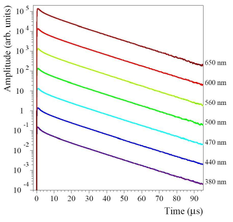
No dependence of pulse shape on the wavelength of emitted light under the laser excitation was observed.
2.4.4 Study of scintillation decay time for particles and quanta at different wavelength of emission spectra
The pulse shape for particles and quanta at different wavelength were measured by the single photon counting method. The CWO–7 crystal scintillator was optically connected to EMI9256KB PMT. The signal from the PMT gives the start signal to the time-digital converter (Time Analyzer, Canberra, Model 2143). Scintillation light from the CdWO4 crystal entered through the diaphragm 10 mm in diameter and passed through interference filters (Edmund Scientific Co.) with central wavelength 420, 460, 480, 590 nm to a PMT cooled down to –20∘ C (Product for Research, inc, USA). The PMT operating at single electron counting mode generated stop signals for the converter. The time scale of the time-digital converter was calibrated with the help of an ORTEC Model 462 Time Calibrator.
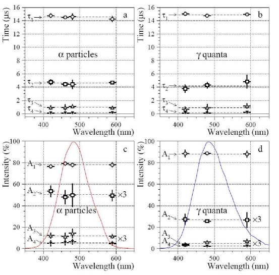
The pulse shapes of CdWO4 scintillator for particles (241AmPuCm source) and quanta (137Cs) measured by the single electron counting method with the 480 nm filter are depicted in Fig. 11. Fit of the obtained forms by sum of four exponential components gives values of the decay constants and their intensities (Table 4) similar to that obtained with the help of the transient digitizers. The results of the measurements with the different filters are presented in Fig. 12. No dependence of decay times on wavelength of the emission spectra both for particles and quanta was observed.
| Type of irradiation | Decay constants (s) and relative intensities | |||
|---|---|---|---|---|
| (A | (A | (A | (A | |
| particles | ||||
| rays | ||||
According to ref. [19], the spectral composition of the light emitted by CdWO4 should contain two different parts, one in the blue-green region, the other in the yellow region. In our measurements the latter can hardly be recognized over the tail of the blue-green component. The time distribution of the emitted light does not change significantly in the wavelength region from 380 to 650 nm under laser excitation nor under and irradiation in the region of 420–590 nm.
2.4.5 Dependence of pulse shape on temperature
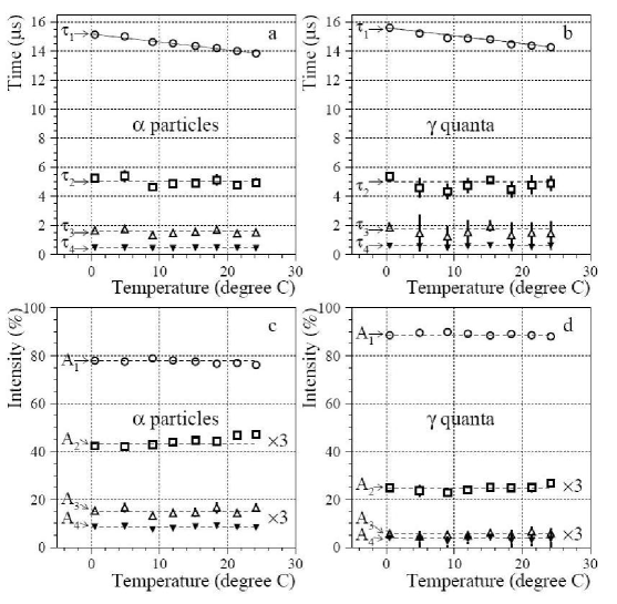
Temperature dependence of the pulse shape for rays and particles was checked in the range C. The CWO–4 crystal was optically connected to EMI9256KB PMT operating at –1000 volts. The scintillation crystal and the PMT were kept at the same temperature. The pulse shape was recorded by the 12 bit 20 MS/s transient digitizer. The crystal was irradiated by rays from 60Co source and particles from 241Am source. Forms of signals for rays and particles have been obtained as a result of summation of several thousand individual pulses. The values of the time constants and their intensities were obtained by fitting of the forms. The sum of four exponential functions has been taken as model for the description of scintillation signals.
Temperature dependence of the decay time constants and their intensities are presented in Fig. 13. The decay component depends on the temperature as –0.055(3) s/∘C for particles and as –0.048(3) s/∘C for quanta. It should be noted, that the intensities of this component both for and signals remain constant: for particles and for quanta.
The temperature dependence of the averaged decay time is shown in Fig. 14, a. The averaged decay time decrease with temperature as s/∘C for particles and s/∘C for quanta, which is in an agreement with results reported by Melcher et al. [23]. This dependence is mainly due to the temperature dependence of the decay component.
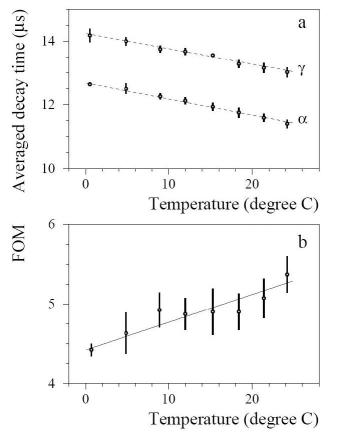
The factor of merit of pulse-shape discrimination between particles and quanta was calculated for the data accumulated in the temperature interval of C. The weight function (see subsection 2.4.2) was constructed by using pulse shapes for particles and quanta measured at room temperature. As one can see in Fig. 14, b, the factor of merit is slightly improved with increase of temperature. It could be explained by the increase of the difference between scintillation decay times under and excitation with increasing of temperature.
3 CONCLUSIONS
Scintillation properties of CdWO4 crystals were studied. The energy resolution 7.0% and 3.9% for the 662 and 2615 keV lines was obtained with large ( mm) CdWO4 crystal scintillator. Small crystal ( mm) showed an even better energy resolution: 6.8% and 3.4% for the 662 and 2615 keV lines, respectively.
The absolute photon yield of CdWO4 crystal scintillators was estimated to be ( photons per 1 MeV of energy deposit (under ray irradiation). This result was obtained by the analysis of the measurements of energy resolution. At least the lower border of this estimation is in agreement with the results of [26, 32]. In our opinion, more accurate measurements are necessary to determine the absolute light yield of CdWO4.
Spectra of the low energy and X-ray lines (6 keV of 55Fe, 18 keV of Neptunium L line and 60 keV quanta from 241Am source) were measured, which demonstrates possibility to apply CdWO4 crystal scintillators to search for double electron capture in 106Cd. Non-proportionality in the scintillation response observed in the present work is in agreement with that reported by other authors.
The energy dependence of the ratio was measured with beam produced by accelerator. Behaviour of the dependence is in an agreement with that reported in [11]. The ratio increases linearly in the energy interval MeV.
A difference in long-wavelength part of the emission spectra for rays and particles was observed, however we can not exclude that this effect is due to different absorption of scintillation light emitted under and irradiation.
Transmissivity of CdWO4 crystals was measured and considerable scattering of light was observed. This data indicates a presence of a substantial amount in our CdWO4 crystal of optical heterogeneity whose sizes are comparable or exceed the scintillation light wavelength.
Four components of scintillation decay (, , and s) and their intensities under particles and quanta irradiation were measured with different CdWO4 crystal scintillators by using different methods: transient digitizers with 20 MHz and 125 MHz sampling frequency as well as single electron counting method. The difference in the scintillation pulse shapes for particles and quanta is mainly due to difference in the intensities of the different decay components.
Clear discrimination between particles and rays was achieved using the optimal filter method.
No dependence of the pulse shape of the CdWO4 fluorescence light on wavelengths was observed in the range 380–650 nm under laser excitation as well as under particles and quanta irradiation in the range 420–590 nm.
Temperature dependence of the decay constants and intensities of CdWO4 pulse shape for rays and particles was investigated in the temperature range of C. Clear temperature dependence of the s component at the level of s/∘C was observed for particles and quanta, while the intensities of this component both for and signals remain constant. The pulse-shape discrimination improved slightly with increasing of temperature.
4 ACKNOWLEDGEMENTS
It is very pleasant to express gratitude to the personnel of the LABEC laboratory of the Sezione di Firenze of INFN, Prof. P.A. Mando, Dr. M. Chiari, Dr. L. Giuntini for the opportunity to carry out the measurements with a beam of alpha particles. The authors would like to thank Prof. M. Pashkovskii and Dr. M. Batenchuk from the Ivan Franko National University (Lviv, Ukraine) for providing one of the CdWO4 crystals used in the present study. We are grateful to Prof. A. Vinattieri from the Physics Department of Florence University for lending of the interference filters and the device for the single electron counting measurements.
References
- [1] V.I. Tretyak and Yu.G. Zdesenko, At. Data Nucl. Data Tables 61 (1995) 43; 80 (2002) 83; Yu.G. Zdesenko, Rev. Mod. Phys. 74 (2002) 663; J.D. Vergados, Phys. Rept. 361 (2002) 1; S.R. Elliot and P. Vogel, Ann. Rev. Nucl. Part. Sci. 52 (2002) 115; S.R. Elliot and J. Engel, J. Phys. G: Nucl. Part. Phys. 30 (2004) R183; A.S. Barabash, Phys. At. Nucl. 67 (2004) 438. F.T. Avignone, G.S. King, and Yu.G. Zdesenko, New J. Phys. 7 (2005) 6. H. Ejiri, J. Phys. Soc. Japan 74 (2005) 2101.
- [2] H.V. Klapdor-Kleingrothaus et al., Mod. Phys. Lett. A 16 (2001) 2409.
- [3] F. Feruglio, A. Strumia and F. Vissani, Nucl. Phys. B 637 (2002) 345; 659 (2003) 359.
- [4] C.E. Aalseth et al., Mod. Phys. Lett. A 17 (2002) 1475.
- [5] Yu.G. Zdesenko, F. A. Danevich and V. I. Tretyak, Phys. Lett. B 546 (2002) 206.
- [6] H.V. Klapdor-Kleingrothaus et al., Phys. Lett B 586 (2004) 198.
- [7] Yu.G. Zdesenko et al., Proc. 2-nd Int. Symp. Underground Phys., Baksan Valley, 1987 – Moscow, Nauka, 1988, p. 291.
- [8] F.A. Danevich et al., Phys. Rev. C 62 (2000) 045501.
- [9] F.A. Danevich et al., Phys. Rev. C 68 (2003) 035501.
- [10] F.A. Danevich et al., Phys. At. Nucl. 59 (1996) 1.
- [11] F.A. Danevich et al., Phys. Rev. C 67 (2003) 014310.
- [12] G. Bellini et al., Phys. Lett. B 493 (2000) 216; Eur. Phys. J. C 19 (2001) 43.
- [13] F.A. Danevich et al., Nucl. Instrum. Meth. A 556 (2006) 259.
- [14] A. Alessandrello et al., Nucl. Phys. B (Proc. Suppl.) 28A (1992) 233; Phys. Lett. B 420 (1998) 109.
- [15] S. Pirro et al., Nucl. Instrum. Meth. A 559 (2006) 361.
- [16] F.A. Kröger, Some Aspects of the Luminiscence of Solids, Elsevier Publ. Co., Amsterdam, 1948.
- [17] R.J. Moon, Phys. Rev. 73 (1948) 1210.
- [18] R.H. Gillette, Rev. Sci. Instr. 21 (1950) 294.
- [19] M.J.J. Lammers et al., Phys. Stat. Sol. 63 (1981) 569.
- [20] B.C. Grabmaier, IEEE Trans. Nucl. Sci. 31 (1984) 372.
- [21] E. Sakai, IEEE Trans. Nucl. Sci. 34 (1987) 418.
- [22] I. Holl et al., IEEE Trans. Nucl. Sci. 35 (1988) 105.
- [23] C.L. Melcher et al., IEEE Trans. Nucl. Sci. 36 (1989) 1188.
- [24] F.A. Danevich et al., Instr. Exp. Res. 32 (1989) 1059.
- [25] D.R. Kinloch et al., IEEE Trans. Nucl. Sci. 41 (1994) 752.
- [26] P. Dorenbos et al., IEEE Trans. Nucl. Sci. 42 (1995) 2190.
- [27] A.Sh. Georgadze et al., Instr. Exp. Technique 39 (1996) 191.
- [28] S.Ph. Burachas et al., Nucl. Instr. Meth. A 369 (1996) 164.
- [29] T. Fazzini et al., Nucl. Instrum. Meth. A 410 (1998) 213.
- [30] Y. Eisen et al., Nucl. Instr. Meth. A 490 (2002) 505.
- [31] G.M. Onyshchenko et al., Nucl. Instr. Meth. A 537 (2005) 394.
- [32] M. Moszynski et al., IEEE Trans. Nucl. Sci. 52 (2005) 3124.
- [33] G.B. Beard and W.H. Kelly, Nucl. Phys. 16 (1960) 591.
- [34] A.A. Pavlyuk, Ya.V. Vasiliev, L.Yu. Kharchenko, F.A. Kuznetsov, Proceedings of the. APSAM-92, Asia Pacific Society for Advanced Materials, Shanghai, 26- 29 April 1992, 1993, p. 164.
- [35] C.W.E. Eijk, Nucl. Instr. Meth. A 41 (2001) 244.
- [36] S. Agostinelli et al. (GEANT4 Collab.), Nucl. Instr. Meth. A 506 (2003) 250; http://geant4.web.cern.ch/geant4/.
- [37] P. Dorenbos et al., Radiat. Meas. 24 (1995) 355.
- [38] E.P. Sysoeva et al., IEEE Trans. Nucl. Sci. 43 (1996) 1282.
- [39] http://labec.fi.infn.it
- [40] J.B. Birks, Theory and Practice of Scintillation Counting, Pergamon Press, London, 1964.
- [41] H.C. van de Hulst, Light Scattering by Small Particles, Yohn Wiley & Sons, New York, Chapmen & Hall, Ltd., London, 1957.
- [42] G. Pasquali et al., Proc. of the IWM2005 Workshop, November 28th – December 1st 2005, Catania, Italy.
- [43] G. Pasquali et al., submitted to Nucl. Instr. Meth. A.
- [44] E. Gatti and F. De Martini, Nuclear Electronics 2, IAEA, Vienna, 1962, p. 265.
- [45] T. Fazzini et al., Proc. Int. Workshop on Tungstate Crystals, Roma, Oct. 12-14, 1998, eds. S. Baccaro, B. Borgia, I. Dafinei, E. Longo, University degli Studi La Sapienza, 1999, p. 243.