Lifetime of 19Ne∗(4.03 MeV)
Abstract
The Doppler-shift attenuation method was applied to measure the lifetime of the 4.03 MeV state in 19Ne. Utilizing a 3He-implanted Au foil as a target, the state was populated using the 20Ne(3He,Ne reaction in inverse kinematics at a 20Ne beam energy of 34 MeV. De-excitation rays were detected in coincidence with particles. At the 1 level, the lifetime was determined to be 11 fs and at the 95.45% confidence level the lifetime is 11 fs.
pacs:
26.30.+k, 23.20.-g, 25.55.-e, 26.50.+x, 27.20.+nI Introduction
The 15O(Ne reaction leads to the initial breakout from the hot CNO cycles that operate in Type I x-ray bursts, which are thermonuclear runaways on accreting neutron stars in binary star systems Wiescher et al. (1999). Recent calculations have suggested that if the rate of this reaction were below a certain threshold, the periodic x-ray bursts observed from some accreting neutron stars would not occur Fisker et al. (2004). Hence the rate of this reaction is of considerable importance. However, direct measurements at the relevant energies would require (radioactive) 15O beams of high intensity not presently available. Since the first theoretical investigation of this reaction Wagoner (1969), experimental data on the radiative and widths of excited states in 19Ne have been used to better constrain its rate. As was pointed out first in Ref. Langanke et al. (1986), at temperatures below 0.6 GK the reaction rate is dominated by resonant capture to the first state above the -emission threshold, lying at an excitation energy of 4.03 MeV.
The decay widths of the 4.03 MeV state in 19Ne have until recently remained elusive. Its width, , has been experimentally unobservable on account of its small value. All published attempts to measure the -decay branching ratio B have yielded only upper limits Magnus et al. (1990); Laird et al. (2002); Davids et al. (2003); Rehm et al. (2003); Davids et al. (2003); Visser et al. (2004). Therefore the reduced width of the analog state at 3.91 MeV in 19F Mao et al. (1995, 1996) has been used to estimate the width. Early theoretical estimates of the reaction rate assumed the radiative widths of the 19Ne and 19F analog states to be equal Wallace and Woosley (1981); Langanke et al. (1986). Despite attempts to measure the radiative width of the state in 19Ne itself that resulted in lower and upper limits Davidson and Roush (1973); Hackman et al. (2000), the analog state has been the most reliable source of experimental information on the radiative width. With the recent report of the first measurement of the lifetime of the 4.03 MeV state, as well as more precise determinations of the excitation energies of this and other states in 19Ne Tan et al. (2005), the experimental situation has improved dramatically. We report here a second successful measurement of the lifetime of the 4.03 MeV state in 19Ne, using a different reaction in which the recoil velocity was higher, allowing for a more precise lifetime determination.
II Experiment and Analysis
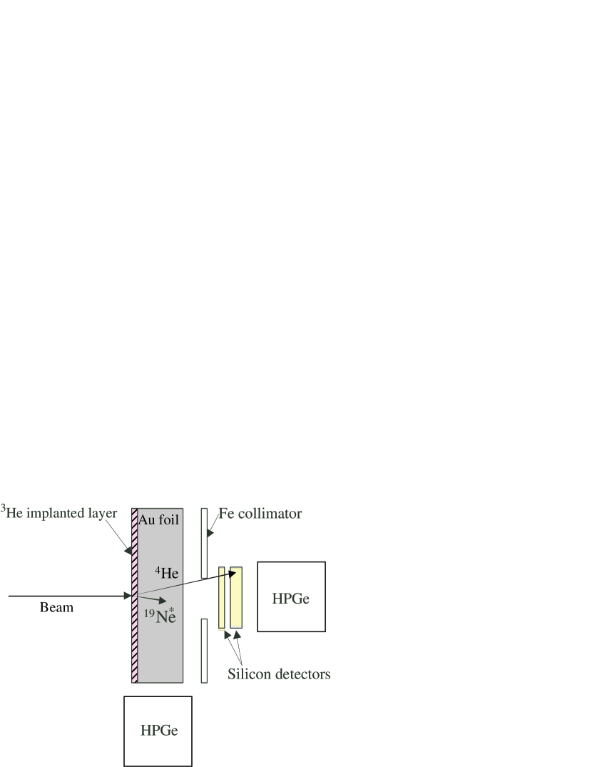
The experiment was performed at the TRIUMF-ISAC facility using the Doppler-shift attenuation method. A 34 MeV 20Ne beam was incident on a 3He-implanted Au foil target, populating the level of interest via the 3He(20Ne,Ne reaction. The 20Ne beam and recoiling 19Ne nuclei were stopped in the Au foil. The average beam intensity was 10 particle nA. A schematic depiction of the experimental setup is shown in Fig. 1. De-excitation rays were detected in coincidence with particle ejectiles using an 80% high-purity germanium (HPGe) coaxial detector placed at 0∘ with respect to the beam axis. The lifetime was determined from a lineshape analysis of this -ray energy spectrum. A second HPGe detector placed at 90∘ was used as a reference detector to measure the unshifted peak energies. The detectors were located 9 cm from the target. The energy calibration of the HPGe detectors was performed using a 56Co source whose highest energy ray is 3.2 MeV. This calibration was extrapolated linearly to higher energies. The energies of rays from the source were measured before and after the experiment and were found to differ by less than 1 keV.
The scattering chamber was designed with a cold trap to ensure a clean target surface and also to prevent losses of the implanted 3He. This was achieved using a narrow differential pumping aperture followed by a copper cylinder enclosing the path of the beam to the target. The copper cylinder was cooled using liquid nitrogen. To avoid any condensation of impurities on the surface of the target, the copper cylinder was not in direct contact with the target ladder. Indirect contact of the cold copper cylinder with the copper target ladder was achieved using BeCu fingers mounted on a boron nitride plate, which provided electrical isolation as well. This arrangement maintained a temperature difference between the copper cylinder and the target ladder. In this way the target was cooled below room temperature to ensure that 3He did not diffuse out when heated by bombardment with a beam power of up to 0.3 W. Moreover, the colder surfaces surrounding the target foil and the beam path in front of it reduced the buildup of carbon and other contaminants on the target itself during the experiment.
The target foil was prepared at the Université de Montréal by implanting 30 keV 3He ions into a 12.5 m thick Au foil, yielding an areal number density of 61017 cm-2. The implantation region was 0.1 m thick. Similar implanted foils prepared at the Chalk River laboratories were used in earlier femtosecond lifetime measurements Keinonen et al. (1981). The foil was found to contain some surface deposits of carbon after the implantation process. The concentration of 3He in the foil was monitored via yields of elastically scattered 3He and was found to remain unchanged during the experiment. The beam was collimated to a 2 mm diameter spot on the target.
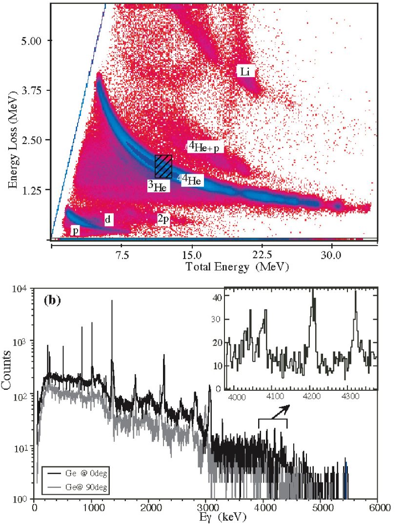
The particles were identified by means of the energy loss (E) and total energy (E) correlation using a silicon detector telescope. The telescope consisted of a 25 m thick silicon detector for E measurement and a 500m silicon detector for E measurement. Both the detectors were standard, circular, ORTEC transmission-type detectors with an active area of 150 mm2. The detector telescope subtended a solid angle of 360 msr and was centered about the beam axis, allowing the detection of particles with scattering angles less than 20∘. The E-E particle identification spectrum obtained in coincidence with at least one HPGe detector is shown in Fig. 2(a). A wide range of particle energies arose from fusion-evaporation reactions with the carbon contaminant on the foil. The region in which particles from the 4.03 MeV level in 19Ne can be found is marked by the hatched band. The -ray energy spectrum obtained from the HPGe detectors in coincidence with particles falling within the hatched energy band is shown in Fig. 2(b). Several peaks arising from fusion evaporation products can be identified. The inset shows the E 4 MeV region from the 0∘ HPGe detector, revealing several distinct peaks. The two peaks observed between 4.0 and 4.1 MeV in the 0∘ detector are inconsistent with known rays from 19Ne, while those at 4.2 and 4.3 MeV are consistent with known 19Ne transitions Doppler shifted appropriately for recoils moving with .
The ray energy spectrum from the 0∘ HPGe detector measured in coincidence with particles in two different total energy (Eα) ranges is shown in Fig. 3. The particles corresponding to 4.03 MeV excitations in 19Ne falling within the angular acceptance of our setup have total energies between 10 and 13.5 MeV. Examining the spectrum with a gate on Eα between 11 and 13 MeV (a slightly smaller range than that expected in order to improve the signal/noise ratio), shown in Fig. 3(a), we see two peaks corresponding to Doppler-shifted rays from 19Ne. The peak observed at 4.2 MeV in the 0∘ HPGe detector corresponds to the Doppler-shifted direct transition from the 4.03 MeV level to the ground state of 19Ne, and the peak at 4.32 MeV is consistent with the Doppler-shifted 4.14 MeV de-excitation ray from the 4.38 MeV level to the 238 keV level in 19Ne. Gating on particles from 13-14 MeV, shown in Fig. 3(b), we see little evidence for Doppler-shifted rays from the 4.03 MeV and 4.38 MeV states, just as we would expect.
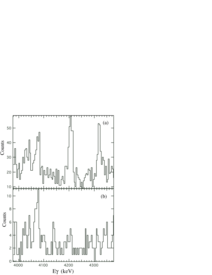
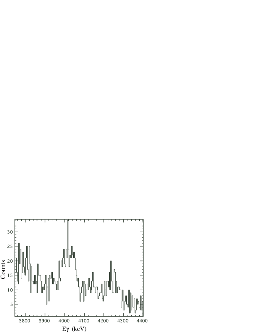
Figure 4 shows the energy spectrum from the 90∘ HPGe detector in coincidence with particles having energies between 11 and 13 MeV. The spectrum shows a peak at 4.03 MeV, consistent with the direct transition from the 4.03 MeV state to the ground state, which is Doppler-broadened because of the large angular acceptance of the HPGe detector. The absence of a sharp peak at 4.2 MeV indicates that there are no rays from a long-lived, stopped contaminant in the region of interest.
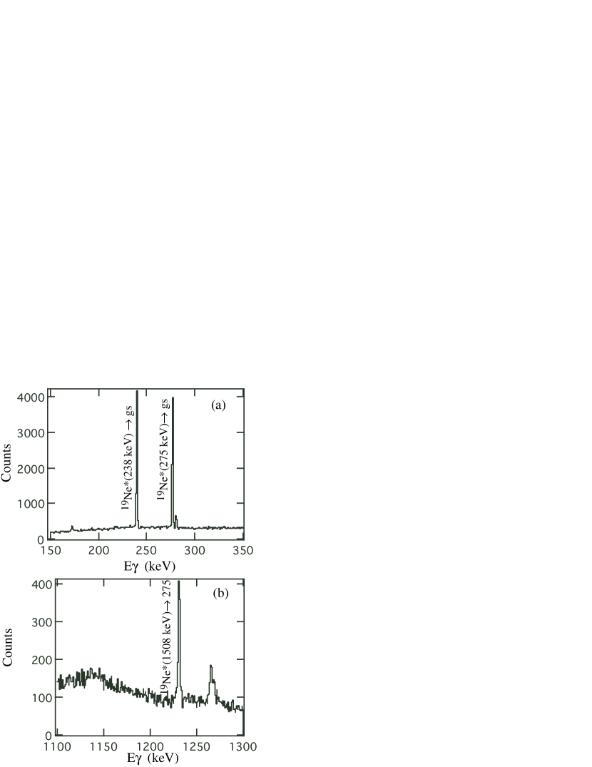
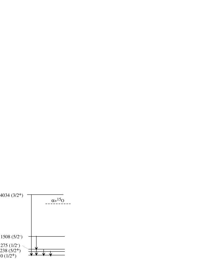
Looking at the low energy range of the -ray spectrum from the 0∘ HPGe detector obtained in coincidence with all particles, shown in Fig. 5(a) and (b), clear signatures for population of the first three excited states in 19Ne are also apparent. Gamma rays from the first excited state appear at 238 keV. The 275 keV ray visible in Fig. 5(a) is from the de-excitation of the second excited state in 19Ne. Higher-lying states in 19Ne also decay through these two levels with finite decay branches. One such decay branch is from the 1508 keV state in 19Ne which decays by the emission of a 1233 ray to the 275 keV level, as can be seen in Fig. 5(b). This branch can be confirmed by looking at the Eα spectrum gated on the 275 and 1233 keV rays, depicted in Fig. 6(a) and (b), respectively. The particle energy spectrum shows a peak at 17 MeV when gated on either the 275 or 1233 keV rays, which corresponds to the population of the third excited state in 19Ne, lying at 1508 keV. Additionally the Eα peak at 20 MeV due to the second excited state in 19Ne is seen to be clearly separated from the peak corresponding to the third excited state. For clarity, a level diagram indicating the 19Ne states and transitions discussed in the paper is shown in Figure 7.

The response function of the experimental setup includes the effects of kinematic broadening arising from the finite angular acceptance of the silicon detectors as well as Doppler broadening due to the finite opening angle of the HPGe detector. The intrinsic lineshape of the HPGe detector, measured using a 3.2 MeV ray from the de-excitation of a 56Fe level populated in 56Co electron capture, was included in the response function. The line width due to the intrinsic resolution (3.5 keV FWHM at 4 MeV) is much narrower than the Doppler broadening observed in the measurement. The detection efficiency as a function of the emission angle of a 4 MeV ray for the angular range subtended by the 0∘ HPGe detector was taken into account by a GEANT4 simulation Geant4 Collaboration et al. (2003) appropriate for the geometry of the setup. The effects of different -ray angular distributions were investigated and found to have a negligible influence on the calculated lineshape. We estimate the 1 uncertainty in the relative detection efficiency to be 5%, which is the result of uncertainties in the geometry of the setup.
Experimental data exist on the stopping powers of heavy ions in Au at this recoil energy. These data have been used to constrain theoretical stopping power calculations. We used the parametrization of Ziegler Ziegler (2004). This result was compared to calculations based on the measurements of the Chalk River group Forster et al. (1976). The difference between the two stopping powers is less than 10%; based on this we estimate a 1 stopping power uncertainty of 5%. The analysis also took into account the change in stopping power in the 3He-implanted region of the Au foil following the prescription outlined in Ref. Alexander et al. (1981).
The lifetime was determined from a minimization using the lineshape calculated with the computer program described in Ref. Forster et al. (1979), taking into account the initial velocity distribution of the 19Ne recoils, the intrinsic lineshape of the HPGe detector, and the swelling of the 3He-implanted target layer. Apart from the lifetime, the free parameters in the search were the overall normalization, the flat background level and the centroid position. We allowed the centroid position to be a free parameter in order to account for any possible shift which could arise due to energy calibration uncertainties.
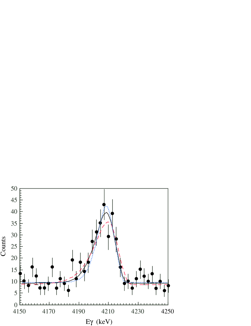
The minimization led to a lifetime for the 4.03 MeV level in 19Ne of fs at the 1 level; this uncertainty is the result of several contributions, the most important of which are stopping power ( 1 fs), relative detection efficiency ( 1 fs), statistics ( 2 fs), and the peak centroid position, which dominates the remaining uncertainty. At the 2 level, fs. The lineshapes corresponding to the best fit and the 2 upper and lower limit lifetimes are shown in Fig. 8. This value is in excellent agreement with the first lifetime measurement Tan et al. (2005), which yielded 13 fs (1). The higher precision of the present result arises from the fact that the 19Ne recoil velocity and the corresponding Doppler shift in this experiment were much larger than those in the measurement of Ref. Tan et al. (2005), which used the 17O(3He,Ne reaction at 3 MeV. The decay in flight produced a clearly asymmetric lineshape, with a long low-energy tail compared with the more sharply rising edge on the high energy side of the peak in Fig. 8. Reducing the angular acceptance of the detectors would improve the precision of the lifetime determination at the cost of statistics, but this is impractical given the low yield observed in the present experiment. Efforts are underway to reduce the experimental uncertainty by using cleaner target foils to reduce background and finding the optimum beam energy for the reaction to increase yield.
III Summary and Conclusions
The lifetime of the 4.03 MeV state in 19Ne was measured via the Doppler shift attenuation method. Populating the state using the 3He(20Ne,Ne reaction at a beam energy of 34 MeV, we stopped the recoils in the 3He-implanted Au target foil and detected de-excitation rays in coincidence with particles. The substantial Doppler shift of the recoils allowed a relatively precise determination of the lifetime. At the 1 level, the lifetime was determined to be 11 fs; at the 95.45% confidence level = 11 fs. The lifetime reported here agrees well with both the measurement of Ref. Tan et al. (2005) and the lifetime of the 19F analog, = 9(5) fs Tilley et al. (1995), further bolstering the evidence that isospin is a good symmetry in the T = 1/2, A = 19 system Davids et al. (2003).
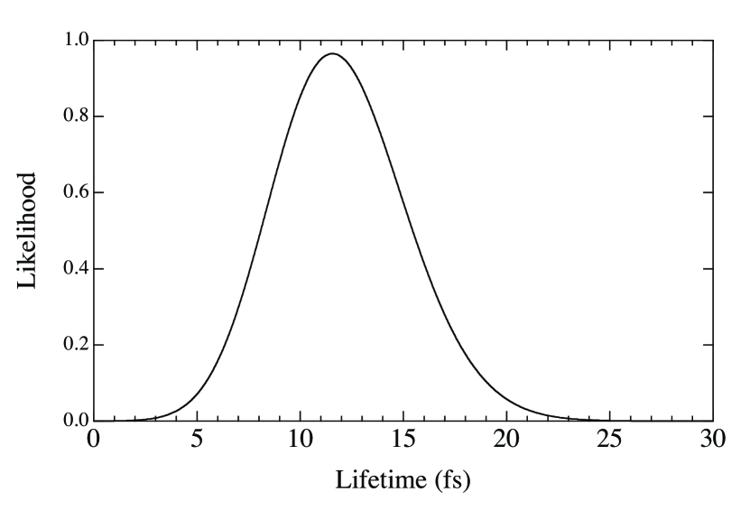
By combining the present lifetime determination with that of Ref. Tan et al. (2005), we can tighten the experimental constraints on the radiative width of the 4.03 MeV state. Using the information given in the Tan et al. paper Tan et al. (2005) and that from the present measurement, it is possible to construct the joint likelihood for the lifetime, taking into account the data of both experiments. This joint likelihood is shown in Fig. 9 and peaks around 12 fs. When the two experiments are combined, the lifetime is constrained to lie within 3 and 22 fs at the 99.73% confidence level.
Despite the fact that the -decay branching ratio Bα is presently constrained experimentally only by an upper limit, the two reported measurements of the lifetime of the 4.03 MeV state in 19Ne and the upper limit on Bα reported in Ref. Davids et al. (2003) allow us to place an experimental upper limit on . Using the 3 upper limit on Bα and the 3 lower limit on , at the 99.73% confidence level eV. This implies a 3 upper limit on the resonance strength, and thereby the 15O(Ne reaction rate at T GK, approximately 3 times smaller than the 3 upper limit quoted in Ref. Davids et al. (2003). Since the experimental upper limit on the rate has decreased, the conclusion reached in that work that this reaction probably plays no significant role in classical novae remains valid, consistent with the conclusions drawn in Refs. Wiescher et al. (1999); Iliadis et al. (2002). Measurements of the -decay branching ratios of the 4.03 and 4.38 MeV states will be required to more precisely identify the importance of the 15O(Ne reaction in x-ray bursts.
Acknowledgements.
This work was generously supported by the Natural Sciences and Engineering Research Council of Canada. TRIUMF receives federal funding via a contribution agreement through the National Research Council of Canada. RK is grateful to A. C. Shotter for helpful discussions and BD would like to thank R. H. Cyburt for enlightening discussions on statistics.References
- Wiescher et al. (1999) M. Wiescher, J. Görres, and H. Schatz, J. Phys. G 25, R133 (1999).
- Fisker et al. (2004) J. L. Fisker, J. Görres, M. Wiescher, and B. Davids, Astrophys. J., to be published; arXiv electronic preprint astro-ph/0410561 (2004).
- Wagoner (1969) R. V. Wagoner, Astrophys. J. Suppl. Ser. 18, 247 (1969).
- Langanke et al. (1986) K. Langanke, M. Wiescher, W. A. Fowler, and J. Görres, Astrophys. J. 301, 629 (1986).
- Magnus et al. (1990) P. V. Magnus, M. S. Smith, A. J. Howard, P. D. Parker, and A. E. Champagne, Nucl. Phys. A506, 332 (1990).
- Laird et al. (2002) A. M. Laird, S. Cherubini, A. N. Ostrowski, M. Aliotta, T. Davinson, A. di Pietro, P. Figuera, W. Galster, J. S. Graulich, D. Groombridge, et al., Phys. Rev. C 66, 048801 (2002).
- Davids et al. (2003) B. Davids, A. M. van den Berg, P. Dendooven, F. Fleurot, M. Hunyadi, M. A. de Huu, K. E. Rehm, R. E. Segel, R. H. Siemssen, H. W. Wilschut, et al., Phys. Rev. C 67, 012801(R) (2003).
- Rehm et al. (2003) K. E. Rehm, A. H. Wuosmaa, C. L. Jiang, J. Caggiano, J. P. Greene, A. Heinz, D. Henderson, R. V. Janssens, E. F. Moore, G. Mukherjee, et al., Phys. Rev. C 67, 065809 (2003).
- Davids et al. (2003) B. Davids, A. M. van den Berg, P. Dendooven, F. Fleurot, M. Hunyadi, M. A. de Huu, R. H. Siemssen, H. W. Wilschut, H. J. Wörtche, M. Hernanz, et al., Phys. Rev. C 67, 065808 (2003).
- Visser et al. (2004) D. W. Visser, J. A. Caggiano, R. Lewis, W. B. Handler, A. Parikh, and P. D. Parker, Phys. Rev. C 69, 048801 (2004).
- Mao et al. (1995) Z. Q. Mao, H. T. Fortune, and A. G. Lacaze, Phys. Rev. Lett. 74, 3760 (1995).
- Mao et al. (1996) Z. Q. Mao, H. T. Fortune, and A. G. Lacaze, Phys. Rev. C 53, 1197 (1996).
- Wallace and Woosley (1981) R. K. Wallace and S. E. Woosley, Astrophys. J. Suppl. Ser. 45, 389 (1981).
- Davidson and Roush (1973) J. M. Davidson and M. L. Roush, Nucl. Phys. A213, 332 (1973).
- Hackman et al. (2000) G. Hackman, S. M. Austin, T. Glasmacher, T. Aumann, B. A. Brown, R. W. Ibbotson, K. Miller, B. Pritychenko, L. A. Riley, B. Roeder, et al., Phys. Rev. C 61, 052801(R) (2000).
- Tan et al. (2005) W. P. Tan, J. Görres, J. Daly, M. Couder, A. Couture, H. Y. Lee, E. Stech, E. Strandberg, C. Ugalde, and M. Wiescher, Phys. Rev. C 72, 041302(R) (2005).
- Keinonen et al. (1981) J. Keinonen, H.-B. Mak, T. K. Alexander, G. C. Ball, W. G. Davies, J. S. Forster, and I. V. Mitchell, Phys. Rev. C 23, 2073 (1981).
- Geant4 Collaboration et al. (2003) Geant4 Collaboration, S. Agostinelli, J. Allison, K. Amako, J. Apostolakis, H. Araujo, P. Arce, M. Asai, D. Axen, S. Banerjee, et al., Nucl. Instrum. Methods Phys. Res. A 506, 250 (2003).
- Ziegler (2004) J. F. Ziegler, Nucl. Instrum. Methods Phys. Res. B 219-220, 1027 (2004).
- Forster et al. (1976) J. S. Forster, D. Ward, A. H. R., G. C. Ball, G. J. Costa, W. G. Davies, and I. V. Mitchell, Nucl. Instrum. Methods Phys. Res. 136, 349 (1976).
- Alexander et al. (1981) T. K. Alexander, G. C. Ball, W. G. Davies, and I. V. Mitchell, Journal of Nuclear Materials 96, 51 (1981).
- Forster et al. (1979) J. S. Forster, T. K. Alexander, G. C. Ball, W. G. Davies, I. V. Mitchell, and K. B. Winterbon, Nuclear Physics A 313, 397 (1979).
- Tilley et al. (1995) D. R. Tilley, H. R. Weller, C. M. Cheves, and R. M. Chasteler, Nucl. Phys. A595, 1 (1995).
- Iliadis et al. (2002) C. Iliadis, A. Champagne, J. José, S. Starrfield, and P. Tupper, Astrophys. J. Suppl. Ser. 142, 105 (2002).