Measurement of Change of decay rate
in Be and Au
Abstract
We have measured the possible change of the decay rate of implanted
into hosts of natural beryllium and natural gold. No difference between the
decay rates in the two hosts is observed within the experimental
precision of 0.12%. This result implies that change of the decay rate of
implanted in different materials cannot be simply expected from the
electron affinity difference consideration lonely and the lattice structure of the host materials
should be taken into account.
PACS:23.40.-s, 71.20.-b
Electron-capture decay rates depend sensitively on the density of atomic electrons within the nucleus. Thus, physically and chemically environmental factors such as pressure, chemical form, magnetic fields, etc. that can alter electron densities, may affect electron-capture decay rates. Since the change of nuclear decay in different environments has fundamental significance, as well as application in nuclear physics, geology and condensed matter physics, such a study is of current interests. is the lightest radioactive nucleus that decays by electron capture with a half-life of 53 days, thereby, it is a good candidate for studying perturbation of nuclear decay rates. Furthermore, the study of decay rate changes for has particular significance to the solar neutrino problem, where there is a large discrepancy between theoretical predictions and experimental determinations of the solar neutrino flux.
The decay rate sensitively depends on chemical environment at the nucleus. Changes of decay rates of nuclei in different compounds have been measured at normal pressure and a maximum change is about 1.5%. Moreover, this decay rate is also sensitive to physical environments, such as high pressure and host materials in which the nucleus is located. Liu et al. have measured a large increase, up to 1%, of the decay rate of under a high pressure of 400 kilobars. Recently Norman et al. measured the decay rate of implanted into hosts of lithium fluoride, gold, graphite, boron nitride, tantalum, lithium, and so on. It has been found that the decay rate varies by as much as 0.72% from one host to another. Qualitatively, it has been reported that if a atom is implanted in a medium having high electron affinity (EA), as a result of its interaction with nearby atoms of such a medium, the atom would lose a significant fraction of its 2s electrons. The decay rate of in a high-EA medium is thereby smaller than that in a low-EA medium. Thus, we would expect that implanted in natural gold (EA=2.308eV ) should decay slower up to 0.7% than that in natural beryllium (EA= 0.19eV ) according to the result of Ray et al., in which difference up to 0.72% between the decay rates of implanted in Au and in was observed.
In this Letter, we report our measurement of possible change of the decay rate of implanted into metal foils of natural beryllium and natural gold. The nuclei are produced by bombarding a 500 -thick foil of lithium fluoride (LiF) with a 3.2 MeV proton beam, with average current 5 A, from the 5SDH-2 tandem accelerator at CNNC Radiation Metrology and Measurement Center, China Institute of Atomic Energy. Thus, nuclei produced by the reaction 7Li (p, n) with recoil energy 1.0 MeV in the forward direction are implanted into beryllium and gold foils placed immediately behind the LiF target. The implantation for each foil lasts about 15 h. As a result of such implantations, atoms are expected to be randomly located in the interstitial lattice space of the host media beryllium and gold.
By electron capture the nucleus decays to the 3/2- ground state of 7Li directly with a branching ratio of 89.5% and to the first excited state with a ratio of 10.5%, which decays subsequently to its ground state by emitting a 478-keV gamma ray. In this experiment, two high-purity coaxial germanium (HP Ge) detectors are used to measure the 478-keV gamma-ray photons. The implanted beryllium and gold foils are separately mounted about 2 cm away from the endcups of two HP Ge detectors. The two detectors, each is surrounded by a graded shield of organic glass-Al-Cu-Pb, are located four meters away from each other to avoid cross detection of the gamma-ray photons from the two sources. The gamma-ray spectra are accumulated with a KODAQ data acquisition system.
In order to decrease as much effect originated from the decays of short-lived isotopes produced in bombarding as possible, we wait for about 40 days to start to acquire experimental data. The two gamma-ray spectra are acquired for successive one-hour intervals and recorded on a computer hard disk. Then this is followed by next interval automatically. After such a measurement has lasted about 70 days, we exchange the positions of the two sources, while keeping other measurement conditions unchanged, and we measure for another 50 days. We extract from each time bin the net peak area of the 478 keV gamma-ray and its time information after dead-time correction and time calibrations with the time provided by Hongkong observatory. Figure 1 shows a typical gamma-ray spectrum in a one-hour counting period from the gold sample. The ratio method, as to be described below, is employed to draw the possible difference in the decay rate of nuclei implanted in beryllium and gold. Let and denote the numbers of 478 keV gamma-ray photons measured by the corresponding detectors within an interval of to (+1) days. Similarly, let and denote the numbers of 478 keV gamma-ray photons measured by the corresponding detectors within an interval of 0 to =1 days. Let and denote the decay rate of the nuclei in the beryllium and gold samples respectively and let . Then
| (1) |
where , , and is the difference in the decay rate.

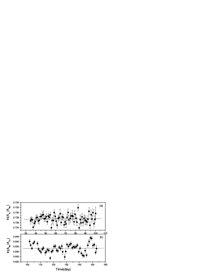
Figures 2(a) and 2(b) show the experimental data of versus and corresponding linear fit lines before and after exchanging the source positions, respectively. The value is determined by the slope in each case. The value of derived from Figs. 2(a) and 2(b) are and , respectively. By fitting the exponential decrease of versus time for the two cases, we obtain the weighted value for the decay rate of implanted in gold and in beryllium. Then using equal to 0.012986, we obtain the values of % before the source exchange and ()% after the source exchange. This result indicates that within our experimental precision, the large change in the decay rate of in Be and Au is not observed and an upper limit 0.12% of this change can be set.
In conclusion, we have performed the first-ever measurement of the possible change in the decay rate of implanted in natural beryllium and natural gold. According to the large difference of electron affinity between beryllium and gold, a large change of 0.7% in the decay rate of in the two cases is expected. However, within our measurement precision ( and 0.12% for the measurement before and after the source exchange respectively) no difference between the decay rates of implanted in beryllium and in gold is observed. This result implies that except electron affinity the lattice structure of the host medium in which the atom sits has to be taken into account.
Our experimental result also indicates that the difference between the average electron numbers in -shell (2 state) of atoms in the two samples is smaller than our measurement precision of %, since the -shell capture rate should essentially remain unchanged in different environments. According to the calculation of Ray et al., which has taken the lattice structure into account, the average number of 2 electrons of atoms implanted in natural beryllium sample should be equal to 0.443 while that in the gold sample should be 0.416. Thus, from Hartree’s calculation that the ratio of the square of the beryllium 2 electronic state wave function (2 electrons) to that of 1 state wave function at the nucleus (r=0) is 3.31%, the decay rate of in natural beryllium should be faster than that in gold by 0.045%. However, this value is beyond the reach of our measurement precision.
Acknowledgement
We are grateful to Amlan Ray at Variable Energy Cyclotron Centre of India for useful discussion. We also acknowledge Da-Qing Yuan and Chao-Fan Rong for using their gamma-ray spectroscopy laboratory.
References
- [1] Emery G T 1972 Ann. Rev. Nucl. Sci. 22 165
- [2] Dostal K P, Nagel M and Pabst D 1977 Z. Naturforsch. 32a 345
- [3] Bahcall J N 1996 Astrophys. J. 467 475
- [4] Kerr R A 1999 Science 286 882
- [5] Johlige H W, Aumann D C and Born H J 1970 Phys. Rev. C 2 1616
- [6] Wolfenstein L and Krastev PI 1997 Phys. Rev. D 55 4405
- [7] Chauhan B C and Dev S 1997 J. Phys. Soc. Jpn. 66 917
- [8] Junghans A R et al. 2002 Phys. Rev. Lett. 88 041101
- [9] Huh C-A 1999 Earth Planet. Sci. Lett. 171 325
- [10] Liu L-G and Huh C-A 2000 Earth Planet. Sci. Lett. 180 163
- [11] Jaeger M et al. 1996 Phys. Rev. C 54 423
- [12] Ray A et al.1999 Phys. Lett. B 455 69
- [13] Norman E B et al.2001 Phys. Lett. B 519 15
- [14] Souza D J et al. 2002 J. Nucl. Sci. Technol. Suppl. 2 (August) 470
- [15] Lide D R (Ed.) 2002 CRC Handbook of Chemistry and Physics (Florida and: CRC Press 2002 83rd) vol 83 pp 10-147
- [16] Firestone R B et al. 1996 Table of Isotopes CD-ROM (New York and: John Wiley & Sons Inc. 1996 8th.) vol 8 p 272
- [17] Ray A et al. 2002 Phys. Lett. B 531 187
- [18] Ray A et al. 2002 Phys. Rev. C 66 012501(R)
- [19] Hartree D R and Hartree W 1935 Proc.Proc. Roy.R. Soc. (London) A 150 9