Self-localization of laser induced tumor coagulation
limited by heat
diffusion through active tissue
Abstract
We analyze the necrosis growth due to thermal coagulation induced by laser light absorption and limited by heat diffusion into the surrounding live tissue. The tissue is assumed to contain a certain tumor in the undamaged tissue whereof the blood perfusion rate does not change during the action. By contrast, the normal tissue responds strongly to increase in the tissue temperature and the blood perfusion rate can grow by tenfold. We study in detail the necrosis formation under conditions typical for a real course of thermal therapy treatment, the duration of the action is taken about 5 minutes when a necrosis domain of size about or above 1 cm is formed. In particular, if the tumor size is sufficiently large, it exceeds 1 cm, and the tissue response is not too delayed, the delay time does not exceed 1 min, then there are conditions under which the relative volume of the damaged normal tissue is small in comparison with the tumor volume after the tumor is coagulated totally.
I Laser induced heat diffusion limited tissue coagulation
Thermal coagulation of living tissue caused by local heating due to laser light absorption is one of the novel thermotherapy techniques of tumor treatment which is currently undergoing clinical trials (see, e.g., I3 ). Thermal coagulation is used to form a necrosis domain of desired form for the removal of the malignant tissue. So mathematical modeling of the necrosis growth is required, first, to find out the physical limitations and the basic characteristics of the treatment and, second, to optimize the therapy course. However, living tissue is extremely complex in structure, thereby, for the adequate theoretical model to be developed and for the mathematical modeling of the given process to be implemented reliably typical limit cases should be singled out and studied individually.

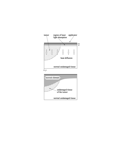
One of these cases is the laser induced heat diffusion limited tissue coagulation we9 ; we10 . It is characterized by the following features (Fig. 1). Absorption of laser light delivered into a small internal region of living tissue causes the temperature to attain such high values (about or above C) that lead practically to immediate thermal coagulation in this region. Heat diffusion into the surrounding live tissue causes its further coagulation, giving rise to the growth of the necrosis domain. In this case heat diffusion plays a significant role in the necrosis growth because the necrosis size exceeds the depth of laser light penetration into the tissue. Therefore, the temperature distribution inevitably has to be substantially nonuniform and for the tissue to coagulate at peripheral points heat diffusion should cause the temperature to grow at these points. The latter property singles out the specific mode of thermal coagulation under discussion from other possible types of thermotherapy treatment and that is why we refer to the necrosis growth under the given conditions as to thermal tissue coagulation limited by heat diffusion. In particular, it turns out that the optimal implementation of this thermotherapy mode is characterized by the formation of necrosis domains of size cm and by the treatment duration of – min (where cm2/sec is the tissue temperature diffusivity) we9 ; we10 .
Heat diffusion in the live tissue is affected substantially by blood perfusion causing the heat sink CH80 . Thus, the characteristics of the spatial distribution and the dynamics of the blood perfusion rate should have a substantial effect on the necrosis growth limited by heat diffusion. Therefore, in modeling this therapy mode one has to take into account the tissue response to the temperature growth which can locally give rise to a tenfold increase in the blood perfusion rate Song84 . The latter effect, in particular, is responsible for a substantially nonuniform distribution of the blood perfusion rate and visually manifests itself in a red ring (“hyperemic ring”) appearing around the necrosis region during the thermotherapy treatment.
In the previous papers we1 ; we2 ; we3 ; we4 ; we5 ; we6 ; we7 ; we8 basing on the free boundary description we have developed a model for the heat diffusion limited thermal tissue coagulation and studied the properties of the corresponding necrosis growth. In particular, we have shown that the given mode of thermal coagulation comprises two stages, fast and slow. At the former the necrosis domain grows fast and its size attains values of order cm. The latter is characterized by a substantially slower growth of the necrosis domain. Exactly at this stage the necrosis growth is governed by the competition between the heat diffusion into the surrounding undamaged tissue and the heat dissipation caused by blood perfusion. Therefore, a sufficiently strong tissue response to temperature variations which, in turn, gives rise to an essential local increase in the blood perfusion rate has to affect the necrosis formation. Namely, the higher is the local blood perfusion rate, the smaller is the size of the necrosis domain.
Inside tumors the blood vessels are abnormal, in particular, they have lost the ability to expand substantially in the response to tissue heating. As a result, inside a tumor the blood perfusion rate can increase no more then twice under strong local heating, although in the unaffected tissue the blood perfusion rate inside a tumor typically exceeds the perfusion rate in the normal tissue Song84 . So, if the normal tissue exhibits the strong response to temperature variations then the boundary of the necrosis domain will penetrate deeper insider the tumor than in the normal tissue during the thermal therapy course. Therefore in this case, first, the relative volume of the damaged normal tissue can be less than the volume of the tumor after its coagulation. Second, when the necrosis boundary reaches the tumor boundary the necrosis growth should slow down. At first glance it would seem that the necrosis growth is mainly confined to the tumor space. Exactly this phenomenon is the subject of the present paper and will be called the self-localization of the necrosis growth in active living tissue with a tumor.
II Free boundary model of the necrosis growth
We study the necrosis formation caused by laser light absorption in living tissue with a tumor applying to the following model we9 ; we1 ; we2 ; we3 ; we4 ; we5 ; we6 ; we7 ; we8 . The laser light absorption causes heat generation and, as a result, thermal coagulation of the tissue. For simplicity sake the heat generation rate is considered to be a beforehand given function of the spatial coordinate that is independent of the tissue state and the time (certainly, for and is the beginning of the action on the tissue). Besides, we assume the heat generation rate to be localized in a small region adjacent to the applicator whose thickness is substantially less than the characteristic size cm of the necrosis domain formed during a typical course of thermotherapy treatment.
In modelling heat propagation in the tissue we single out three regions: the necrosis domain , where the blood perfusion rate is equal to zero
| (1) |
the undamaged tumor tissue , and the undamaged (live) normal tissue . Inside the necrosis domain the tissue temperature obeys the heat diffusion equation for solids:
| (2) |
where and are the specific heat and density of the tissue, and is the cellular tissue heat conductivity. In the undamaged tissue (including also the undamaged part of the tumor) the temperature is governed by the bioheat equation (see also, BOOK ):
| (3) |
Here is the temperature of arterial blood in systemic circulation, is the value of the blood perfusion rate locally averaged over spatial scales
| (4) |
where and is the mean ratio of the individual length to radius of blood vessels forming peripheral circulation, the cofactor takes into account the counter-current effect WJ85 ; WXZE97 , and the factor allows for the renormalization of the heat conductivity due to blood flow. It should be pointed out that the scale is exactly the minimal scale on which equation (3) is justified BOOK and the characteristic size of the necrosis domain formed during this mode of thermal therapy can be estimated as :
| (5) |
Finding the relationship between the averaged and true blood perfusion rates, and , we have to take into account that the scale of averaging in its turn depends on the local value of (expression (4)). This dependence enables us to specify this relationship in the form BOOK :
| (6) |
where is also a constant of order unity. Equation (6) should be subjected to a certain boundary condition at the interface of the necrosis domain because it makes no sense to average the blood perfusion rate over the necrosis domain impermeable to blood. The physical layer separating the necrosis domain and the undamaged tissue where the local vascular network is not damaged is complex in structure and contains a spatial increase of the blood perfusion rate from zero to the value in the undamaged tissue. In order to avoid the problem of analyzing the blood perfusion rate in this layer we take into account the following simplifying circumstance. On one hand, the typical size of the necrosis domain formed during a thermal therapy course and the characteristic length of temperature variations are of the same order about 1 cm. So, particular details of spatial variations in the blood perfusion rate on scale much less than 1 cm are not the factor. On the other hand, the damaged part of the vascular network located inside the necrosis domain is most probable to be made up of an artery and vein having supplied previously this region with blood as a whole and of shorter vessels formed by their branching. Therefore, the region containing the vascular network part in which blood flow is remarkably disturbed because of the necrosis formation does not exceed substantially the necrosis domain. The latter enables us not to make difference between the given layer and the interface and to deal with a sharp jump of the blood perfusion rate at the necrosis interface. The desired boundary condition imposed on the averaged blood perfusion rate meets the requirement that the normal gradient of the averaged blood perfusion rate be equal to zero at the interface :
| (7) |
We note that the adopted boundary condition will not hold if a large vessel goes through the necrosis domain. However, the probability of this event is small enough and this case should be analyzed individually.
Now let us specify the tissue response to temperature variations. Only the undamaged normal tissue is considered to respond to local heating, inside the undamaged tissue of the tumor the blood perfusion rate maintains the initial value assumed for simplicity sake to be the same in the normal tissue and the tumor
| (8) |
Blood vessels can expand only to a certain extent as the temperature grows. So when it becomes high enough, , the blood perfusion rate attains a large but finite value and remains approximately constant. Keeping in mind this feature we describe the normal tissue response to local heating by the equation:
| (9) |
Here is the delay time of the tissue response and the function is of the form
| (10) |
where and the temperature at which the blood vessels exhaust their ability to expand is estimated as –C.
Equations (2) and (3) governing the evolution of the tissue temperature should be completed by the boundary conditions relation the temperature field and the necrosis interface . To solve this problem we have developed the free boundary model that assumes the temperature and the heat flux to have no jumps, i.e. the temperature distribution meets the following boundary conditions
| (11) |
and the normal velocity of the interface is given by the expression
| (12) |
Here the constant and the function
| (13) |
is actually a convenient approximation of the Arrhenius dependence of the thermal coagulation rate . The available experimental data J94 for the temperature dependence of the exposure time enable us to estimate the value of as C (C for pig liver at C). Below in numerical calculations the dependence (13) will be taken in the form
| (14) |
where the temperature is in degrees Celsius.
III Results of numerical simulation. Self-localization of the necrosis growth in living tissue with a tumor
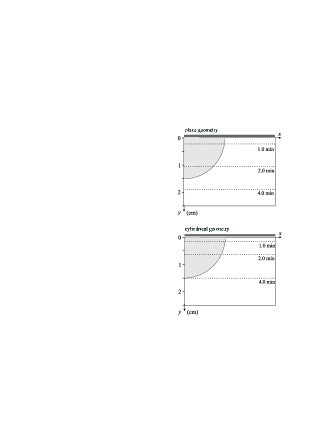
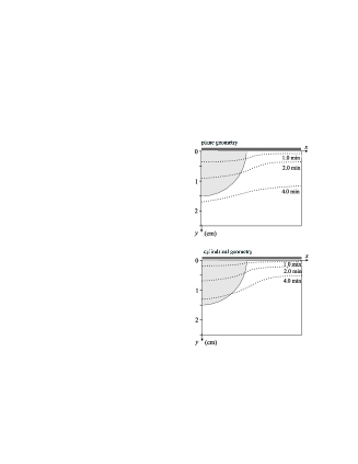
The stated model has been used to analyze numerically the necrosis growth in the case shown in Fig. 2. The laser light absorption near the applicator gives rise to the heat generation rate uniform along the -axis and decreasing with as , where the characteristic thickness of the laser light absorption layer is chosen less then the characteristic size cm of the necrosis domain formed during the thermal therapy mode under consideration, . The intensity of laser light has taken such values that the tissue temperature directly near the applicator, , be about 80 to 90 ∘C ( for large tumor and for small tumor). We have studied two opposite cases specified by the relation of the scale (see expression (5)) and the tumor size . For a small tumor, , the self-localization phenomenon cannot come into being and this case is presented for illustration only. For a large tumor, , there are conditions under which the self-localization is pronounced as is demonstrated directly in the present section. Besides, we have considered the -region of plane geometry as well as cylindrical one in order to compare the necrosis growth for applicators of different geometry, the surface plane applicator and internal cylindrical one. In the latter case the radius of the applicator also was assumed to be small in comparison with the scale .
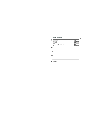
In numerical simulation we have used the following typical values of the thermal conductivity W/cmK, the heat capacity J/g K, and the density g/cm3 of the tissue, as well as set the blood perfusion rate min-1 and the factors , . Figure 3 demonstrates the necrosis growth in living tissue without response. In this case the existence of the tumor has no effect on the necrosis growth. A sufficiently small tumor also cannot substantially affect the necrosis growth, as is seen in Fig. 4 exhibiting the necrosis growth in living tissue with strong response without delay. The tumor radius was chosen equal to cm the tissue response to heating was assumed to be strong, , and without delay, . This case has been considered because under such conditions the effect of large tumor on the necrosis growth is most pronounced.
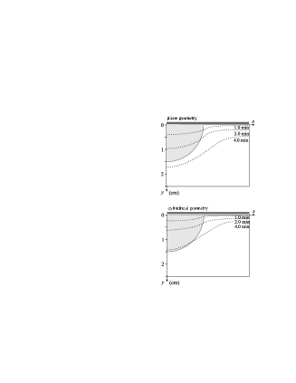
When the tumor size is sufficiently large, , and the tissue response is not too delayed, the self-localization phenomenon comes into being as demonstrated in Figures 5–6. Namely, Fig. 5 exhibits the necrosis growth in living tissue containing a tumor of size of cm where the tissue response is strong, , and not delayed, . Exactly this situation matches the conditions under which the self-localization is most pronounced. When the tissue response is delayed the effect of the tumor existence is weakened as it is demonstrated in Fig. 6, where we set min.
IV Closing remarks
Concluding the paper we would like to state once more the obtained result. Namely, we have analyzed the necrosis growth due to thermal coagulation induced by laser light absorption and limited by heat diffusion into the surrounding live tissue. The tissue is assumed to contain a certain tumor where the blood perfusion rate does not change during the action. The latter certainly concerns only the undamaged part of the tumor. By contrast, the normal tissue responds strongly to increase in the tissue temperature and, as a result, the blood perfusion rate can grow by tenfold.
We studied in detail the necrosis formation under conditions typical for a real course of thermal therapy treatment, the duration of the action has been taken about 5 minutes when the necrosis domain of size about or above 1 cm is formed. In particular, we have shown that if the tumor size is sufficiently large, it exceeds 1 cm, and the tissue response is not too delayed, the delay time does not exceed 1 min, then there are conditions under which the relative volume of the damaged normal tissue is small in comparison with the tumor volume after the tumor is coagulated totally.
Acknowledgements.
This work was supported by STCU grant #1675.References
- (1) Laser-Induced Interstitial Thermotherapy, G. Müller and A.Roggan, eds. (SPIE Optical Engineering Press, Bellingham, Washington, 1995).
- (2) I. A. Lubashevsky, A. V. Priezzhev, and V. V. Gafiychuk.“Laser induced heat diffusion limited tissue coagulation as a laser therapy mode”. In: Laser-Tissue Interaction XI: Photochemical, Photothermal, and Photomechanical, D. D. Duncan, J. O. Hollinger, S. L. Jacques, Eds., Proc. SPIE 3914, 66–74 (2000).
- (3) I. A. Lubashevsky, V. V. Gafiychuk, and A. V. Priezzhev. “Laser Induced Heat Diffusion Limited Tissue Coagulation: Problem and General Properties”, e-print: physics/0101002.
- (4) M. M. Chen and K. R. Holmes, “Microvascular contributions in tissue heat transfer”. Ann. N. Y. Acad. Sci., 335, 137–154 (1980).
- (5) C. W. Song, A. Lokshina, I. G. Rhee, M. Patten, and S. H. Levitt. “Implication of blood flow in hyperthermia treatment of tumors”. IEEE Trans. Biom. Eng., BME-31 (1), 9–15 (1984).
- (6) I. A. Lubashevsky, A. V. Priezzhev, V. V. Gafiychuk, and M. G. Cadjan. “Free-boundary model for local thermal coagulation”. In: Laser-Tissue Interaction VII, S. L. Jacques, Editor, Proc. SPIE 2681 81–91 (1996).
- (7) I. A. Lubashevsky, A. V. Priezzhev, V. V. Gafiychuk, and M. G. Cadjan. “Dynamic free boundary model for laser thermal tissue coagulation”. In: Laser-Tissue Interaction and Tissue Optics II, H. J. Albrecht, G. Delacrétaz, T. H. Meier, R. W. Steiner, and L. O. Svaasand, Editors, Proc. SPIE 2923, 48–57 (1996).
- (8) I. A. Lubashevsky, A. V. Priezzhev, V. V. Gafiychuk, and M. G. Cadjan. “Local thermal coagulation due to laser–tissue interaction as irreversible phase transition”. J. Biomed. Opt. 2(1), 95–105 (1997).
- (9) I. A. Lubashevsky, A. V. Priezzhev, and V. V. Gafiychuk. “Free boundary model for local thermal coagulation. Growth of a spherical and cylindrical necrosis domain”. In: Laser–Tissue Interaction VIII, S. L. Jacques, Editor, Proc. SPIE 2975, 43–53 (1997).
- (10) I. A. Lubashevsky and A. V. Priezzhev. “Laser induced heat diffusion limited tissue coagulation. I. Form of the necrosis boundary caused by random temperature nonuniformities”. In: Laser-Tissue Interaction, Tissue Optics, and Laser Welding III, G. Delacrétaz, L. O. Svaasand, R. W. Steiner, R. Pini, and G. Godlewski, Editors, Proc. SPIE 3195, 143–150 (1998).
- (11) I. A. Lubashevsky, A. V. Priezzhev, and V. V. Gafiychuk. “Effective interface dynamics of heat diffusion limited by thermal coagulation”, J. Biomed. Opt., 3(1), 102–111 (1998).
- (12) I. A. Lubashevsky and A. V. Priezzhev. “Laser induced heat diffusion limited tissue coagulation. II. Effect of random temperature nonuniformities on the form of a spherical and cylindrical necrosis domain”. In: Laser–Tissue Interaction IX, S. L. Jacques, Editor, Proc. SPIE 3254, 61–68 (1998).
- (13) I. A. Lubashevsky and A. V. Priezzhev. “Effect of the blood vessel discreteness on the necrosis formation during laser induced thermal coagulation limited by heat diffusion”. J. Biomed. Opt. 4(2), 248–255 (1999).
- (14) V. V. Gafiychuk and I. A. Lubashevsky. Mathematical Description of Heat Transfer in Living Tissue, (VNTL Publishers, Lviv, 1999); e-print: adap-org/9911001,9911002.
- (15) S. Weinbaum, L. X. Xu, L. Zhu, and A. Ekpene. “A new fundamental bioheat equation for muscle tissue: Part I–Blood perfusion term”, ASME J. Biomech. Eng. 119, 278–288 (1997).
- (16) S. Weinbaum and L. M. Jiji. “A new simplified bioheat equation for the effect of blood flow on local average tissue temperature”, ASME J. Biomech. Eng. 107, 131–139 (1985).
- (17) S. L. Jacques. “Laser–tissue interactions: Photochemical, photothermal, and photomechanical”, Surgical Clinics of North America 72(3), 531–558 (1992).