Many-body Effects in Angle-resolved Photoemission: Quasiparticle Energy and Lifetime of a Mo(110) Surface State
Recent investigations of strongly correlated electron systems have questioned the validity of one of the most fundamental paradigms in solid state physics - Fermi liquid theory. The latter picture is based on the existence of ”quasiparticles”, or single-particle-like low energy excitations which obey the exclusion principle and have lifetimes long enough to be considered as particles. Strictly speaking, the quasiparticle concept is restricted to zero temperature and a narrow region around the Fermi level [1], but its usefulness often continues to finite temperatures, and energies away from the Fermi level [2]. Indications for possible non-Fermi-liquid behavior have been found in some organic one-dimensional conductors [3] and in the normal state of high temperature superconductors [4]. A whole variety of experimental techniques have been employed in the search for such behavior, including resistivity measurements [5], infra-red spectroscopy [6], scanning tunneling spectroscopy [7] and time-resolved two-photon photoemission [8]. Angle-resolved photoemission (ARPES) has an advantage, in that the energy and lifetime of the photo-hole are directly observable in the experiment. ARPES in principle measures the quasiparticle spectral function [9]:
| (1) |
where represents the energy of the state in the Hartree potential and is the quasiparticle self-energy reflecting many body interactions. Thus, momentum resolved self-energies are directly accessible in the experiment and as such, ARPES represents a crucial experimental probe for the presence or absence of Fermi liquid behavior. Furthermore, complications connected to the lifetime of the photoelectron (in three-dimensional systems) may be overcome in quasi low-dimensional systems. Indeed, there have already been several photoemission studies, which quantitatively compare peak widths to calculated lifetimes for metallic surface states [10] and two-dimensional states in layered materials [11].
When considering the lifetime of the valence hole, there are three main decay mechanisms: electron-electron scattering, electron-phonon scattering and impurity (defect) scattering. In three-dimensional systems, the electron- electron interaction contributes to the total width or inverse lifetime with the term . This scattering rate does not depend on the form of the interaction, but it may depend on the shape of the Fermi surface. If the scattering process is two-dimensional, then the quadratic energy (temperature) dependence is modified by a logarithmic factor [12]. Previous attempts to observe this term for ”prototypical” Fermi liquids, free-electron-like metals, have failed because is often too small. Indeed, with estimated values of for , contributes less than 5 meV variation through the whole band for the surface states studied [10]. Further, the temperature dependent contribution is negligible, being of the order of 0.1 meV at room temperature. The electron-phonon scattering contribution to the inverse lifetime is given by:
| (2) |
where is the Eliashberg coupling function, and and are the Fermi and Bose-Einstein functions. This term monotonically increases with energy over the region (for T=0), where is the cutoff of the phonon spectrum. The exact functional form is slightly dependent on the phonon spectrum. The temperature dependence of is approximately linear at higher temperatures, with slope , where is the electron-phonon coupling constant. For most metals 20 meV100 meV, and falls between 0.1 and 1.5. Impurity scattering is elastic provided that the energy gap between the impurity ground state and its lowest excited state is large compared with the hole energy. To a first approximation, it is proportional to the impurity concentration, but independent of energy or temperature. Thus, at sufficiently low temperature in any real system, it will be the dominant decay mechanism for the hole close to . If the scattering mechanisms are independent, the total scattering rate is given by . Virtually all temperature dependence in reflects the term, whilst any energy dependence falls within two distinct regions. In a narrow region (at T=0) the only significantly varying term is . Any measurable change out of that region, is most likely due to the term. The real part of the quasiparticle self-energy is also known for all three mechanisms of scattering. Of these, electron-phonon coupling is the only term which is able to alter the quasiparticle mass in a narrow energy region around the Fermi level.
In the present paper, we report a detailed study of the lifetime effects associated with photo-excitation of a surface state on the Mo(110) surface. The electronic structure of this surface has already been the subject of several studies [13]. Our own tight-binding calculations in the slab formulation reproduce the presence of -derived surface states or resonances in the mid- region of the and lines, and indicate a bandwidth of the order of 1-2 eV. It is well established that the line-shape or width of a photoemission peak associated with excitation from such states reflects the lifetime of the photo- hole and is independent of any momentum broadening associated with the photoelectron [14]. However, the fact that bulk states are available for the surface state photo-hole to scatter into, means that the three-dimensional decay rates are applicable even in the present case.
The experiment was carried out at the National Synchrotron Light Source, using the VUV undulator beamline U13UB, which is based on a 3-m normal incidence monochromator. The photon energy used in the study was 15.16 eV. The electron analyzer was a Scienta SES-200, which uses a two dimensional micro-channel plate as a detector. The detector collects simultaneously a wide energy window and a wide angular window () of excited photoelectrons. This greatly reduces the time needed for data acquisition. The spread of the UV light was less than 2 meV, and the energy resolution of the electron analyzer was around 5 meV. The combined energy resolution therefore makes a minimal contribution to widths observed in this study. The angular resolution is of the order of which contributes 10 to 15 meV to the width of the studied state. The sample was cleaned by oxidation cycles (at 1400 K in an oxygen atmosphere of Pa), followed by flashes to K. The sample temperature was measured with a W-WRe thermocouple. The base pressure was Pa, more than 90 % of which was hydrogen.
Fig. 1 shows a typical spectrum of the Mo(110) surface, taken along the symmetry line, with the sample held at 70 K. The state shown in the figure corresponds to the surface resonance which closes the elliptical hole Fermi orbit around [13]. The spectrum in the inset that shows a narrow energy region close to the Fermi level was taken in a short time interval, 1 min. Such short measuring intervals are required because, as shown later, there is a significant influence from adsorption of residual gasses. Important qualitative observations can be made directly from the spectrum shown in the inset: the state sharpens up on going towards the Fermi level, and there is an obvious change in band velocity near . We focus our attention on these observations. Quantitative analysis is performed by taking slices through the spectrum, either at constant emission angle, or at constant energy to obtain the spectral intensity as a function of energy or emission angle, respectively. The two methods are equivalent in the sense that they provide information on the same A. However, analyzing the data by taking slices at constant energies is often more convenient because it is not affected by the Fermi distribution in the same way that slices at constant angle are. In our analysis we have used ”horizontal” cuts (at constant energy) to extract the dispersion, and ”vertical” cuts (at constant angle) to extract the width of the quasiparticle peak.
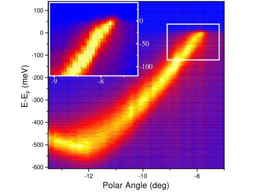
Fig. 2 shows representative spectra obtained by slicing the spectral intensity in the ”vertical” direction. Spectra are divided by an experimentally determined Fermi cut-off. The analysis was performed by fitting the spectra to Lorentzian peaks with a small linear background.
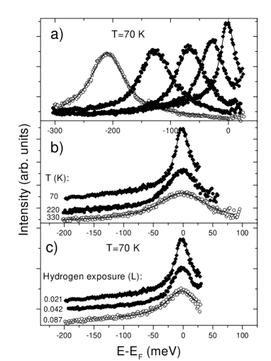
The high-energy limit for the fitting interval was always below . Influences of the three experimental parameters; binding energy (a), temperature (b) and hydrogen exposure (c) are shown, one being varied, as the others two are kept constant. The trends are obvious; the width increases with all three parameters.
The energy dependence of the quasiparticle peak width is shown in Fig. 3. The error bars are statistical uncertainties from the fits to peaks such as those shown in Fig. 2(a). The peak width shows a minimum at , a sharp increase within the interval meV, followed by a slower increase at higher binding energies. The same behavior is also found for the angular peak width (”horizontal” cuts). At the same temperature, all spectra show the same dependence, offset by a constant value dependent on the surface impurity level. The sharp increase in width near the Fermi level reflects the electron-phonon scattering. This is confirmed if we compare the experimental points with calculated from equation (2), using a theoretical for bulk molybdenum [15]. Both the range and magnitude of the increase agree well with calculation, suggesting that the theoretical bulk electron-phonon coupling constant from ref. [15], , applies also to the surface studied here. Further, the surface Debye energy is similar to the bulk value ( meV), in accordance with recent measurements of surface phonon dispersions [16]. To obtain the agreement between the experimental points and the theoretical curve it was necessary to shift the latter uniformly by 26 meV. We attribute this difference to impurity scattering. The observation that the experimental points shift uniformly by the same amount is an indication that the impurity scattering is independent of energy. If we subtract the calculated electron- phonon contribution from the total width, we are left with a monotonic increase in binding energy that can be fitted with a parabola. This component is the electron-electron scattering term, with coefficient . Within the Born approximation, . Our result is consistent with the on-site Coulomb repulsion eV predicted for molybdenum [17] if we equate with the bandwidth of the surface state ( eV). It should be noted that for such a rapidly dispersing band with negative velocity, the measured energy width could be smaller than the intrinsic width [18]. We estimate that in our case this ”compression” of the spectral width is less than 7 %, which has minor consequences on our results.
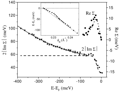
Also shown in Fig. 3 is the real part of the quasiparticle self-energy. Experimentally, it is obtained by subtracting a straight line from the measured dispersion of the quasiparticle peak (as shown in the inset of Fig. 3). In this energy range the straight line represents a good approximation for the dispersion of the ”non-interacting” system (i.e. the system without electron- phonon coupling). It is chosen to have the same as the quasiparticle dispersion (Luttinger theorem for interacting fermions) and to match the quasiparticle dispersion in the range meV. This procedure gives only the electron- phonon term for the real part of the self-energy. The component reflecting the electron-electron interaction stays hidden in the dispersion of the ”non- interacting” system. Also shown is the electron-phonon contribution to the real part of the self-energy, obtained via the Kramers-Kronig transformation of the calculated imaginary part.
Fig. 4 shows the width of the quasiparticle peak as a function of temperature (a) and as a function of hydrogen exposure (b) for two different binding energies. In Fig. 4(a), we also show electron-phonon contributions calculated from (2), and shifted up by 26 meV. The excellent agreement with the experimental points confirms that the temperature dependence reflects the electron-phonon interaction. Linear fits to the experimental data points produce different values for the electron-phonon coupling constant for the two binding energies: (at ) and (at meV), compared to the theoretical value of 0.42.
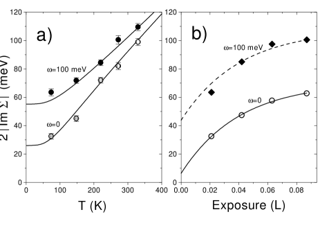
The observation that the width of the quasiparticle peak always has a significant constant term indicating the presence of impurity scattering lead to further investigation. It is known that this surface state is very sensitive to hydrogen adsorption. Fig. 4(b) shows how the width changes with the exposure to residual hydrogen. Note that it saturates with exposure . If the scattering rate is proportional to the concentration of adsorbed particles, the experimental points become a measure of the concentration. Since the number of free adsorption sites decays exponentially with exposure, the concentration of adsorbed atoms as a function of exposure should change as , where is the adsorption probability and () is the initial (saturation) concentration. The width of the quasiparticle peak can be fitted with the same dependence (lines). It is notable that extrapolation to zero exposure results in a residual width of meV at . Electron-phonon coupling contributes with meV for T=70 K. However, we should also note that there is some uncertainty in the initial coverage due to the change in adsorption conditions between flashing the sample and the measurement.
In conclusion, we have analyzed the dispersion and the width of the Mo(110) surface state and isolated different mechanisms for scattering of the quasiparticle. For the first time it has been possible to isolate the electron- electron, electron-phonon and electron-impurity scattering contributions to the quasiparticle lifetime. The electron-electron contribution is shown to be an order of magnitude higher than that for -derived states. Our study shows that ARPES offers the possibility of momentum resolving the electron-phonon contribution to the real and imaginary parts of the self-energy.
This work is supported by the U.S. Department of Energy (DOE) under Contract No. DE-AC02-98CH10886.
REFERENCES
- [1] D. Pines and P. Nozières, The Theory of Quantum Liquids (Benjamin, New York, 1969).
- [2] G. Grimvall, The Electron-Phonon Interaction in Metals (North- Holland, New York, 1981).
- [3] C. Bourbonnais et al, J. Phys. Lett. 45 L-755 (1984); B. Dardel et al, Europhys. Lett. 24, 687 (1993).
- [4] C. G. Olson et al, Phys. Rev. B 42, 381 (1990).
- [5] M. Gurvitch and A. T. Fiory, Phys. Rev. Lett. 59, 1337 (1987).
- [6] G. A. Tomas et al, Phys. Rev. Lett. 61, 1313 (1988).
- [7] J. Li, et al, Phys. Rev. Lett. 81, 4464 (1998).
- [8] W. Nessler et al, Phys. Rev. Lett. 81, 4480 (1998).
- [9] In sudden approximation ARPES intensity is given by I, where A is the spectral function, is the Fermi function and M is a slowly varying term containing matrix elements for photoemission.
- [10] B. A. McDougall, T. Balasubramanian and E. Jensen, Phys. Rev. B 51, 13891 (1995); T. Balasubramanian et al, Phys. Rev B 57, R6866 (1998); P. Hofmann et al, Phys. Rev. Lett. 81, 1670 (1998).
- [11] R. Claessen et al, Phys. Rev. Lett. 69, 808 (1992).
- [12] C. Hodges, H. Smith and J. W. Wilkins, Phys. Rev. B 4, 302 (1971).
- [13] K. Jeong, R H. Gaylord and S. D. Kevan, Phys. Rev. B 38, 10302 (1988); K. Jeong, R H. Gaylord and S. D. Kevan, Phys. Rev. B 39, 2973 (1989).
- [14] J. B. Pendry in Photoemission and the Electronic Properties of Surfaces, B. Feuerbacher, B. Fitton and R. F. Willis (Wiley, New York, 1978).
- [15] S. Y. Savrasov and D. Y. Savrasov, Phys. Rev. B 54, 16487 (1996).
- [16] J. Kröger, S. Lehwald and H. Ibach, Phys. Rev. B 55, 10895 (1997).
- [17] W. A. Harrison, Electronic Structure and the Properties of Solids (W. H. Freeman & Co, San Francisco, 1980).
- [18] E. D. Hansen, T. Miller and T. -C Chiang, Phys. Rev. Lett. 80, 1766 (1998).