Spontaneous curvature-induced pearling instability
I Introduction
Vesicles naturally exist in different shapes. The most common shapes are spherical and oblates like vesicles, toroidal vesicles also exist under some particular conditions [1, 2]. Tubular vesicles are also common in nature, but they are not an equilibrium solution for the elastic energy [3]. One of the most fascinating phenomena are the shape changes of cells and the manifold of local instabilities of the cell plasma membrane in particular and the vesicles membranes in general. Under physiological condition the cell (vesicle) exhibits the familiar biconcave shape (the discoide form). Removal of some particular proteins leads to the echinocyte form, or to the stomatocyte form [4]. The interest of vesicles as model system of living cells, for drug delivery or for different industrial usage…etc. is the origin of several fundamental studies over a couple of decades [5]. Local instabilities play an essential role for material transport process between cellular compartments or through the plasma membrane. A most important example is the transport of newly synthesized membranes to the cell envelope, and it involves three steps: a) budding of vesicles, b) their fission from a parent membrane, c) their fusion with the target compartment. Tubular vesicles when excited by mean of different techniques (UV radiation, optical tweezers…), exhibit curious behavior similar to the Rayleigh instability in liquid column. This behavior is known as “pearling” [6, 7, 8]. After an exposition to radiation, tubules become a succession of spheres, separated by narrow necks. In the case of UV radiation, a long time radiation induces a separation of the spheres [7]. When the tubules are excited by optical tweezers, the shape of the spheres depends on the intensity of the laser and a long time exposition of the optical tweezers on the tubules induces a separation of the spheres separated by narrow necks [6]. Also it was found that vesicles can suffer the same pearling instability when subjected to a change of the water Ph between the inside and the outside of the vesicle [9].
In this article we study an instability that suffer tubular vesicles made of a water soluble amphiphile. This instability is similar to the ones cited above. However, the tubules are not excited via an irradiation, but the interlayer distance may be affected by incorporating a solvent of the aliphatic chaines of the amphiphiles into the layer. In Section II we will describe the experiment and in section III we present the theoretical model of the instability observed and finally a conclusion is presented.
II Experiment
It is known that Alkanes are more or less good solvents, depending on their chain length, for the aliphatic chain of the surfactant called AOT (sodium di-2-ethylhexyl-sulfosuccinate) at the oil-water interface[10]. AOT is a water-soluble surfactant, it forms micelles when dissolved in water up to a concentration equal to 30 mmol/L. Beyond this “solubility limit”, AOT forms spherical and multilamellar vesicles [12]. These vesicles are found to be opaque when observed through a microscope and present different layer when observed under a phase contrast micropscope. We believe they are onions like vesciles. However at low salinities and at low AOT concentrations, it forms unilamelar sphericals and prolate vesicles as well as dumbell shape vesicles. At high salinities (we investigated the range 0.1-0.175 mol/L), we found that AOT in brine forms tubular vesicles [12]. These tubular vesicles can be very long and can be of several order of magnitudes of their diameter. There are two ways of preparing this phase: Either solubilising AOT in a very short alkane (Heptane), and drying up the solution before adding water on the residue left after the evaporation of Heptane; or mixing first AOT and salt in a ration 1:3 in weight, then pouring gently the appropriate quantity of water to the AOT/Salt mixture to have the desired (AOT/Salt) concentrations. For the purpose of this work we used the second procedure, so we can avoid any kind of mixing of the two alkanes. If we prepare a low salinity solution by dissolving the amount of AOT in water and adding brine to bring the solution to the desired salinity, no vesicle is observed under the phase contrast microscope. The formation of these vesicles is probably due to an electrostatic effect between the amphiphiles heads [18]. In this article we will not describe the phase observed when changing salinities or amphiphiles concentration. Instead we will focus on a particular effect of dodecane on these tubules obtained at high salinities. An instability is studied and the wavelength selected at the onset of the instability is measured as a function of the radius of the unperturbed tubules. The selection is found to be a result of the incorporation of the oil into the layer of the tubule which causes the spontaneous curvature to deviate from zero.
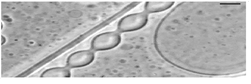
The vesicles are observed through an inverted phase contrast microscope ( objective, Nikon diaphot 200). The cell where the vesicles are observed is a 1-mm- thick glass cell, the gap between these two glass slides is also 1-mm- thick. The AOT-brine solution is left for a couple of days, so that any shape due to macroscopique flow -after transfering the solution from a test tube to the observation cell- desapears, though no shape transformation was observed after the transfer of the vesicle dispersion into the observation cell. The brine salinities used corresponding to the minimun of oil-water interfacial tensions and are 0.175 mol/L for dodecane and 0.075 mol/L for decane and an AOT concentration of 7.5 mmol/L and 4 mmol/L respectively. The CMC of the same system without salt is around 2.5 mmol/L. At 0.075 mol/L of NaCl, the stable shapes are essentially prolate and spheres; and at 0.175 mol/L of NaCl, the shapes are cylinders. This shape transformation from low salinities to high salinities is probably du to an electrostatic effect, leading to shape transformation) [13, 14]. We observed the effect of dodecane on stable tubules at 0.175 mol/L (at this salinity the Dodecane-Brine interface tension is minimum) [10]. The introduction of the oil into the cell does not perturbe hydrodynamically the solution and it is a noninvasive way to introduce the drop into the solution. To avoids instabilities such as Marangoni effect, the oil is introduced through a less than diameter orifice. The drop, with a volume of the order of , travells through the glasse by capillarity before it reaches the solution. The orifice is situated at the lateral wall of the cell, that is between the lower glass slide and the piece separating the two slides. The drop is then directly introduced into the solution. Due to the weak solubility of dodecane in water, and its slow diffusion, it takes several hours for the instability to start. Most probably the oil travells inside the solution after being incorporated in the swollen micelles. After the oil reaches the tubules situated far enough from the point where the drop was introduced, tubules become unstable, by forming pearls similar to the ones observed in Ref. [6, 9]. We have not noticed any shape changes when the gap between the horizontal walls of the observation cell is much less than 1 . Up to now, we are not able to know why this effect depends on the gap between the cells-walls where the vesicles are being observed. A sinusoidal instability is initiated, and develops to a peristaltic state, with a reduction in fluctuations. However large cylinders are stables, this will be clarified in the following model. The structure observed is periodic and typical necks between the pearls are apparent. In the case of thick walled vesicles, the pearls don’t disconnect. However tubules with thin layers, cut into separated spheres at the end of the instability. The time over which the spheres diconnect is still unknown. This kind of state is shown in (fig. 1).
Bar-Ziv and Moses [6] suggested, after an experiment where a tubular phospholipidic membrane was excited using an optical tweezers, that, the instability appears because of a change in the surface tension induced by the tweezers, by analogy to the Rayleigh instability of a column of liquid [15, 16, 17]. The surface energy should be the source of instability, however as the surface energy increases the linear analysis show that the instability develops only longwave modulations without any wavelength selection. We will show in the following that in the case of our experiment surface tension is not sufficient to select a well-defined periodic spatial modulation of tubular membranes. Instead, in our case an extra length scale becomes important in the problem. We introduce a simple model which introduce the physical meaning of this length scale.
Figure (2), shows the dependance of the dimensionless number () as a function of the initial radius of the tubule . The wavelength length at the onset hence , is measured whenever a tubule starts to suffers the instability. The dispersion in the data in figure 2 is probably due to the fact that the wavelength is not measured exactly at the onset with an error of the order of 2 seconds (that is after the establishement of the perstaltic state). The oil molecules reaches the tubules in a random way, so the onset is also random. The finer tubules start to suffer the instability earlier than the larger ones, this fact explains also the dispersion in the data of figure 2.
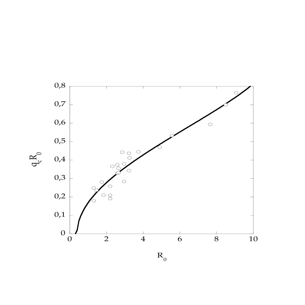
The characteristic slope of the experimental values of figure 2, represents an extra length (of the order of ) in the problem. The inverse of this length may be interpreted as a spontaneous curvature , measuring a symmetry breaking between the two sides of the membrane by incorporating different amount of oil molecules (in some sense is the analog of chirality in liquid crystals).
III Theory
One may models this by regarding a membrane layer as composed of two different layers separated by a distance and with different values of surface tension and surface elements one with and the other and . Therefore one has an energy of the form . This energy would include all the elastic contributions (curvature and surface tension of the whole layer) [18]. Expanding now both surface elements in powers of , with , one gets
Here is the mean curvature and is a geometrical neutral surface element between the two layers. Finally, adding a “bending” energy of a tube, the free energy is [20]:
| (1) |
Where is the rigidity, the “effective” surface tension of the bilayer and is the spontaneous curvature of the layer and is null when the bilayer is symmetric, or when the neutral surface of the layer is in the middle of the layer [19]. Here the spontaneous curvature is not introduced as a parameter, but as an assymetry in the layer and a shift of the neutral surface from the middle of the layer. Also the surface tension is expressed as combination of the different mechanical parameters of the mambrane.For an axisymmetric surface characterized by a local radius the mean curvature is
| (2) |
on the other hand the surface element is Here
Let be the radius of a non perturbed tube; as in [15] we put
| (3) |
(the square root appears because of conservation of the volume of the tube, note that cannot hold a value diffrent from zero) into the free energy getting a power expansion in :
| (4) |
here
is a dimensionless number, and the coefficient of the quadratic term is
| (5) | |||||
| (6) | |||||
| (7) |
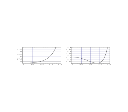
The linear instability appears for . Geometrically, the transition allows us to express the control parameter as a function of the pertubation wavenumber . For , has a minima at . The cylinder is stable for low values of , however, as one increases up to a critical value (), the tube suffers a long wavelength instability, without wave number selection. For , has a minima for . A short wavelength instability appears as soon as see figure (3). From the expresion (7) one get explicitly the threshold of the instability, hence the wave vector of the most instable mode (), and the value of the controle parameter () at the onset of the instability.
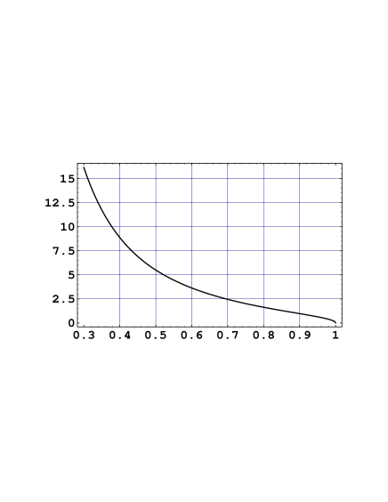
In the figure (4), one can see that the control parameter vanishes as soon as the radius of the tube is equal to the spontaneous curvature, i.e. when the tubules layer are symmetrical. From 7 and imposing one gets the critical wavenumber and the threshold. Being the first
| (8) |
Note that there are two different solutions, however the sign represents a situation where and this instability is possible only if takes negative values***Note that this negative surface energy makes instability even for ., however, the selected wavenumber in this case has a negative “mean slope”, that is, is a line essentially perpendicular to the experimental line given in figure 2. The critical surface tension for the case is
| (9) |
The fit represented in figure 2 was done using the expression (8), i.e. we tried , the fit gives (instead of 1.4) and this value is in the range of value measured for vesicles. In figure (3) the value of at the is of the order of 2.
At the critical point , the sign of the coefficient of the fourth power term , is positive for all values of such that . Therefore this peristaltic instability is supercritical (a second order transition). It is interesting to note that for large tubular radius there is no instability, as we can see in the expression ( 8) where becomes a complex number. This characteristic is indeed observed in the experiment [12]. On the other hand, for small tubules radius, the instability grows for , however this is not possible because of the conservation of volume, in other words a homogeneous growth of the is forbidden. In conclusion in both cases: i) for small tubular radius: (experimentally we get ) and for ii) large tubular radius (experimentally ) the tubules are stables.
IV conclusion
The oil when incorporated in the membrane of the vesicle, is distributed assymtrically between the different layers composing the membrane, i.e. between the outer layer and the inner layer, giving rise to a non-zero spontaneous curvature. Also a possible scenario would be that when the swollen micelles reach the tubular vesicles, they load their amphiphiles at the outer surface and a symetry bearking occures between the outer and the inner layers. A microscopic description of the distribution of molecules in terms of the volume fraction of oil introduced into the alipahtic part of the layer, is necessary to understand this effect on the spontaneous curvature of the tubules [21]. One can wonder of how it is possible to vary the spontaneous curvature, without changing the length of the surfactant molecule? As for AOT monolayers at the planar interface between the oil and brine phases, this can be done by changing the brine salinity [22], but in the range of the salinity one can probe, the Debye length changes from 0.456 nm to 0.608 nm, which is not sufficient to change the spontaneous curvature of the tubular vesicles radius in the range 5 to 20 . This can be perfomed using optical tweezers to bring proteins to a particular region of the membrane.
In conclusion, tubules can be excited by different manners: from UV radiations to laser tweezers; the latter represents a powerfull tool to control the onset of the instability and the size of the pearls nucleated [6]. However, it is found that the incorporation of another molecule in this case an alkane in the bilayer, a more or less good solvent for the aliphatic chain of the surfactant, the tubules suffer an instability similar to the raleigh instability. Therefore, the non-zero spontaneous curvature is created by the incorporation of oil molecules (dodecane) into the membrane of vesicles, inducing a short wavelength instability to appear. In our model a surface tension of the whole layer is necessary but not sufficient to describe the peristaltic states in fluid tubular membranes. However it is important to introduce an effective surface tension (which is probably created by the incorporation of oil molecules between the aliphatic tails of the surfactant) composed of the true surface tension of the outer and inner layer which suffer elongation and compression respectively and the rigidity of the layer as well as its thickness.
acknowledgments
We are very grateful to X. Michalet and J. Meunier for helpful discussions and critical comments. Laboratoire de Physique Statistique is asscié au CNRS, aux Universités Paris VI et Paris VII.
REFERENCES
- [1] R. Lipowsky, Nature 349, 475 (1991).
- [2] X. Michalet, D. Bensimon and B. Fourcade, Phys. Rev. Lett. 72, 168 (1995).
- [3] W. Harbich, H.J. Deuling and W. Helfrich, J. Physique 38, 727 (1977).
- [4] H. -P. Duwe, P. Eggl and E. Sackmann, Angew. Chem. 166, 1 (1989).
- [5] J. Madox, Nature, 363, 205 (1993).
- [6] R. Bar-Ziv and E. Moses,Phys. Rev. Lett. 73, 1392 (1994)
- [7] H. Ringsdorf, B. Schlarb and J. Venzmer, Angew. Chem. 27, 113 (1988).
- [8] E. Sackmann, H.-P. Duwe, K. Zeman and A. Zilker, in Structure and Dymanics of Nucleic Acids, Proteins and membranes, E. Clementi and S. Chin Eds. (Plenum, New York, 1986).
- [9] E. Farge and P.F. Devaux, Biophys. J. 61, 347 (1992).
- [10] Y. Hendrikx, H. Kellay and J. Meunier, Europhys. Lett. 25, 735 (1994).
- [11] O. Gosh and C. A. Miller, J. Phys. Chem. 91, 4528 (1987).
- [12] Sahraoui Chaïeb, PhD. Thesis Université Pierre et Marie Curie, Paris VI, (1995).
- [13] L. Miao, B. Fourcade, M. Rao, M. Wortis and R.K.P. Zia, Phys. Rev. A 43, 6843 (1991).
- [14] U. Seifert, K. Berndl and R. Lipowsky, Phys. Rev. 44, 1182 (1991).
- [15] S. Chandrasekhar, Hydrodynamic and Hydromagnetic Instabilities (Dover, New York 1981).
- [16] R. Granek and Z. Olami, J. Phys. II (France) 5, 1349 (1995).
- [17] R.E.Goldstein, P. Nelson, T. Powers and U. Steife, J. Phys. 6, 767 (1996); T. Powers and R. E. Goldstein preprint cond-mat/9609289.
- [18] B.W. Ninham and D.F Evans, Faraday. Chem. Soc. 81, 1 (1986); J.N. Israelachvili, D.J. Mitchell and B.W. Ninham, J. Chem. Soc. Faraday Trans. 2, 1525 (1976).
- [19] W. Helfrich, Z. Naturforsch. 28A, 693 (1973).
- [20] During the submission of this article, R. Granek communicated to us that a spontaneous curvature was introduced to explain an instability in swollen cylindrical micelles, see R. Granek, Langmuir 12, 5022 (1996).
- [21] B. Fourcade (private communication).
- [22] H. Kellay, PhD. Thesis, Université d’Orsay (1993).