Infrared Spectrum and STM images of Cyclohexene-2-Ethanamine: First Principle Investigation
Abstract
We have investigated the structure of cyclohexene-2-ethanamine molecule both theoretically and experimantally. Theoretical investigation is based on a first principle technique Density Functional Theory (DFT) using plane wave basis sets and ultrasoft pseudo-potentials while the experimental technique is infrared (IR) spectroscopy. Exchange-correlation potential of DFT was approximated in the frame of both local density approximation (LDA) and generalized gradient approximation (GGA) schemes. Vibrational properties of this molecule are given by the assignments in the range for wavenumber . Stable equilibrium structure of the molecule was also obtained by using LDA and GGA. Obtained optimized geometrical structure was used to calculate vibrational properties and STM images. A remarkable agreement was obtained between theory and experiment, especially in the symmetric and asymmetric vibrations of NH groups.
Keywords: Cyclohexene-2-ethanamine, First Principle, IR spectrum, STM images.
pacs:
31.15.Ar, 31.15.Ew, 33.20.Ea, 71.15.MbI Introduction
The design and synthesis of strong organic bases have long been an active field of research Hibbert ; Staab ; Alder1 ; Alder2 . Infrared spectroscopy is a valuable tool in order to obtain information about the molecular structure and properties of the molecules. This technique is used widely in qualitative and quantitative molecular analysis. IR spectrum of interatomic vibrations can be used as structural probes for determining weak changes of structure or chemical bonding in molecules. Cyclohexene-2-ethanamine molecule consists of cyclohexene group attached to the carbon of ethylamine . There are previous works on the cyclohexene and ethylamine structures. Some studies showed that the lowest energy conformations of cyclohexene are in a half-chair form and a boat structure. Basically, the cyclohexene ring can interconvert from one twisted form to the other over the boat conformation with symmetry Rodin . The point symmetry group for trans-ethylamine ion is whereas there is no such symmetry for gauge-ethylamine Zeroka . Cyclohexene-2-ethanamine (CyHEA) has also important industrial applications, that is used as chemical intermediate in rubber industry. They demonstrated prototypical non-conjugated olefinic substrate CyHEA which was not only a highly active substrate but also a mechanism-based inhibitor for DBM. CyHEA was also used as a substrate and oxidizing agent for Ru complex. Sirimanne and May reported that dopamine -monooxygenase (DBM) catalyzed stereo-selective allylic hydroxylation of CyHEA Sirimanne . CyHEA was first synthesized by İzgi et al. Izgi and some of IR and NMR properties of this compound were reported by them.
Density functional theory(DFT) is a widely used and very precise ab initio technique which is used to provide vibrational frequencies of organic compounds perfectly Handy1 ; Handy2 ; Stephens ; Devlin ; Lee1 ; Lee2 . The vibrational modes and STM images of this molecule have not been investigated by an ab initio theoretical method. In this study, the molecule has been investigated by using planewave pseudopotential calculation based on DFT. Exchange-correlation potential of DFT scheme was taken into account within the LDA and GGA which are the commonly used approximations and both are used in calculation process. The stable conformation of the molecule is obtained by following a relaxation procedure within the framework of DFT under periodic boundary conditions.
II Method
The normal modes and STM images of the molecule were calculated with both LDA and GGA by using the freely available DFT program PW-SCF (Plane Wave Self Consistent Field) pwscf which uses plane wave basis sets for electronic wavefunctions. For all calculations, we have used Perdew-Zunger PZ and Perdew-Burke-Ernzerhof PBE exchange-correlation parameterizations for LDA and GGA, respectively and Vanderbilt Vanderbilt ultrasoft pseudopotentials. The electronic wavefunctions were expanded in terms of plane waves with kinetic energy cut-off up to 25 Ry. The special k-points of the molecule in the cubic cell is selected as gamma point. The lattice constant of cubic cell is 20 bohr(au).
For experimental work, the pure Cyclohexene-2-ethanamine in liquid form was obtained from Aldrich Chemical Co., USA and was used without further purification. The IR spectra of the molecule in liquid form was recorded to be in the range of using Perkin Elmer FT-IR 2000 spectrometer with a resolution of .
III Results and Discussions
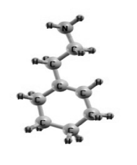
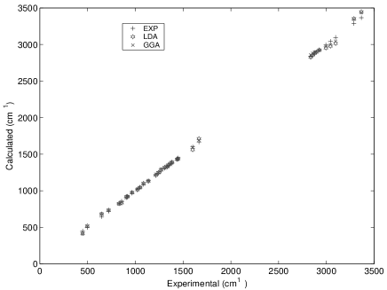
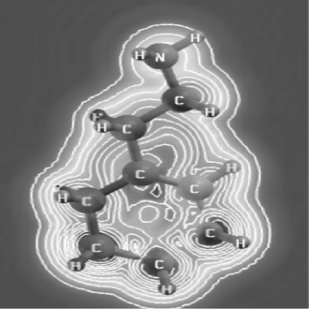
The calculated stable structure of CyHEA is shown in Fig.1 which was drawn by XCrySDen (Crystalline Structures and Densities) program xcrysden . The vibrational assignments and frequencies of cyclohexene-2-ethanamine was reported experimantally by İzgi et al. Izgi . The spectral properties of the molecule were evaluated through the calculated vibrational frequencies of the free ligand molecule. The calculated and experimental infrared spectra data of the molecule are given in Table.1. The experimental, GGA and LDA results are also compared in Fig.2.
| mode | Experimental Izgi | Calculated | |||
|---|---|---|---|---|---|
| Assignments | LDA | GGA | |||
| 3366s | 3447 | ||||
| 3288s | 3353 | ||||
| 3097vw | 3015 | ||||
| 3043m | 2979 | ||||
| 2995m | 2954 | ||||
| 2926vs | 2925 | ||||
| 2894vw | 2896 | ||||
| 2877vw | 2880 | ||||
| 2857vs | 2856 | ||||
| 2836vs | 2827 | ||||
| 1666 m | 1712 | ||||
| 1600mb | 1562 | ||||
| 1505vw | - | ||||
| 1473vw | - | ||||
| 1448vw | 1439 | ||||
| 1438s | 1431 | ||||
| 1384w | 1391 | ||||
| 1370vw | 1376 | ||||
| 1344m | 1352 | ||||
| 1334w | 1328 | ||||
| 1307w | 1320 | ||||
| 1269m | 1285 | ||||
| 1242w | 1248 | ||||
| 1215w | 1212 | ||||
| 1136m | 1131 | ||||
| 1086w | 1101 | ||||
| 1066w | - | ||||
| 1049w | 1045 | ||||
| 1022w | 1014 | ||||
| 966w | 976 | ||||
| 919m | 921 | ||||
| 906vw | 919 | ||||
| 857w | 834 | ||||
| 829m | 824 | ||||
| 800m | - | ||||
| 720sh | 733 | ||||
| 647w | 680 | ||||
| 497vw | 518 | ||||
| 448w | 417 | ||||
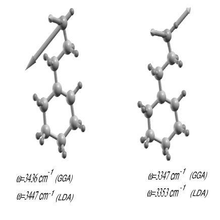
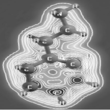
The strong N-H asymmetric and symmetric stretch bands seen in Table.1 are due to the contribution of ethylamine (see Fig.4). C-H stretch bands between are attributed to cyclohexene group and the very strong C-H stretch bands at and result from ethylamine. The very strong bands are attributed to the attachment of ethylamine and cyclohexene and appear between . Most of the modes below the arise from cyclohexen. If the vibrational assignments of the molecule involving these groups are investigated, it is seen that the assignments obtained for the molecule also involve the group frequencies. Furthermore, the observed medium broad band appears at is an N-H bending band as well as a group frequency. There is also a good agreement between the experimental and the theoretical vibrational frequencies in the region of except some GGA and LDA results. The ground state energy of the molecule was obtained to be -128.66 ryd and -128.53 ryd for GGA and LDA, respectively. Finally, we examined the electronic properties by using calculated STM images for cyclohexene-2-ethanamine. In Fig.4 and Fig.5 which were drawn by using XCrySDen, we calculated the STM images at constant current and bias voltage -2.5 eV and 2.5 eV, respectively. These results supply a microscopic model for STM images and can serve as a source for STM experiments for organic molecules.
IV Conclusion
The experimental and the theoretical investigation of CyHEA molecule have been performed successfully by using FT-IR and density functional theory calculations. For all calculations, it is shown that the results of GGA and LDA methods are in excellent agreement with all experimental findings. Thus, density functional theory (DFT) methods are suitable for the calculation of ground state properties and potential energies. Hence, DFT is an excellent method for calculating vibrational spectra and STM images from first principles.
V ACKNOWLEDGEMENTS
We thank G.Gokoglu and T. Boz for improving our paper English.
References
- (1) F. Hibbert, Acc. Chem. Res. 17, 115 (1984).
- (2) H. A. Staab, T. Saupe, Angew. Chem. 100, 895 (1988); Angew. Chem. Int. Ed. Engl. 27, 865 (1988).
- (3) R.W. Alder, Chem. Rev. 89, 1215 (1989).
- (4) R.W. Alder, Tetrahedron 46, 683 (1990).
- (5) S. Rodin-Bercion, L. Lespade, D. Cavagnat, J.C. Cornut, J. Mol. Struct. (Theochem) 526, 343 (2000).
- (6) D. Zeroka, J.O. Jensen and A.C. Samuels, J. Mol. Struct. (Theochem) 465, 119 (1999).
- (7) S.R. Sirimanne and S.W. May, J. Am. Chem. Soc. 110, 7560 (1988).
- (8) T. İzgi, Ö. Alver, C. Parlak, M.T. Aytekin and M. Ṣenyel, Spectrochemica Acta A:Molecular and Biomolecular Spectroscopy , accepted (2006).
- (9) A. Kokalj, Comp. Mat. Sci. 28, 155 (2003),www.xcrysden.org/.
- (10) S. Baroni, A. Dal Corso, S. de Gironcoli and P. Giannozzi; http:// www.sissa.it/cm/PWcodes.
- (11) N.C. Handy, P.E. Maslen, R.D. Amos, J.S. Andrews, C.W. Murray and G.J. Laming, Chem. Phys. Lett. 197, 506 (1992).
- (12) N.C. Handy, C.W. Murray and R.D. Amos, J. Phys. Chem. 97, 4392 (1993).
- (13) P.J. Stephens, F.J. Devlin, C.F. Chabalowski and M.J. Frisch, J. Phys. Chem. 98, 11623 (1994).
- (14) F.J. Devlin, J.W. Finley, P.J. Stephens and M.J. Frisch, J. Phys. Chem. 99, 16883 (1995).
- (15) S.Y. Lee, B.H. Boo, Bull. Korean Chem. Soc. 17, 754 (1996).
- (16) S.Y. Lee, B.H. Boo, Bull. Korean Chem. Soc. 17, 760 (1996).
- (17) D. Vanderbilt, Phys. Rev. B 41, 7892 (1990).
- (18) J.P. Perdew and A. Zunger, Phys. Rev. B 23, 5048 (1981).
- (19) J.P. Perdew, K. Burke and M. Ernzerhof, Phys. Rev. Lett. 77, 3865 (1996).