[
Molecular Dynamics Simulation of Folding and Diffusion of Proteins in Nanopores
Abstract
A novel combination of discontinuous molecular dynamics and the Langevin equation, together with an intermediate-resolution model, are used to carry out long (several s) simulation and study folding transition and transport of proteins in slit nanopores. Both attractive () and repulsive () interaction potentials between the proteins and the pore walls are considered. Near the folding temperature and in the presence of the proteins undergo a repeating sequence of folding/partially-folding/ unfolding transitions, while decreases with decreasing pore sizes. The opposite is true when is present. The proteins’ effective diffusivity is computed as a function of their length (number of the amino acid groups), temperature , the pore size, and the interaction potentials . Far from , increases (roughly) linearly with , but due to the thermal fluctuations and their effect on the proteins’ structure near , the dependence of on in this region is nonlinear. Under certain conditions, transport of proteins in smaller pores can be faster than that in larger pores.
PACS: 87.15.Aa, 83.10.Mj, 87.15.Cc, 87.15.Vv, 87.83.+a
]
Proteins’ importance to biological systems cannot be overstated [1]: as enzymes they catalyze and regulate cells’ activities; tissues are made of proteins, while as antibodies proteins are a vital part of the immune system. Proteins with globular structure fold into compact and biologically active configurations, and an important problem is understanding the mechanisms by which they attain their folded structure, and factors that contribute to the folding [2, 3, 4]. Such understanding is important due to debilitating illnesses, such as Alzheimer’s and Parkinson’s diseases, that are believed to be the result of accumulation of toxic protein aggregates [5, 6, 7, 8], as well as to the industrial production of enzymes and therapeutic proteins based on the DNA recombinant method [9].
While the three-dimensional (3D) structure of native proteins is controlled by their amino acid sequence [2, 3, 4], their transport properties and the kinetics of their folding depend on the local environment. But, whereas protein folding in dilute solutions under bulk condition, typically used in in vitro studies, is relatively well-understood, the more important problem of protein folding in a confined medium is not. The environment inside a cell in which proteins fold is crowded, with the volume fraction of the crowding agents (such as RNA) may be 0.2-0.3. Thus, even in the absence of interactions between proteins and other cellular molecules, their movement inside the cell is limited. The limitation affects proteins’ stability. Experiments indicated [10] that confinement often stabilizes the proteins’ native structure [11], denatures them in the limited space of the cage model, first suggested by Anfinsen [2, 3, 4], and accelerates folding relative to that in bulk solutions. Studies of proteins of different native architectures in cylindrical nanpores indicated [12] that, in vivo folding is not always spontaneous; rather, a subset of proteins may require molecular chaperones.
Protein (enzyme) immobilization using porous solid support, via adsorption, encapsulation, and covalent linking, has been used for a long time [13, 14]. Such practical applications as biocatalysis [15] and biosensors also entail not only better understanding of the folding in confined media, but also transport of proteins in such media. At the same time, protein purification using nanoporous membranes is also gaining attention [16]. SiC nanoporous membranes [17] allow [18] diffusion of proteins up to 29000 Daltons, but exclude larger ones. Despite the fundamental and practical significance of transport of protein in confined media, there is currently little understanding of the phenomenon.
The goal of this paper is twofold. First, we use molecular dynamics (MD) simulation to study protein folding and stability in slit nanopores. Second, we utilize a novel combination of MD simulation and the Langevin equation (LE) to study protein transport in the nanopores. To our knowledge, our combination of the MD simulation and the LE has never been proposed before, nor has there been any simulation of transport of proteins in nanopores. For such important practical applications as membrane purification, biocatalysis, and sensors, the transport of proteins in nanopores is of utmost importance. A slit nanopore is a reasonable model for the type of pores that one encounters in such applications [15, 16, 17, 18] and, despite its simplicity, it might also be a reasonable model for the pores in biological membranes.
Some Monte Carlo [19] and MD [5, 20] simulations of proteins’ behavior in nanopores were reported before. In particular, Lu et al. [5] and Cheung et al. [20] studied folding of proteins in spherical pores of different radii. Cheung et al. studied the phenomenon as a function of the volume fraction of a crowding agent, which they modeled by a bed of hard spheres with repulsive interaction with the proteins. While a spherical pore may be a suitable model for the cavity of GroEl-GroES complex, it is not so for the pores of membranes, biocatalysts, and sensors that are of prime interest to us. Instead, the slit (and cylindrical) pores are more appropropriate. Moreover, for the types of applications that we consider, the pore space consists of interconnected channels, which is completely different from what Refs. [5,20] considered.
In addition, the protein model that we use (see below) is, in our opinion, much more realistic than what the previous investigations [5, 20] utilized. For example, they used a simplified model for the amino acids that was based on two united atom (UA) beads. Moreover, the side chains of the amino-acid residues were not explicitly considered. The model that we utilize represents the amino acids using four UA beads (see below), while the side chains are also considered explicitly, hence honoring the proteins’ structure much more realistically.
We simulate de novo-designed family of proteins [21], which consists of only 4 types of amino acids in their 16-residue sequence, simplified further [22] to a sequence of hydrophobic (H) and polar (P) residues. Using periodicity in the H-P sequence of the 16-residue peptide , we made 3 other sequences with lengths , 23 and 30 residues. As the four proteins have similar native structures, the differences in their behavior is attributed to their lengths. The simulations indicated that they all fold into an -helix.
The proteins are modeled by an intermediate-resolution model [23, 24, 25, 26], with several changes described below. Every amino acid is represented by four UA groups or beads. A nitrogen UA represents the amide N and hydrogen of an amino acid, a Cα UA represents the -C and its H, and a C UA for the carbonyl C and O. The fourth bead represents the side chains, all of which are assumed to have the same diameter as CH3. All the backbone bond lengths and bond angles are fixed at their ideal values, and the distance between consecutive Cα UA is fixed according to experimental data.
To carry out long and efficient simulations, we use discontinuous MD (DMD) [27]. This allowed us to carry out 5 s MD simulations, one order of magnitude longer than the previous simulations. The forces acting on the beads are the excluded-volume effect, and attraction between bonded and pseudobonded beads, between pairs of backbone beads during HB formation, and between hydrophobic side chains. Nearest-neighbor beads along the chain backbone are covalently bonded, as are the Cα and R UAs. Pseudobonds are between next-nearest neighbor beads along the backbone to keep the backbone angles fixed; between neighboring pairs of Cα beads to maintain their distances close to the experimental data, and between side chains and backbone N and C UAs to hold the side-chain beads fixed relative to the backbone. All of this keep the interpeptide group in the trans configuration, and all the residues as isomers, as required.
The potential between a pair of bonded beads, separated by a distance , is given by, , for, , and , and, for . Here, is the ideal bond length, and is the tolerance in the bond’s length [23, 24, 25, 26]. The hydrophobic (HP) interactions between the side chains and the H in the sequence, if there are at least 3 intervening residues between them, is given by, and 0 for, , , and , respectively, where is the HP side-chains’ diameter.
The HB interaction may occur between the N and C beads with at least 3 intervening residues, but each bead may not contribute to more than one HB at any time, with the range of the interaction being about . The HBs are stable when the angles in N-H-O and C-O-H, controlled by a repulsive interaction between each of the N and C beads with the neighboring beads of the other one, are almost . Thus, if a HB is formed between beads Ni and Cj, a repulsive interaction between neighbor beads of Ni, namely, Ci-1 and Cαi, with Cj is assumed, and similarly for the neighbor beads of Cj, namely, Nj+1 and Cαj, with the Ni bead.
An N or C bead at one end of the protein has only one neighbor bead in its backbone, instead of 2. Hence, controlling the HB angles will be limited, causing the HBs with one of their terminal constituents to be less restricted and, thus, more stable than the other HBs. This may cause formation of non--helical HBs in a part of the protein between the N and C beads, and of semistable structures that influence the results. To address this problem, assume that the N-terminal bead, N1, has a HB with Cj. For , bead Ci-1 does not exist to have a repulsive interaction with Cj and help control the HB angles. So, we use Cα1. Not only can we consider the repulsion between this bead and Cj, but also we define an upper limit for their distance so as to control the motion freedom of N1 and Cj that constitute the beads in the HB. The potential of such interactions is given by, and for , , , and, , respectively.
Two H atoms have chemical bonds with the nitrogen in the proteins’ N-terminal, and are free to rotate around the N1-Cα1 bond, while at the same time satisfying the constraints on the angles between the chemical bonds of N1. Thus, if a HB is formed, one of the two H atoms lies in the plane of N, O and C, such that the angles in N-H-O and C-O-H are as close to as possible. Hence, we force the maximum distance between Cα1 and Cj to be the same as the maximum distance between Cαi and Cj in the usual HBs, and similarly when the C-terminal Cℓ has a HB with Ni. This allows us to control the angles in a HB that contains N1. The dependence ( is dimensionless) of (in ), obtained from separate MD simulations (the details will be given elsewhere), is, for N1-Cαj, and for Cℓ-Cαi.
There is also hard-core repulsion between two unbonded beads that have no HB and HP interactions. At the same time, interactions between a pair of beads, separated along the chain by 3 or fewer bonds, are more accurately represented by those between the atoms themselves, not the UAs. Thus, we developed a variant of the previous models [23, 24, 25, 26] to account for such interactions: the beads are allowed to overlap by up to 25% of their bead diameters, while for those separated by 4 bead diameters the allowed overlap is 15% of their diameters.
We use a slit nanopore, modeled as the space between two 2D structureless carbon walls in the plane between , with periodic boundary conditions in the and directions. The interaction between the walls and the protein beads is, and for, , , , , and , respectively, where is the distance between the center of a bead and the walls. For all the beads, , so chosen to represent realistically the competition between protein folding and its beads’ interaction with the walls. To estimate and , the energy and size parameters between the C atoms in the walls and various beads were calculated using Lorentz-Berthelot mixing rules, , and , where N, Cα, C and R. Then, using separate simulations (details will be given elsewhere), the interaction potential between different beads was estimated. The distances at which UCX and its second derivative were zero were taken as and . The results (in ) are, , and 3.31, and , and 1.12, for N, Cα, C, and R, respectively.
We now describe how the effective diffusivity of the proteins is computed. To do so, one must take into account the effect of the solvent on the motion of the proteins. In the previous works the solvent’s effect on the proteins’ motion was included only implicitly by the HP attraction between the side chains. While this might be appropriate for studying the folding, it is not so for computing , since the solvent’s viscosity strongly and directly affects . To explicitly include the solvent effect, we have developed the following model which, to our knowledge, is new.
We first carry out DMD simulation for a time period . Suppose that the speeds of the proteins’ center of mass (CM) at the beginning and end of the period are, respectively, and . Since the solvent’s viscosity affects the proteins’ velocity in the pore, but the time scale over which this effect is important is much different from , we apply, at the end of the time period , the Langevin equation (LE) to the proteins’ CM to correct their velocity due to the presence of the solvent’s molecules. To do so, we represent a protein as a particle with a mass and an effective radius equal to its radius of gyration . Then, the force F on its CM is given by,
| (1) |
The discretized LE is given by,
| (2) |
where, , is a Gaussian random force (with zero mean and variance ), and is the speed after applying the LE (acting as the for the next LE application). Thus,
| (3) |
which yields , the velocity of the proteins corrected for the solvent effect. The DMD simulation is then continued for another time period using as the , the LE is applied again to correct the proteins’ velocity at the end of the period, and so on.
We now present the results of our simulations. Consider the case of attractive interaction potential between the proteins and the pore’s walls. A folded state attaches itself to the walls only through its end groups, while unfolded ones may completely attach themselves to the walls. Thus, with the decrease in the average potential energy of the unfolded states is larger than the corresponding decrease for the folded one. Hence, compared to the bulk, the unfolded states in the pore with a are more stable than the folded one, in qualitative agreement with Refs. [4,20] for spherical cavities.
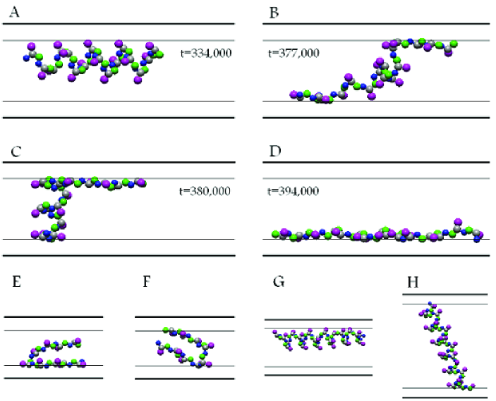
Figure 1 shows a sequence of events for a protein of size in a pore of size nm at . The protein changes its state from completely folded to a partially folded to an unfolded one which is completely attached to the pore’s wall (frame D). Due to , the transitions occur easily and repeatedly, even after a long time. Note that, in moving from B to C, the set of deformed helical HBs changes, hence indicating rapid dynamics of the HB formation and deformation near .
Also shown in Figure 1 are protein configurations (frames E and F with pore size nm) for at . In these pores, a protein of length does not attain its native state at low . Instead, it has a U shape with its two sides attached to the walls; it has 4 HBs, only one of which is helical (the native state has 5 helical HBs), and more of its atoms are close to the walls than those in the folded state. Although the potential energy of such unfolded states is roughly the same as one in the folded one, entropic effects which favor the unfolded states are also important. Upon further cooling at below the apparent folding temperature , the protein becomes trapped in the U shape without enough kinetic energy to overcome the energy barrier to attain a folded state. Thus, such configurations do not represent truly folded states.
We also find that decreases with the pore size, which is due to the attractive interaction energy between the protein and the walls. The decrease in is indicative of the more stable unfolded states (or less stable folded state).
The opposite (namely, increasing with decreasing pore size) is true if the interaction is purely repulsive, . In that case, proteins are even more confined in the pores. Thus, the number of possible unfolded states and, hence, their total entropy, will be smaller [28]. But, due to its compact configuration, confinement affects the entropy of the folded state less strongly, implying that, in the presence of the folded state is more stable than that in the bulk.
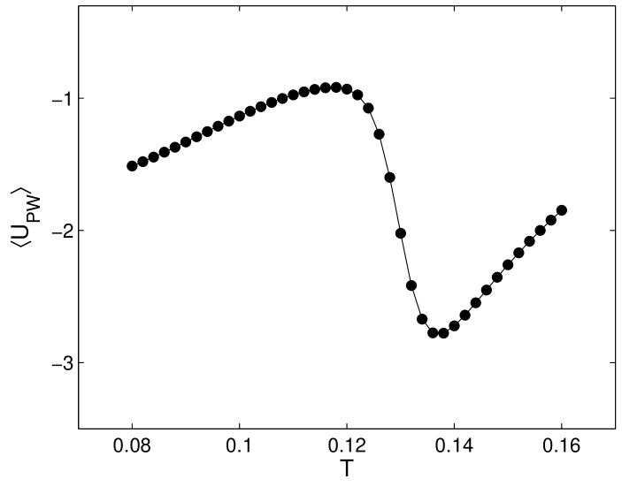
Figure 2 presents the average interaction energy of the proteins with the walls, computed by the weighted histogram analysis method [29]. Cooling the proteins at increases , as well as the average of the helical HBs (which is, however, very small). Near Tf is nonnegligible, and the proteins can only laterally attach themselves to the walls, hence decreasing . By lowering further, nearly the entire -helix is formed, and increases again. Thus, Figure 2 indicates that, not only does the interaction with a nanopore disturb folding, but also folding to a definite structure disturbs the proteins’ interaction with the pore.
Before describing the results for the effective diffusivities, we should point out that, over the temperature range that we have simulated, the proteins resemble a prolate ellipsoid. Thus, they become elongated in very small nanopores, while they can “stand up” in larger pores, such that their ends touch the pores’ walls, if they are long enough. Such configurations are also shown in Figure 1. Thus, transport in small pores may actually be faster than in larger pores, a counterintuitive, but important result. Such phenomena can complicate further delineation of the dependence of on the various important parameters of the system.
Our simulations indicate that transport of the proteins in the planes (parallel to the pore’s walls) is Fickian, so that, after a sufficiently long time (which depends on the proteins’ length, the pore size, and ) the mean-square displacements (MSDs) of the proteins’ CM vary linearly with the time, hence yielding an effective diffusivity for the proteins. In contrast, the MSDs in the direction perpendicular to the pore’s walls saturate at a finite time.
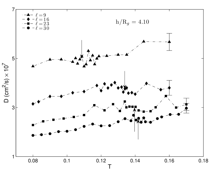
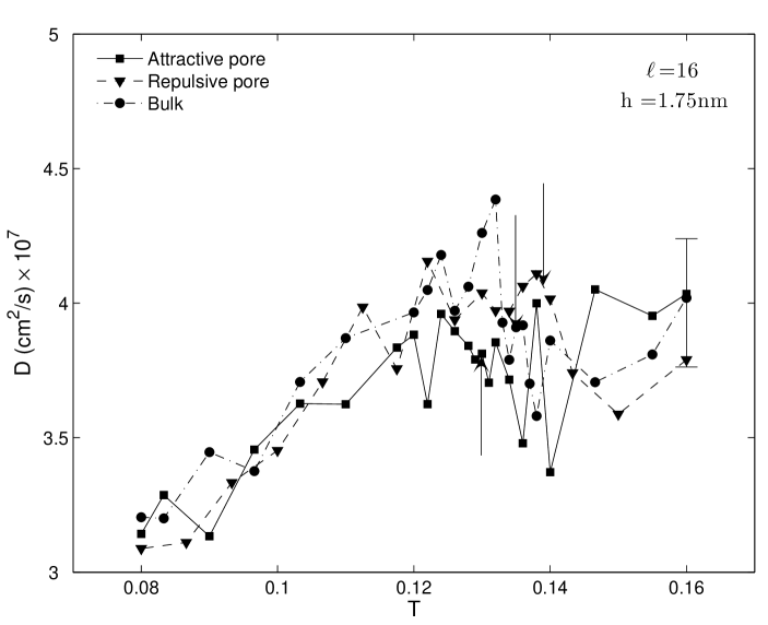
Figure 3 presents the proteins’ effective diffusivity in the planes, in a pore with fixed . As increases and is approached, also increases, since the proteins’ decreases. Far from (where changes little) varies roughly linearly with . However, near , there are strong thermal fluctuations due to which strongly varies with . Consequently, no longer varies linearly with . Increasing the proteins’ length decreases , as expected.
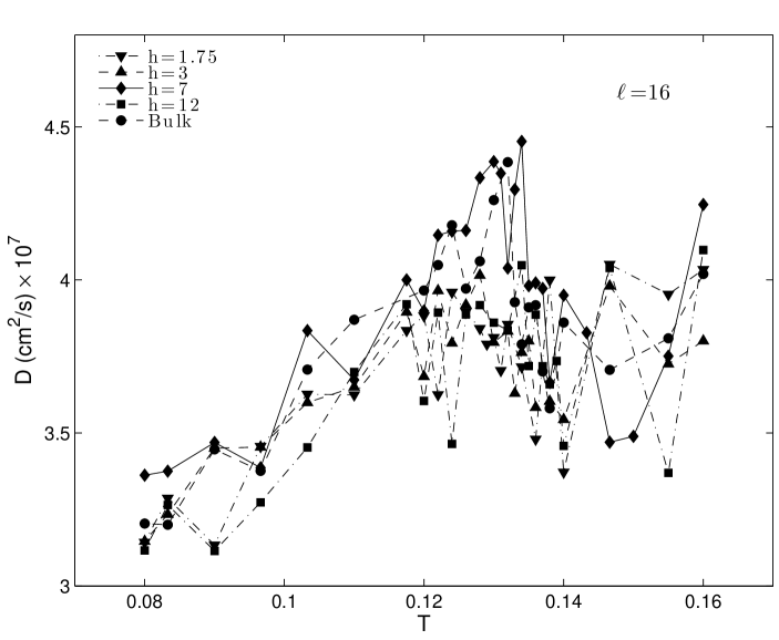
Figure 4 presents the diffusivities for a protein of fixed length, , in four different pores, and compares the results with those in the bulk. Note that, the for the bulk state and those for the pores are not the same. Therefore, due to the strong fluctuations of and in the region around , the pore and bulk diffusivities cannot be directly compared. The comparison is possible mainly at temperatures far from (both below and above ). As Figure 4 indicates, at least for the (taking into account the numerical uncertainties.
The results shown in Figures 3 and 4 were for an attractive potential between the proteins and the pores’ walls. Figure 5 compares the diffusivities for a protein of length in a pore of size nm for both attractive and repulsive interaction potentionals , and compares them with the bulk values. Generally speaking, for the diffusivities with are larger than those with .
In our current work, we are carrying out extensive simulations for computing the effective diffusivity as a function of the strength of the interaction potential between the proteins and the walls, its sign (attractive versus repulsive), and other relevant parameters. The results will be presented in a future paper.
We thank M. R. Ejtehadi, C. K. Hall, and M. D. Niry for useful discussions. The work of LJ was supported by Iranian Nanotechnology Initiative.
REFERENCES
- [1] Branden, C.; Tooze, J. Introduction to Protein Structure; Garland Publishing: New York, 1998.
- [2] Anfinsen, C. B. Science 1973, 181, 223.
- [3] Klimov, D. K.; Thirumalai, D. Phys. Rev. Lett. 1996, 76, 4070.
- [4] Mirny, L.; Shakhnovich, E. Annu. Rev. Biophys. Biomol. Struct. 2001, 30, 361.
- [5] Lu, S.; Liu, Z.; Wu, J. Biophys. J. BioFAST 2006, 105, 071761.
- [6] Kirschner, D. A.; Abraham, C.; Selkoe, D. J. Proc. Natl. Acad. Sci. USA 1986, 83, 503.
- [7] Conway, K. A.; et al. Proc. Natl. Acad. Sci. USA 2000, 97, 571.
- [8] Lynn, D. G.; Meredith, S. C. J. Struct. Biol. 2000, 130, 153.
- [9] Thomas, J. G.; Ayling, A.; Baneyx, F. Appl. Biochem. Biotechnol. 1997, 66, 197.
- [10] Eggers, D. K.; Valentine, J. S. Prot. Sci. 2001, 10, 250.
- [11] Brinker, A.; et al. Cell 2001, 107, 223.
- [12] Tagaki, F.; Koga, N.; Takada, S. Proc. Natl. Acad. Sci. USA 2003, 100, 11367.
- [13] Bickerstaff, G. F. Immobilization of Enzymes and Cells; Humana Press: Totowa, NJ, 1997.
- [14] Lei, C.; et al. J. Am. Chem. Soc. 2002, 124, 11242.
- [15] Dadvar, M.; Sahimi, M. Chem. Eng. Sci. 2003, 58, 4935.
- [16] Avramescu, M.-E.; Borneman, Z.; Wessling, M. Biotechnol. Bioeng. 2003, 84, 564.
- [17] Elyassi, B.; Sahimi, M.; Tsotsis, T. T. J. Memb. Sci. 2007, 288, 290.
- [18] Rosenbloom, A. J.; et al. Biomed. Microdev. 2004, 6, 261.
- [19] Ping, G.; et al. J. Chem. Phys. 2003, 118, 8042.
- [20] Cheung, M. S.; Klimov, D.; Thirumalai, D. Proc. Natl. Acad. Sci. USA 2005, 102, 4753.
- [21] Regan, L.; DeGrado, W. F. Science 1988, 241, 976.
- [22] Guo, Z.; Thirumalai, D. J. Mol. Biol. 1996, 263, 323.
- [23] Takada, S.; Luthey-Schulten, Z.; Wolynes, P. G. J. Chem. Phys. 1999, 110, 11616.
- [24] Smith, A. V.; Hall, C. K. Proteins 2001, 44, 344.
- [25] Smith, A. V.; Hall, C. K. Proteins 2001, 44, 376.
- [26] Nguyen, H. D.; Marchut, A. J.; Hall, C. K. Prot. Sci. 2004, 13, 2909.
- [27] Smith, S. W.; Hall, C. K.; Freeman, B. D. J. Comput. Phys. 1997, 134, 16.
- [28] Thirumalai, D.; Klimov, D. K.; Lorimer, G. H. Proc. Natl. Acad. Sci. USA 2003, 100, 11195.
- [29] Ferrenberg, A. M.; Swendsen, R. H. Phys. Rev. Lett. 1989, 63, 1195.