Present address: ]Van der Waals-Zeeman Instituut, Universiteit van Amsterdam, Valckenierstraat 65, 1018 XE Amsterdam, The Netherlands
Spatially resolved photo ionization of ultracold atoms on an atom chip
Abstract
We report on photo ionization of ultracold magnetically trapped Rb atoms on an atom chip. The atoms are trapped at 5 K in a strongly anisotropic trap. Through a hole in the chip with a diameter of 150 m two laser beams are focussed onto a fraction of the atomic cloud. A first laser beam with a wavelength of 778 nm excites the atoms via a two photon transition to the 5D level. With a fiber laser at 1080 nm the excited atoms are photo ionized. Ionization leads to depletion of the atomic density distribution observed by absorption imaging. The resonant ionization spectrum is reported. The setup used in this experiment is not only suitable to investigate BEC ion mixtures but also single atom detection on an atom chip.
pacs:
03.75.Be, 32.80.Rm, 39.90.+dI Introduction
With microfabricated current conductors on a chip complex magnetic field geometries can be constructed in which atoms can be trapped and manipulated. Today, clouds of ultra cold atoms and Bose-Einstein condensates are routinely trapped on such atom chips and a variety of geometries have been demonstrated such as waveguides, spatial and temporal beam splitters, double well potentials, and periodic lattices (for a review see Fortágh and Zimmermann (2007)). It is now conceivable to develop atom chip applications as for instance sensors for rotation, acceleration, or gravitational forces gradients. The precision with which condensates and ultra cold thermal clouds can be positioned at the chip surface may also be exploited for surface probing and matter wave microscopy Lin et al. (2004); Wildermuth et al. (2005); Günther et al. (2005a). The distinct suitability of atom chips for generating strongly anisotropic trapping potentials also offers unique possibilities to study the fundamental properties of the trapped quantum gas in the crossover regime from three to one dimension. In very elongated chip traps the phase coherence of a Bose-Einstein condensate breaks up giving rise to phase fluctuations Esteve et al. (2006). If, in addition, the gas is strongly diluted and consists of only several ten atoms the regime of a strongly interacting one dimensional Tonks-Girardeau gas may be reached Paredes et al. (2004); Kinoshita et al. (2004). Similar experiment with fermionic atoms in the 1D regime Wonneberger (2006) may also be possible Aubin et al. (2006). Such one dimensional quantum gases are of great interest in fundamental many particle physics. Finally, if methods can be found for carrying out experiments with only few or even single atoms on a chip fascinating perspectives open up for engineered quantum entanglement and quantum information processing. Single atom detection on a chip is thus a promising challenge with significant progress during the last year when optical resonators have been successfully used to detect atoms by optical spectroscopy Teper et al. (2006); Haase et al. (2006); Takamizawa et al. (2006). Alternatively, atoms can be detected by ionization and subsequent ion detection Campey et al. (2006). This approach is followed with this paper.
A second, still very young field of research is the investigation of atom-ion-mixtures Cote et al. (2002); Ciampini et al. (2002); Massignan et al. (2005). In the polarizing electric field of an ion the atoms are expected to form a bubble with yet unknown properties. With many ions implanted in the dilute quantum gas a system appears which has not been studied even theoretically. A planar chip trap for ions has already been demonstrated Seidelin et al. (2006) and its integration on an atom chip seems feasible. In such combined traps for atoms and ions the magnetic field generating conductors trapping the atoms could simultaneously be used as electrodes for generating the electric field for trapping the ions. Together with the highly flexible geometries that can be realized on atom chips, atom ion systems offer fascinating new opportunities for constructing and investigating novel types of quantum systems.
In this paper we demonstrate the controlled production of cold ions by photo ionization of ultracold neutral atoms on an atomchip. The ions are generated in a small spatially resolved region given by the focus of the ionizing laser beams. The generated ions with kinetic energies corresponding to temperatures of only a few millikelvin may in principle be trapped in planar ion traps and subsequently cooled by thermalization with a surrounding atomic gas. Furthermore, spatially resolved photo ionization with close to 100% efficiency allows for single atom detection on a chip. With a suitable ion optics the ions can be extracted from the chip and detected with an electron multiplier (CEM). Such a detector has recently been described in Stibor et al. (2007).
In the next two chapters we describe a scheme for fast spatially resolved photo ionization and its implementation on the chip. Chapter three and four presents the experimental observations and concludes the paper.
II Ionization scheme
In our setup 87Rb atoms are prepared in the 5S ground state.
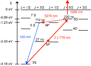
The atoms are ionized by a resonant two photon transition into the 5D excited state and subsequent excitation to the continuum with a second laser (see Fig. 1). The two photon transition is driven by a grating stabilized continuous wave single mode laser diode with a wavelength of 778.1066 nm. The transition strength is enhanced by the intermediate 5P level which is off resonant to the laser by only 2 nm. The 5D level with a lifetime of 220 ns gives rise to a resonance line width of approximately 500 kHz Nez et al. (1993). The rate of the two photon transition depends quadratically on the light intensity which leads to an improved spatial resolution and to a reduction of unwanted stray light effects. This is important since we aim at a fast ionization of the directly illuminated atoms on a time scale of several microseconds while all other atoms should not be affected by the light on a time scale of seconds, which is set by the lifetime of the condensate. For a two photon transition stray light must thus only be suppressed to a level of 0.1% in contrast to a single photon transition which requires a suppression stronger by three orders of magnitude.
Furthermore, a hyperfine resolving optical excitation scheme can be exploited to improve the spatial resolution of the ionization: for atoms prepared in the 5S, F=2, mF=2 hyperfine state and a laser tuned in resonance with the F=1, mF=1 hyperfine state ionization only occurs if the two states are coupled by an additional radio frequency. Due to the Zeeman effect resonant coupling only occurs at a distinct magnetic field that depends on the detuning of the radio frequency. In a magnetic field gradient ionization then takes place only at a well defined position which, in addition, can be varied by simply tuning the radio frequency. At atom chips large field gradients can be easily constructed and a resolution of below 100 nm appears feasible. Thus, there is no principle obstacle for resolving an atomic distribution on a scale that is comparable to the healing length of a condensate or the inter atomic distance in a Tonks gas. This scheme also further suppresses stray light effects and it should be possible to detect atoms in a gas without affecting their neighbors.
For ionizing from the 5D level we use a continuous wave fiber laser near 1080nm with a Gaussian single mode beam profile and a spectral width of smaller than 1 nm. Photons at this wavelength are sufficiently energetic to bridge the binding energy of the 5D level of 0.99 eV corresponding to a wavelength of 1250 nm. In principle the photon energy of the diode laser near 778 nm is sufficient to ionize the excited atoms, however, efficient ionization requires higher power than available with the diode laser.
In order to reach a high ionization efficiency the ionization rate from the 5D state to the continuum should be at least on the order of the spontaneous decay rate to the ground state. The ionization cross section from the 5D level is reported to be 25 Mb Duncan et al. (2001) for light near 1250 nm and drops to Mb for the shorter wavelength of the fiber laser near 1080 nm Duncan et al. (2001). For the moderate intensities discussed here, ionization is well described by a simple rate model which relates the intensity and the ionization rate by:
| (1) |
Consequently, an ionization rate of ns) requires an intensity of W / m2. The laser beams in our experiment are focussed to a radius of m. The ionization shall take place within this radius and hence the intensity has to be reached at . For a Gaussian beam with this properties this results in a total power of about 4.8 W which is easily available with a low cost commercial fiber laser.
The two photon transition can also be treated in a rate model, given that its rate is significantly slower than ionization rate from the 5D level Ackerhalt and Eberly (1976). Then, Grove et al. (1995). For a beam radius of m a laser power of only 6 mW already results in an excitation rate of s).
The laser light not only excites the atoms, it also gives rise to light induced dipole potentials which have a strong impact on the atomic motion. The most important contribution in Rb is related to the resonance between the 5S ground state and the exited 5P3/2 level (D2-line). The diode laser is 2 nm blue detuned relative to this transition and leads to a repulsive potential barrier with a hight of 5 K for the above parameters. In contrast, the fiber laser is red detuned by 300 nm and generates an attractive potential of 10 K. Together both effects almost cancel with a residual attractive net potential which drags the atoms into the ionization volume.
Figure 2
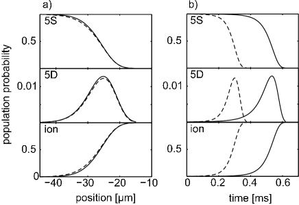
shows a simulation of the ionization process including the center of mass motion of the atom in the optical dipole potential. The laser powers are set to 6 mW and 5 W for the diode laser and the fiber laser, respectively. In (a) the occupation of the involved states is plotted versus the position of the atom which starts with a small but final velocity (14 m/s) at a distance of 60 m from the focus of the lasers (located at ). At a distance of approximately 40 m from the center of the laser focus the probability for excitation in the 5D state grows significantly. The solid line shows the simulation with a beam radius of m for both lasers. If the beam radius of the fiber laser is expanded to 60 m (dashed line) the results in (a) remain almost unchanged. Since for this set of parameters the two photon transition forms a bottleneck for ionization the internal dynamics starts not until the atom reaches the light of the diode laser. However, widening the focus of the fiber laser results in earlier and faster ionization (dashed lines in Fig.2 (b) due to the attractive dipole potential of the fiber laser which accelerates the atoms into the ionization volume. A trivial way of increasing the ionization speed and the spatial resolution not shown in the simulation is simultaneously decreasing the beam radius of both laser beams. Here all numbers are given for trapped and cooled 87Rb atoms, however similar schemes can be found also for other species Hurst et al. (1979).
III Ionization on the atomchip
The atomchip used in this experiment is described in detail elsewhere Günther et al. (2005a). It carries a conductor geometry which allows for transporting condensates or thermal atoms to different locations at the chip in a controlled way. At these locations the atoms can be brought in contact with specialized trapping geometries such as narrow wave guides, double well potentials or periodic lattice potentials Günther et al. (2005b) each individually addressable by the transport system. Instead of actively transporting the atoms it is also possible to couple atoms into a waveguide and let them propagate ballistically Fortágh et al. (2003). To this scenario we have added a region where the atoms are ionized by the lasers. A sketch of the setup is shown in Figure 3.
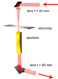
Both laser beams are overlapped outside the vacuum chamber and focussed on to the chip with a lens of mm focal length located inside the vacuum chamber. The beams with a waist of m are aligned perpendicular to the surface of the atom chip and pass the chip through a small hole with a diameter of 150 m. With a Rayleigh length of the beams of 3.6 mm and 2.9 mm, respectively, the intensity variation with the distances of the atoms to the chip surface can be neglected. The same holds for the slightly different positions of the foci due to the chromatic abberation of the lens. By passing the beams through a small hole in the chip instead of an alignment parallel to the chip surface stray light is minimized. In addition the atoms can be ionized very near to the surface, where narrow trapping potentials can be realized with structures on a spatial scale of m and below. Behind the chip the laser beams leave the chamber through an antireflection coated window.
Also conceivable is guiding the laser beams through an optical fiber. However, at the end facette of the fiber the beams rapidly diverge which makes it hard to control stray light.
IV Experimental observations
In the experiment 87Rb atoms in the state are prepared in a magnetic micro trap at a temperature of 5 K. The power of the diode and the fiber laser beams are 8 mW and 2 W, respectively. The wavelength of the diode laser is stabilized to the two photon transition recorded with 87Rb in a vapor cell Kraft et al. (2005).
The atoms are trapped below the hole in the chip at a distance of 350 m from the chip surface. The length of the atomic cloud in the trap amounts to approximately 1 mm. Typical trapping frequencies are Hz in axial direction and Hz in radial direction. Ionization is initialized by exposing the atoms to the laser fields for 10 ms. The remaining atoms are detected by absorption imaging after 1 ms of ballistic expansion.
IV.1 Dipole forces and ionization
Fig. 4 shows
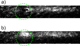
absorption images for the cases that (a) only the diode laser or (b) only the fiber laser was activated. In the first case the density distribution is depleted at the position where the laser illuminates the cloud. With only the fiber laser activated an increase of the density is observed. Both effects are due to the dipole forces of the laser beams.
To investigate this in more detail Fig. 5

shows the integrated atomic density profile at the position of the laser beams. In (a) the diode laser was turned on for 10 ms and tuned in resonance with the two photon transition of 87Rb (solid line). The dashed line shows the density with the diode laser tuned 576 MHz to the red of this transition. For both frequency settings the dipole potential due to the diode laser is almost identical and depletes the density as expected. There is no sign of additional losses or heating while the diode laser is tuned to resonance. This proves that resonant photon scattering does not affect the density on an observable level. Furthermore it shows that the power of the 778 nm diode laser is to low to cause significant ionisation itself.
Fig. (b) shows the integrated density distribution for the analog situation with only the fiber laser turned on for 10 ms. The laser introduces an attractive potential and thus an increase of the local density. Due to the far detuning of the laser no additional heating is expected.
In Fig. (c) both lasers are turned on at the same time. Again the diode laser was tuned 576 MHz red to the two photon resonance (dashed line). The two dipole forces cancel and leave the density profile almost unperturbed. With diode laser tuned in resonance with the two photon transition (solid line in Fig. (c)) the density is significantly depleted. This can not be explained with the dipole potentials which are identical in and out off resonance. Since ions can not be detected by the imaging laser, the additional losses are due to photo ionization which thus can be directly observed spatially resolved in the depletion of the density profile.
IV.2 Resonant ionization Spectrum
Ionization spectra can be recorded by taking images for different detunings of the diode laser. Here, the laser beams were turned on for one millisecond and the atoms were imaged after one millisecond of ballistic expansion. From the losses of atoms in a region of interest around the focus of the laser beams the ionized fraction can be determined (Figure 6).
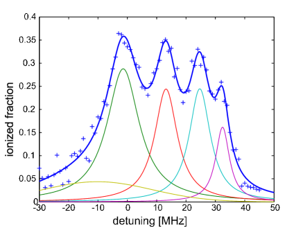
Zero detuning corresponds to the center frequency of the transition as observed in a reference vapor cell. The ionization data (crosses) can be fitted to a combination of four Lorentzian profiles plus one Gaussian profile which models the background (solid line). The four maxima match the expected positions of the transitions , however, width and relative heights deviate from the expected spectrum of free and unperturbed atoms. Both deviations are partly due to a common origin. The ionization was performed in a magnetic trap with an non vanishing magnetic field of about 1 Gauss which is sufficiently strong to split the magnetic sub states by some MHz Baluschev et al. (2000). The Zeeman splitting thus leads to a broadening as well as to a reduction of the peak hight. As the number of sub states increases with increasing this effect is most dominant for the state with its 9 magnetic sub states. Additional broadening may occur due to saturation of the two photon transition and for large ionization rates which reduce the lifetime of the 5D state.
V Conclusion
In summary, we have reported the spatially resolved direct observation of photo ionization of ultracold 87Rb atoms on an atomchip. The atoms are ionized by absorbing three photons from two continuous laser beams. The ionization is detected by observing light induced losses of trapped atoms. Ionization spectra are recorded by scanning across the resonance of the two photon transition from the 5S ground state to the 5D excited state. The combination of two lasers allow for compensating unwanted dipole potentials which may repel the atoms from the ionization region. The setup avoids stray light and is suitable for single atom detection in combination with a sensitive ion detector. Furthermore it may form the starting point for studying ion atom mixtures in a combined micro trap for charged and neutral particles.
Acknowledgements.
We gratefully acknowledge support by the Landesstiftung Baden-Württemberg and by the Deutsche Forschungsgemeinschaft.References
- Fortágh and Zimmermann (2007) J. Fortágh and C. Zimmermann, Rev. Mod. Phys. 79, 235 (2007).
- Lin et al. (2004) Y. J. Lin, I. Teper, C. Chin, and V. Vuletić, Phys. Rev. Lett. 92, 050404 (2004).
- Wildermuth et al. (2005) S. Wildermuth, S. Hofferberth, I. Lesanovsky, E. Haller, L. M. Andersson, S. Groth, I. Bar-Joseph, P. Krüger, and J. Schmiedmayer, Nature 435, 440 (2005).
- Günther et al. (2005a) A. Günther, M. Kemmler, S. Kraft, C. J. Vale, C. Zimmermann, and J. Fortágh, Phys. Rev. A 71, 063619 (2005a).
- Esteve et al. (2006) J. Esteve, J.-B. Trebbia, T. Schumm, A. Aspect, C. I. Westbrook, and I. Bouchoule, Phys. Rev. Lett. 96, 130403 (2006).
- Paredes et al. (2004) B. Paredes, A. Widera, V. Murg, O. Mandel, S. Fölling, I. Cirac, G. V. Shlyapnikov, T. W. Hänsch, and I. Bloch, Nature 429, 277 (2004).
- Kinoshita et al. (2004) T. Kinoshita, T. Wenger, and D. S. Weiss, Science 305, 1125 (2004).
- Wonneberger (2006) W. Wonneberger, Phys. Lett. A 356, 272 (2006).
- Aubin et al. (2006) S. Aubin, S. Myrskog, M. Extavour, L. J. LeBlanc, D. McKay, A. Stummer, and J. H. Thywissen, Nature Physics 2, 384 (2006).
- Teper et al. (2006) I. Teper, Y.-J. Lin, and V. Vuletić, Phys. Rev. Lett. 97, 023002 (2006).
- Haase et al. (2006) A. Haase, B. Hessmo, and J. Schmiedmayer, Optics Letters 31, 268 (2006).
- Takamizawa et al. (2006) A. Takamizawa, T. Steinmetz, R. Delhuille, T. W. Hänsch, and J. Reichel, Optics Express 14, 10976 (2006).
- Campey et al. (2006) T. Campey, C. J. Vale, M. J. Davis, N. R. Heckenberg, H. Rubinsztein-Dunlop, S. Kraft, C. Zimmermann, and J. Fortágh, Phys. Rev. A 74, 043612 (2006).
- Cote et al. (2002) R. Cote, V. Kharchenko, and M. D. Lukin, Phys. Rev. Lett. 89, 093001 (2002).
- Ciampini et al. (2002) D. Ciampini, M. Anderlini, J. H. Müller, F. Fuso, O. Morsch, J. W. Thomsen, and E. Arimondo, Phys. Rev. A 66, 043409 (2002).
- Massignan et al. (2005) P. Massignan, C. J. Pethick, and H. Smith, Phys. Rev. A 71, 023606 (2005).
- Seidelin et al. (2006) S. Seidelin, J. Chiaverini, R. Reichle, J. J. Bollinger, D. Leibfried, J. Britton, J. H. Wesenberger, R. B. Blakestad, R. J. Epstein, D. B. Hume, et al., Phys. Rev. Lett. 96, 253003 (2006).
- Stibor et al. (2007) A. Stibor, S. Kraft, T. Campey, D. Komma, A. Günther, J. Fortágh, C. J. Vale, H. Rubinsztein-Dunlop, and C. Zimmermann, submitted for publication (2007).
- Nez et al. (1993) F. Nez, F. Biraben, R. Felder, and Y. Millerioux, Opt. Comm. 102, 432 (1993).
- Duncan et al. (2001) B. C. Duncan, V. Sanchez-Villicana, P. L. Gould, and H. R. Sadeghpour, Phys. Rev. A 63, 43411 (2001).
- Ackerhalt and Eberly (1976) J. R. Ackerhalt and J. H. Eberly, Phys. Rev. A 14, 1705 (1976).
- Grove et al. (1995) T. T. Grove, V. Sanchez-Villicana, B. D. Duncan, S. Maleki, and P. L. Gould, Phys. Script. 52, 271 (1995).
- Hurst et al. (1979) G. S. Hurst, M. G. Payne, S. D. Kramer, and J. P. Young, Rev. Mod. Phys. 51, 767 (1979).
- Günther et al. (2005b) A. Günther, S. Kraft, M. Kemmler, D. Koelle, R. Kleiner, C. Zimmermann, and J. Fortágh, Phys. Rev. Lett. 95, 170405 (2005b).
- Fortágh et al. (2003) J. Fortágh, H. Ott, S. Kraft, A. Günther, and C. Zimmermann, Appl. Phys. B 76, 157 (2003).
- Kraft et al. (2005) S. Kraft, A. Deninger, C. Trück, J. Fortágh, F. Lison, and C. Zimmermann, Las. Phys. Lett. 2, 71 (2005).
- Baluschev et al. (2000) S. Baluschev, N. Friedman, L. Khaykovich, D. Carasso, B. Johns, and N. Davidson, Appl. Opt. 39, 4970 (2000).