]Author to whom correspondence should be addressed. Electronic mail: myqiang@nju.edu.cn.
Lateral organization in mixed lipid bilayers supported on a geometrically patterned substrate
Abstract
The organization of lipids in biological membranes is essential for cellular functions such as signal transduction and membrane trafficking. A major challenge is how to control lateral lipid composition in supported membranes which are crucial for the design of biosensors and investigation of cellular processes. Here, we undertake the first theoretical study of lateral organization of mixed lipids in bilayers induced by a geometrically patterned substrate, and examine the physical mechanism of patterned substrate-induced structural formation in the supported lipid bilayers. A rich variety of composition segregations of lipids are regulated, and the results can account well for most recent experimental works [Yoon, T. Y. et al. Nat. Mater. 5, 281(2006) and Parthasarathy, R. et al. Langmuir 22, 5095(2006)]. The present study provides a comprehensive understanding of mechanically controlling the spatial organization of membrane components by unifying these experimental evidences.
Keywords: membrane organization; mixed lipid bilayer; patterned substrate
Introduction
Biomembranes are self-assembled bilayers of lipid molecules. Recently, supported membranesackmann1 ; sackmann2 has attracted extensive interest, not only due to its importance in studying the properties and functions of biological membranes, but also due to its potential applications in design and fabrication of biological devices.bayley ; cornell ; Anrather ; Kasemo ; figeys Supported bilayers can maintain the structural and dynamic properties of free bilayer such as the lateral fluidity, share many similarities with the natural membranes, and therefore are widely used as cell-surface models that connect biological and artificial materials.sackmann1 ; sackmann2 To realize the bio-functional supported membranes or design novelly biological devices, we usually need to manipulate the supported membranes in nanometer or micrometer scale.Kasemo For example, one can apply a lateral electric field to rearrange the distribution of lipids in a supported membrane for some related cellular processes.groves3 Additionally, one can also separate a supported membrane into well-defined membrane domains by introducing some barriers into it, and micropattern it by the help of approaches of photolithographic patterning, microprinting, or microfluidic flow patterning.groves1 ; groves4 Supported membrane micropatterning has attracted tremendous attention in recent years because of its potential applications in the investigation of immunobiology, drug discovery, design of biosensors, etc.sackmann2
Most recently, Lee and co-authorsyoon ; yoon1 ; yoon2 presented an experimental approach to manipulate the lipid segregation in a mixed supported membrane by use of the geometrical property of substrate. They studied the lateral organization of lipids in a mixed membrane supported on a substrate with a groove,yoon ; yoon1 and found that, due to different effective molecular shapes of mixed lipids, the two kinds of lipids spontaneously segregate, where the lipids with big spontaneous curvature are preferentially localized at the grooves. In a most recent experimental work, Yoon et al.yoon2 studied the effect of topographical substrate on the lipid raft formation in the supported membrane. In their system, the topography of the substrate is either nanocorrugated or nanosmooth, and the membrane supported on such a substrate will be curved at certain predetermined positions to follow the substrate structure. Because of large bending rigidity of sphingolipid/cholesterol-rich domains, there exists a large free energy when they stay in the curved regions. Thus, the macroscopic rafts which are coarsened by the nanorafts, can emerge in the nanosmooth regions of substrate, whereas there are only nanorafts in the nanocorrugated regions. On the other hand, Parthasarathy et al.groves2006 proposed a curvature-mediated modulation of phase-separated structures in membranes, and they found that, beyond a critical curvature of membrane geometry, the cholesterol-rich lipid-ordered domains stayed in the small-curvature regions, while the cholesterol-poor lipid-disordered domains were preferentially localized in the large-curvature regions due to their different rigidities. Besides the lipid domains in supported membrane systems, it was also found that membrane curvature can induce phase separation in the mixed giant unilamellar vesicles(GUVs) due to the different rigidities of different domains.www ; roux ; bacia
Further research will be needed to uncover and reveal all the possible lipid structures and physical mechanisms behind experimental evidence from lateral organization in supported membranes. As is well-known, it is still difficult to systematically probe and visualize the laterally heterogeneous structures of lipids from tens to hundreds of nanometers in size with current experimental technologies.kraft There is therefore an urgent need to gain greater theoretical insight into the physical picture of how membrane organization is governed. Here, we systematically investigate the lateral organization in a mixed bilayer consisting of symmetric lamella-forming lipids and asymmetric cylinder-forming lipids supported on a geometrically patterned substrate, and explore physical mechanism behind structural formation of lipids by using self-consistent field theory(SCFT). We expect the present study to offer a universal and thorough strategy towards efficiently controlling the lateral phase separation of mixed lipids in bilayers by varying the substrate roughness, in addition to yielding insights into several experimental findings.
Model and Methods
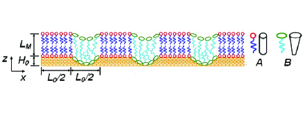
We consider a system composed of lipid species A and lipid species B dispersed in hydrophilic homopolymer solvents,aaa which is supported on a geometrically patterned substrate(see Fig. 1). The headgroup volumes of lipid species A and B are and (, respectively, and their tails have the same length consisting of segments of segment volume .bbb The solvent chain consists of segments with segment volume . In the present problem, we choose without loss of generality. The conformation of the th chain of lipid A, lipid B, and homopolymer solvent can be characterized by continuous space curves , , and , respectively, and all of the chains are completely flexible.gh We assume that the system is translational invariant in -direction,groves2006 then the surface of patterned substrate is characterized by a periodical function , which is only dependent on . In our calculation, we choose as follows,
| (1) |
Here, is the depth of the grooves, is the period of the substrate, and is the number of the periods in the patterned substrate.
To characterize the system, we define the concentration operators of headgroups and tails of -type lipids( denotes lipid species A and B, respectively) and solvents as follows,
| (2) | |||
| (3) |
| (4) |
where is the dimensionless number density of headgroups of -type lipids, and .
We assume that the system satisfies the incompressible condition , and is given by,Matsen1
| (5) |
where is a sufficiently small quantity.
The SCFT method is very powerful to inhomogeneous macromolecular systems (see, for example, reviewsSchmid ; ma33 ; ma22 ) and has been applied to investigate the lipid bilayer or lipid bulk phase behaviors in recent years.leer2 ; leer5 ; Li-Schick ; schick ; schick5 Leermakers and co-authors used a lattice version of SCFT to study the lipid-bilayer system,leer2 ; leer5 where the conformation of lipid chains is described by random-walk and RIS (Rotational Isomeric State) statistics. However, in most of their works, they assumed that the lipid bilayer was laterally homogeneous for computational convenience. On the contrary, Schick and co-authors adopted the off-lattice version of SCFT to study the lipid systems,Li-Schick ; schick ; schick5 where the amphiphilic nature and chain stretching of lipids are still captured. Compared to the lattice version of SCFT used by Leermakers and co-workers, the off-lattice SCFT can account reasonably well for lateral inhomogeneity of the lipid bilayers, although less molecular details are considered. Here, we emphasis on the description of laterally heterogeneous structures and phase separation of lipids due to the interplay among the conformational entropy of lipids, effective molecular shapes of lipids, and substrate geometry. Therefore, the molecular details of lipids are not important, and it will be more reasonable to use the coarse-grained model in the framework of off-lattice SCFT schick5 .
In the framework of self-consistent field theory(SCFT), we first transform a hamiltonian of the lipid system into a coarse-grained field theory description by employing the simple Gaussian model for lipid chains and treating their interactions through mean-field Flory-Huggins form. The partition function of the system is given by
| (6) |
where is a weighted functional integral over all the possible configurations of the th chain of lipids or solvent, and is the probability distributions of molecular conformations for the th chain. is the Flory-Huggins interaction parameter between the hydrophilic components( lipid headgroups or solvents) and the hydrophobic lipid tails, and is the head-head or tail-tail pair interactions of different kinds of lipids. is the surface field of the substrate, and we setBalazs1
| (7) |
where describes the field strength at the substrate surface.
The mean-field free energy of the system is given by,ma33
| (8) |
where is Boltzmann’s constant, is the temperature, and is the effective volume of the system.ma22 and are the overall volume fraction and the tail volume fraction of lipid species A () or B () in the system, respectively. , , and are the single-molecule partition functions of lipid A, lipid B, and solvent, respectively, and they are given by () and . The variables , , and are the concentration functions of -type headgroups, -type tails, and solvents, while , , and are the fields acting on the headgroups, tail segments, and solvent segments, respectively. is a Lagrange-multiplier field which ensures the incompressibility of the system. The fields and densities are then obtained by minimizing the free energy in Eq.(8), and given as
| (9) | |||
| (10) |
| (11) | |||
| (12) | |||
| (13) | |||
| (14) | |||
| (15) | |||
| (16) |
| (17) | |||
| (18) | |||
| (19) | |||
| (20) | |||
| (21) |
where and are the end-segment distribution functions of the -type lipids, and are defined by,ma33
| (22) | |||
| (23) |
They satisfy the modified diffusion equations,
| (24) | |||
| (25) |
where is the statistical segment length. The initial conditions are and . has the analogous definition for the solvent, which satisfies Eq. (24) with the initial condition . The resulting self-consistent equations (9) - (21) together with equations(22)-(25) can be numerically solved by the combinatorial screening method proposed by Drolet and Fredrickson.ma22
We choose the tail volume fractions in lipid species A and B as and , and the other parameters are fixed to be , , , and . Thus, we have , , , and , and with these parameters, lipid species A and B form a lamellar and a hexagonally cylindrical phases in bulk, respectively.Li-Schick This means that lipid species A has a symmetric head/tail shape favoring the packing of flat interfaces, while lipid species B is asymmetric, which prefers to curve the interfaces to the lipid tails. As lipid species A and B are mixed to put onto a hydrophilic substrate, a mixed lipid bilayer may be formed. Here we take the overall volume fractions of lipid species A and B as , and assume that the repulsive head-head/tail-tail interactions between the two kinds of lipids are weak(). The strength of surface field is taken to be . To facilitate the formation of a bilayer structure, we adopt the scenario proposed in Ref. 29 by introducing an external field favoring the lipid tails in the initial stage of the self-consistent calculation, which enforces the bilayer membrane formed along the substrate surface profile. When the bilayer is formed, the external field is turned off and the calculation continues until the equilibrium state of system is reached.
Results and Discussion
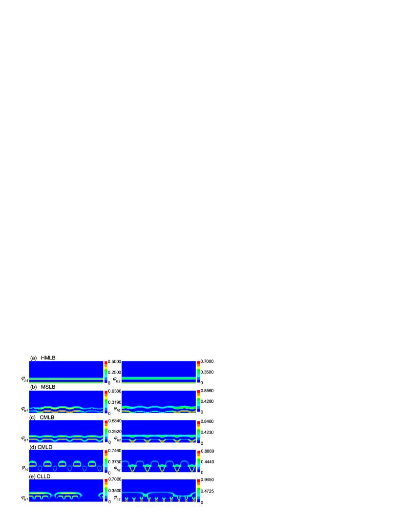
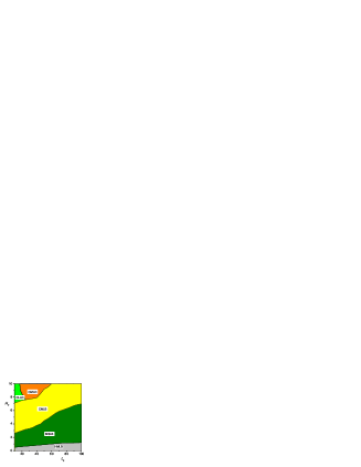
We now examine the formation of all the possible lipid structures by systematically varying the groove depth () and the substrate period (), and the results are summarized in Fig. 2. Figure 3 shows a morphology diagram, and a rich variety of lipid domains can be tuned by varying and . Here, the height of system is fixed to be 40 which is large enough to not influence the morphology of lipid bilayer, and the lateral length ranging from is adjusted to search the stable phases by the total free energy minimization. The substrate period changes from to and the groove depth from to , which are either comparable to or several times larger than the natural size of the lipids(), and therefore, the evolved scale of lateral organization is within the nanometer size(i.e., from tens to hundreds of nanometers).
When is vanishingly small, the perturbation from the substrate geometry is neglectable, and lipids are mixed uniformly on the substrate due to mixing entropy. Thus a homogeneously mixed lipid bilayer(HMLB) state is formed(Fig. 2(a)). With the increase of , the substrate becomes slightly rough, and thus lipid species B will first occupy the grooves to gain more conformational entropy. This is because that, although the substrate is relatively flat, there still exist some perturbations to the lipid bilayer from the substrate where the grooves drive the formation of lipid B-rich islands(nucleation). As lipid B-rich islands initiates from the grooves, a lateral expansion of the islands on the substrate appears, and eventually leads to the formation of macro-segregated lipid bilayer(MSLB) under the help of the lipid A - B interfacial energy (Fig. 2(b)). When the groove depth is increased to modest values where the curvature of grooves becomes comparable to the bulk curvature of lipid species B, both lipid species B and A can get more entropy as they stay in the grooves and on the flat steps, respectively. Thus, a component-modulated lipid bilayer(CMLB) structure appears in this region, as shown in Fig. 2(c).
When is enough large, the groove curvature may be larger than the spontaneous curvature of lipid species B at small or modest region, and the lipid distribution may become more complicated. In this case, if the bilayer structure is still maintained, the bilayer would be curved strongly in the regime of grooves, and thus lipid species B in the upper leaflet must lose plenty of conformational entropy because the curvature of the upper leaflet is in contrast to their bulk curvature. However, lipid species B in the lower leaflet prefers to fill in the grooves, compared to the case if lipid species A fills in the grooves. As a result, the lipid bilayer may be ruptured by the extremely rough substrate: the component-modulated lipid domains(CMLD) appears at modest , and the coalesced long lipid droplets(CLLD) with spreading lipid-A droplets is formed at smaller (see Fig. 3). The formation of the CMLD and CLLD structures in Fig. 2 can be understood by the help of the morphologies of thin films on chemically patterned substrates. Actually, in the CMLD and CLLD structures, the lipid species B almost fill up the grooves, and therefore for lipid species A, the substrate now becomes a chemically patterned one which is composed of alternating more-wettable hard-wall parts and less-wettable lipid-B parts. Thus, the problem can be highlighted by templating of thin films on chemically patterned substrates studied by Kargupta and Sharmakajari . They examined the effects of the periodicity of substrate pattern and the width of the more-wettable stripes on the morphology of thin films, and found that when the patterned substrate periodicity is beyond a critical length, the thin film pattern closely replicates the substrate surface pattern, namely dewetting occurs on every less-wettable parts of the patterned substrate, just as shown in Fig. 2(d) where lipid species A is almost localized on more-wettable hard-surface parts. On the contrary, when the periodicity is smaller than the critical length, the dewetting effect may partially be suppressed, and the lone droplet of thin films spans across the remaining less-wettable sites, exactly like the morphology of lipid-A droplets shown in Fig. 2(e). In real biological systems, it was reported sara that large quantities of membrane fusion pore sites which are concentrated in cone-shaped nonlamellar lipids such as phosphatidylethanolamine(PE), are simultaneously formed at high-curvature regions. This provides further evidence implying the validity of these unexpected structures in Figs. 2(d) and 2(e). Therefore, the lipid heterogeneity by forming CLLD and CMLD structures may highlight the understanding of the relation between the membrane curvature and fusion, fission, or pore formation of membranes. In the large- region, the curvature of the groove become comparable to the spontaneous curvature of lipid species B again, and a CMLB phase is presented there.
Importantly, by changing the position of grooves for given and , we find that at arbitrary spatial distribution of grooves, the above conclusions keep unchanged, independent of distributed positions of grooves. Actually, such a substrate geometry mediated phase-separated behavior of lipids provides a controllable mechanism for spatial sorting of membrane components by varying the roughness of substrates. Furthermore, this is also helpful to understanding how cellular membrane geometry to govern the lipid ordering and domain rafts in real biological systems.
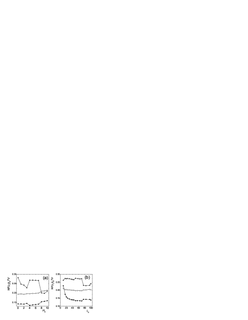
To clarify the underlying mechanism of formed phase behavior of mixed lipids, we examine the competition of the interfacial energy, conformational entropy, and mixing entropy of lipids. Figure 4(a) shows the variation of free energy of the system versus by fixing . When (the substrate is completely smooth), the mixing entropy dominates the lateral organization of the two kinds of lipids, leading to the uniform dispersion of lipids. When is low (e.g., 3), the interfacial energy dominates the distribution of lipids and a MSLB structure appears with lateral growth of lipid B-islands located at grooves. For middle values of (e.g., 7), a CMLB structure is formed by releasing the conformational entropy of lipid species B, while at the same time, the interfacial energy is increased. Such a CMLB structure is mainly contributed to the entropy effect of lipid species B. When is large enough (e.g., 10), the formation of CMLD structure where the two kinds of lipids separate strongly, largely decreases the interfacial energy, while also leads to the loss of mixing entropy. In Fig. 4(b), we show the variation of free energy versus by fixing . The mixed lipid bilayer is either component-modulated structure for small as a result of the release of conformational entropy of lipid species B or a macro-separated bilayer for large where the interfacial energy is greatly decreased.
It is vitally useful to make a comparison between the present work and experimental works.yoon ; yoon1 ; yoon2 ; groves2006 First, the appearance of CMLB structure accurately confirms the experimental results observed in Refs. 11 and 12. We also find that such a CMLB structure formed in the modest curvature regions exactly corresponds to the nanorafts emerged in the nanocorrugated regions in Ref. 13 and the spatial modulated structure of alternating the domains in low curvature and domains in high curvature regions observed in Ref. 14, while the MSLB structure presented in the lower curvature regions is just as the case that the macroscopic domains appear in nanosmooth regions in Ref. 13. Further, our phase diagram describing all the observed morphology regimes, which greatly enriched the lateral organization of membrane component on a geometrically patterned substrate, will be very helpful to experimentally probe the lipid distribution on a wide range of length scales of nanometers.kraft Finally, by comparing the physical models of the experimental works and our theoretical work, we easily find that although our system is not completely identical to those studied by Yoon et al.yoon2 and Parthasarathy et al.groves2006 , the physics of these models is consistent, namely the curved regions offer some barriers for lipid-A domains in the present work and domains in Refs. 13 and 14. This is because the sphingolipid/cholesterol-rich domain and cholesterol-poor domain can reasonably be represented by symmetric lipid A and asymmetric lipid B in our model, respectively. The only difference during the lipid organization is that the free energy barrier in the experimental works is provided by the bending energy of lipid domains, while in our system, comes from the loss of conformational entropy of lipids.
Conclusions
We conducted a thorough investigation in the lateral organization of a two-component lipid bilayer on a geometrically patterned substrate by using self-consistent field theory. Because of their different molecular shapes, the two kinds of lipids prefer to stay in the grooves and flat steps of substrate, respectively. By adjusting the groove depth and the substrate period, we obtained a rich variety of laterally organized lipid structures. We also examine the microscopic mechanism of self-assembly of lipids in the mixed bilayer supported on the geometrically patterned substrate, and find that the resulting phase behavior of mixed lipids is dominated by the competition of the interfacial energy, conformational entropy, and mixing entropy of lipids. The present study confirmed recent experimental results in Refs. 11 and 12, and qualitatively agreed with the experimental findings of Refs. 13 and 14. Most importantly, we also predict, for the first time, the formation of the CLLD and CMLD structures in large curvature regions, which may provide theoretical insight into future experiment with the improvement of visualization techniques towards smaller scales of geometry and help to understand the microscopic mechanism of fusion pore formation in biomembranes.
Acknowledgments
This work was supported by the National Natural Science Foundation of China, No. 10334020, No. 10021001, No. 20674037, and No. 10574061.
References
- (1) Sackmann, E. Science 271, 43(1996).
- (2) Tanaka, M. & Sackmann, E. Nature 437, 656(2005).
- (3) Bayley, H. & Cremer, P. S. Nature 413, 226(2001).
- (4) Cornell, B. A., Braach-Maksvytis, V. L. B., King, L. G., Osman, P. D. J., Raguse, B., Wieczorek, L. & Pace, R. J. Nature 387, 580(1997).
- (5) Anrather, D., Smetazko, M., Saba, M., Alguel, Y. & Schalkhammer, T. J. Nanosci. Nanotechnol. 4, 1(2004).
- (6) Kasemo, B. Surf. Sci. 500, 656(2002).
- (7) Figeys, D. & Pinto, D. Anal. Chem. 72, 330A(2000).
- (8) Groves, J. T., Boxer, S. G. & McConnell, H. M. Proc. Natl. Acad. Sci. U.S.A. 94, 13390(1997).
- (9) Groves, J. T., Ulman, N. & Boxer, S. G. Science 275, 651(1997).
- (10) Groves, J. T. & Boxer, S. G. Acc. Chem. Res. 35, 149(2002).
- (11) Yoon, T. Y., Jeong, C., Kim, J. H., Choi, M. C., Kim, M. W. & Lee, S. D. Appl. Surf. Sci 238, 299(2004).
- (12) Jeong, C., Yoon, T. Y., Lee, S. D., Park, Y. G., & Kwon, H. Mol. Cryst. Lip. Cryst. 434, 625(2005).
- (13) Yoon, T. Y., Jeong, C., Lee, S. W., Kim, J. H., Choi, M. C., Kim, S. J., Kim, M. W. & Lee S. D. Nat. Mater. 5, 281(2006).
- (14) Parthasarathy, R., Yu, C. H. & Groves, J. T. Langmuir, 22, 5095(2006).
- (15) Baumgart, T., Hess, S. T. & Webb, W. W. Nature 425, 821(2003).
- (16) Bacia, K., Schwille, P. & Kurzchalia, T. Proc. Natl. Acad. Sci. U.S.A. 102, 3272(2005).
- (17) Roux, A., Cuvelier, D., Nassoy, P., Prost, J., Bassereau, P. & Goud, B. EMBO J. 24, 1537(2005).
- (18) Kraft, M. L., Weber, P. K., Longo, M. L., Hutcheon, I. D. & Boxer, S. D. Science 313, 1948(2006).
- (19) In real system, the solvent is usually composed of small water molecules. However, in the SCFT framework, if the solvent is modeled as small featureless molecules, one finds that the small molecules are very difficult to be removed from the hydrophobic regions of the lipid bilayers due to large mixing entropy.leer5 Actually, the main goal of the present paper is to reveal the lateral organization of lipid bilayer supported on a patterned substrate, where the solvent only offers a hydrophilic background to maintain the lipid bilayer and could not affect the physics of lipid organization. This means that the conformation of solvent is not important, and therefore, we can use the homopolymer solvents instead of small water molecules, as used by Schick and co-authorsschick ; schick5 .
- (20) Each lipid consists of one hydrophilic headgroup and one hydrophobic tail: the head-tail volume ratio signifies the effective molecular shape of lipids reflecting the head-head and tail-tail interactions, and single tail displays the chain stretching of lipids. Further, at the coarse-grained level, the lipids A and B can reasonably represent cholesterol-rich lipid-ordered () domain and cholesterol-poor lipid-disordered () domainyoon2 , respectively, although the real lipid is usually composed of one headgroup and two tails.
- (21) Li and Schick Li-Schick have compared the SCFT results of the lipid system with the experimental results, and a good agreement was found between them, while the real lipid is not completely flexible.
- (22) Matsen, M. W. J. Chem. Phys. 106, 7781(1997).
- (23) Schmid, F. J. Phys.: Condens. Matter 10, 8105(1998).
- (24) Matsen, M. W. In Soft Matter; Gompper, G., Schick, M., Eds.; Wiley-VCH: Weinheim, 2006.
- (25) Fredrickson, G. H. The equilibrium theory of inhomogeneous polymers; Oxford University Press: Oxford, 2006.
- (26) Meijer, L. A., Leermakers, F. A. M. & Lyklema, J. J. Chem. Phys. 110, 6560(1999).
- (27) Leermakers, F. A. M., Rabinovich, A. L. & Balabaev, N. K. (2003) Phys. Rev. E 67, 011910.
- (28) Li, X. J. & Schick, M. Biophys. J. 78, 34(2000).
- (29) Katsov, K., Müller, M. & Schick, M. Biophys. J. 87, 3277(2004).
- (30) Müller, M., Katsov, K. & Schick, M. arXiv:cond-mat/0609295 (2006).
- (31) Lee, J. Y., Shou, Z. & Balazs, A. C. Phys. Rev. Lett. 91, 136103(2003).
- (32) Kargupta, K. & Sharma, A. Phys. Rev. Lett. 86, 4536(2001).
- (33) Ostrowski, S. G., Van Bell, C. T., Winograd, N. & Ewing, A. G. Science 305, 71(2004).