Force dependent fragility in RNA hairpins
Abstract
We apply Kramers theory to investigate the dissociation of multiple bonds under mechanical force and interpret experimental results for the unfolding/refolding force distributions of an RNA hairpin pulled at different loading rates using laser tweezers. We identify two different kinetic regimes depending on the range of forces explored during the unfolding and refolding process. The present approach extends the range of validity of the two-states approximation by providing a theoretical framework to reconstruct free-energy landscapes and identify force-induced structural changes in molecular transition states using single molecule pulling experiments. The method should be applicable to RNA hairpins with multiple kinetic barriers.
pacs:
82.35.-x,82.37.-j,87.15.-vSingle molecule pulling experiments allow to exert mechanical force on individual molecules such as nucleic acids, proteins and macromolecular complexes Review . By recording force-extension curves it is possible to determine free energies and kinetic parameters of biomolecules and search for intermediates and pathways in biochemical reactions. Over the past years single molecule techniques have been successfully applied to investigate the breakage of molecular bonds in many biological systems such as proteins Mariano , DNA molecules bockelmann97 , RNA molecules Liphardt01 , ligand-receptor binding fritz98 and beyond, e.g. metallic gold nanowires stretched with AFM Agrait01 . Under mechanical load all these structures yield at different values of the applied force in a dynamical process that is stochastic and loading rate dependent. The study of breakage forces under nonequilibrium conditions is known as dynamic force spectroscopy EvansWilliams01 . A detailed comprehension of the rupture kinetics of biomolecular complexes has implications in our understanding of their kinetic stability which is important in enzymatic and/or regulatory processes.
Here we investigate the unfolding/refolding kinetics of RNA hairpins using laser tweezers Liphardt01 ; Smith03 . The RNA sequence and its native structure are shown in Fig.1. To manipulate the RNA hairpin two beads are attached to the ends of the RNA hairpin by inserting two hybrid RNA/DNA handles foot1 . One of the beads is immobilized on the tip of a micropipette while the other bead is captured in the optical trap. By moving the micropipette a force is exerted upon the ends of the RNA hairpin and the force-extension curve (FEC) recorded. In Fig.1 we show the experimental FEC corresponding to a complete cycle of a ramping process where the force is first raised and relaxed afterwards. In the pulling process the molecule is initially in its native folded structure, and the force is increased at a certain rate until the molecule unfolds. If the process is reversed, i.e. the force is decreased at rate , the molecule folds back again (relaxing process) Manosas05 . The unfolding/refolding of the molecule can then be identified as force-extension jumps observed in the FEC.
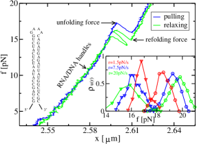
By repeatedly pulling the molecule many times we obtain the distribution of unfolding (refolding) forces, i.e. the force at which the first unfolding (refolding) event occurs along the pulling (relaxing) process (Fig.1). The experimental distribution of the unfolding (u) and refolding (r) forces at different loading rates is shown as an inset of Fig.1.
To model the hairpin we follow Cocco et al. cocco03 and restrict the number of configurations of an base-pair (bp) RNA hairpin to the set of configurations where the first bps are opened and the last are closed (the total number of configurations being ). The end-to-end distance is a well defined reaction coordinate for the unfolding/refolding reaction. We use the variable to label the state of the hairpin; e.g the folded (F) state corresponds to and the unfolded (UF) state to . The stability of each state depends on its free energy, , at a given applied force cocco03 ,
| (1) |
where is the free energy of formation at zero force and is the force-dependent contribution to the free energy. The latter is given by where the functions and are the end-to-end distance and the entropic correction to the free energy of an -bases long single stranded RNA (ssRNA) at force , being the number of bases released after the opening of bps. The latter can be computed as the reversible mechanical work needed to stretch the ends of an -bases long ssRNA a distance ,
| (2) |
where is the FEC of the ssRNA foot2 . The free energy landscape of a hairpin, , as a function of is known to be rugged with different kinetic barriers (or transition states) depending on the sequence and on the applied force Nelson . We use the Mfold prediction Mfold to extract the free energy of the molecule at zero force, . In what follows we take the F state as the reference state for the free energy, i.e. .
The kinetics of unfolding (i.e. the transition between the completely folded (F) and the completely unfolded (UF) states) is an activated process with a force-dependent effective barrier, , measured relative to the F state. The rates of unfolding and refolding, and , can be computed as the first passage rates Zwanzig for a Brownian particle to cross a force-dependent effective barrier :
| (3) |
where is the free energy difference between the F and UF states at force , with and being respectively the Boltzmann constant and the bath temperature, and is an attempt frequency. An analytical expression for can be derived from Kramers theory applied to the dissociation of consecutive bonds under mechanical force in the stationary approximation EvansWilliams01 ; Zwanzig ,
| (4) |
with . The variation in force of the effective barrier gives information about its position along the reaction coordinate:
| (5) |
where and are the distances from the effective barrier to the F and UF states respectively. The location of the barrier along the reaction coordinate is related to the fragility of the molecule which determines how much the unfolding/refolding kinetics is sensitive to the force. To characterize the fragility we introduce the parameter defined as:
| (6) |
corresponds to a brittle structure -e.g. the case of hairpins stabilized by tertiary contacts where the barrier is located near to the F state-, whereas represents a flexible or compliant structure, i.e. molecules that can easily deform under applied force and the barrier is close to the UF state leffler .
The rates (3) are related to the force distributions by the expression -(+) with being the initial force in the pulling (relaxing) process evans . The unfolding (refolding) rates read as , where is the probability that the molecule remains in the F (UF) state along the pulling (relaxing) process until reaching the force , +(-). Note that the experimental FECs show a force jump (Fig.1) when the molecule unfolds or refolds that corresponds to the relaxation of the bead in the trap after the sudden increase or decrease in the RNA extension. To compensate for this effect we shift the value of the folding forces by an amount equal to +(-). From (1,3) and the unfolding (refolding) force distributions, , we can extract the effective barrier as:
| (7) |
| (8) |
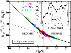
Using polymer theory foot2 we can estimate so the expressions (7,8) have only two unknown parameters, and . We determine by collapsing into a single curve the effective barrier estimates (7) and (8) corresponding to the pulling and relaxing processes at different loading rates. From our data we get in very good agreement with the Mfold prediction, Mfold . In Fig.2 we show the force dependent effective barrier obtained using this method. We then determine the value of the attempt frequency by fitting (7,8) to the prediction by Kramers theory (4). We obtain which is of the order of magnitude of the values reported for other hairpins thirum . The agreement found between the predicted effective barrier (4) and the results from the experiments (7,8) validates our description hence providing a way to estimate the attempt frequency foot5 . The representation of the effective barrier as a function of the applied force reflects two distinct regimes (Fig.2) characterized by different slopes of . These correspond to different locations of the effective barrier (5) and different values of the fragility (6). We define a crossover force as the value at which the extrapolated straight lines corresponding to regimes I and II intersect each other (Fig.2).
A kinetic barrier is characterized by its location and its height . As shown in the inset of Fig.2, the free energy landscape at force shows that there are two barriers corresponding to transition states located at and . At low forces, (regime I), the highest barrier is located at and corresponds to the entropy cost associated to the opening of the four bases loop. Whereas for large forces, (regime II), the kinetics is governed by the barrier located at at the interface between the GC and AU rich regions of the hairpin. In our experiments we observe the two different regimes, I and II, because the crossover force, , is within the experimentally accessible range of rupture forces, (inset of Fig.1), where is the critical force verifying in (1). In order to investigate the unfolding/refolding kinetics over a broader range of forces than those accessible in force-ramp experiments, force-jump experiments pan could be very helpful.
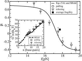
From and we obtain the fragility by using (5,6). In Fig.3 we show the agreement between the fragility obtained from the experimentally measured barrier (7,8) and Kramers theory (4). Finally, our method can be used to experimentally reconstruct the free energy landscape of the molecule from the sole knowledge of the breakage force distribution at different loading rates. We first determine the location of the force dependent transition state from the measured value of (5). Using the saddle point approximation we then identify the effective barrier with the largest contribution to the sum appearing in the r.h.s of (4), , where we have taken foot4 . Finally we apply (1) and extrapolate the free energy to zero force to obtain . In the inset of Fig.3 we show the experimentally reconstructed free energy landscape compared with the results obtained from Mfold Mfold .
Changes in the position of the transition state (5) along the reaction coordinate axis with the force can correspond to two different situations: (i) The location of the transition state does not change with force but the extension does for all configurations as predicted in thirum1 ; (ii) The free energy landscape of the hairpin shows multiple barriers leading to different transition states depending on the value of the force. This is the case considered in the present study. Interestingly the value of varies with force in the situation (ii) but not in (i). Therefore is a good parameter to identify structural changes in the transition state.
A useful analysis of experimental data for molecular rupture is the two-states model mun ; ritort where the position of the kinetic barrier along the reaction coordinate is fixed: and the fragility, , do not depend on the force. In this approximation, is a straight line as a function of the applied force evans . A plot of the experimental data for versus force displays a non-zero curvature (data not shown) indicating a force dependent fragility. Moreover, in two-states systems the dependence of the mean value of the unfolding and refolding forces on the rate can be estimated in the experimental regime where hummerszabo03 :
| (9) |
By fitting the experimental results to (9) we can estimate values for which give . This corresponds to a barrier located in the middle between the F and UF states, in disagreement with the free energy landscape shown in Fig.2. Yet, this value coincides with the average fragility measured over the range of forces (Fig.3) suggesting that fragility estimates obtained by fitting the two-states model to the experimental data correspond to averages of force dependent fragilities over the range of breakage forces explored in the experiments.
We have applied Kramers theory to investigate the kinetics of unfolding and refolding of an RNA hairpin under mechanical force. The analysis of the experimental data for the unfolding and refolding force distributions allows us to determine the location of the force-dependent kinetic barrier, the attempt frequency of the hairpin and the free energy landscape of the molecule. The method should be applicable to hairpins with multiple barriers. The theory presented here may fail to describe the unfolding/refolding of the hairpin at low forces and/or high temperatures, where breathing configurations are relevant thirum1 and the free-energy landscape becomes multidimensional. The presence of force-induced structural changes in molecular transition states is a general feature of biomolecules typically showing a rugged free energy landscape. Proper consideration of the force-dependence of the fragility is crucial to correctly interpret the results from pulling experiments and to relate force unfolding measurements with thermal denaturation experiments.
Acknowledgments. We are grateful to Bustamante and Tinoco labs for kindly providing the facilities where experiments were carried out and S.B.Smith for technical assistance in the tweezers instrument. M.M acknowledges a grant from the University of Barcelona. D.C. is supported by NIH grant GM10840 and F.R by the Ministerio de Educacion y Ciencia in Spain (FIS2004-03545) and Distincio de la Generalitat de Catalunya.
References
- (1) C. Bustamante, Y. R. Chemla, N. R. Forde and D. Izhaky, Ann. Rev. Biochem. 73 , 705 (2004).
- (2) M. Carrion-Vazquez et al., Proc. Nat. Acad. Sci. 96, 3694 (1999).
- (3) U. Bockelmann, B. Essevaz-Roulet and F. Heslot, Phys. Rev. Lett. 79, 4489 (1997).
- (4) J. Liphardt, B. Onoa, S. B. Smith, I. Tinoco Jr, and C. Bustamante, Science 292, 733 (2001).
- (5) J. Fritz, A. G. Katopodis, F. Kolbinger and D. Anselmetti, Proc. Nat. Acad. Sci. (USA) 95, 12283 (1998).
- (6) G. Rubio-Bollinger, S. R. Bahn, N. Agraït, K. W. Jacobsen and S. Vieira, Phys. Rev. Lett. 87, 026101 (2001).
- (7) E. Evans and P. Williams in Physics of Biomolecules and Cells, Les Houches, Eds. H. Flyvberg and F. Julicher, Springer-Verlag (2002).
- (8) S. B. Smith, Y. Cui and C. Bustamante, Methods. Enzymol. 361, 134 (2003).
- (9) Pulling experiments were performed at in 100mM Tris HCl, 8.1pH, 1mM EDTA with a siRNA hairpin that targets the mRNA of the CD4 receptor of the Human Immunodeficiency Virus. For details see D. Collin et al., Nature 437, 231 (2005).
- (10) Experimentally the micropipette is moved at a constant pulling speed. However, above 10pN (where the molecule typically unfolds and refolds) the loading rate is approximately constant and equal to the stiffness of the trap times the pulling speed. For details see M. Manosas and F. Ritort, Biophys. J. 88, 3224 (2005).
- (11) S. Cocco, R. Monasson, and J.F Marko, Eur. Phys. J. E 10, 153 (2003).
- (12) The mechanical response of the ssRNA, and , is described by the worm-like-chain model Bustamante94 with a persistence and contour lengths equal to and nm per base.
- (13) D. K. Lubensky and D. R. Nelson, Phys. Rev. E 65, 031917 (2002) .
- (14) C. Bustamante, J. F. Marko, E. G. Siggia, S. B. Smith, Science 265, 1599 (1994).
- (15) We use the Visual OMP from DNA software to predict free energy values (at , in 0.1M NaCl).
- (16) R. Zwanzig, Nonequilibrium Statistical Physics, 1st Ed. (Oxford University Press, 2001), Chapter 4.
- (17) The fragility is directly related to the parameter () introduced in J. E. Leffler, Science 117, 340 (1953) to characterize the resemblance of the transition state to the reactant and product of a chemical reaction.
- (18) E. Evans and K. Ritchie, Biophys. J. 72, 1541 (1997).
- (19) D. Thirumalai and C. Hyeon, Biochemistry 44, 4957 (2005).
- (20) is not the real attempt frequency of the RNA molecule but has contributions from the setup (handles and bead). Yet these can be shown that do not to change the order of magnitude of its value (M. Manosas et al., unpublished).
- (21) P.T.X. Li et al., Biophys. J. 90, 250 (2006).
- (22) In the experimentally accessible range of rupture forces (around ), the condition for is verified. Therefore only the term of the sum appreciably contributes to in (4)
- (23) C. Hyeon and D. Thirumalai, Proc. Natl. Acad. Sci. USA 102, 16789 (2005); Biophys. J, doi:10.159/biophysj.105.078030 (2006).
- (24) V. Muñoz, P.A. Thompson, J. Hofrichter, and W. A. Eaton, Nature 390, 196 (1997).
- (25) F. Ritort, C. Bustamante and I.N.Tinoco Jr, Proc. Natl. Acad. Sci. USA 99, 13544 (2002).
- (26) G. Hummer and A. Szabo, Biophys. J. 85, 5 (2003).