Probing minimal scattering events in enhanced backscattering
of light
using low-coherence induced dephasing
Abstract
We exploit low spatial coherence illumination to dephase time-reversed partial waves outside its finite coherence area, which virtually creates a controllable coherence volume and isolates the minimal number of scattering events from higher order scattering in enhanced backscattering (EBS, also known as coherent backscattering) of light. We report the first experimental evidence that the minimum number of scattering events in EBS is double scattering in discrete random media, which has been hypothesized since the first observation of EBS of light. We discuss several unique characteristics and potential applications of low-coherence EBS in weakly scattering random media.
pacs:
42.25.Dd, 42.25.Kb, 42.25.Ja.Enhanced backscattering (EBS), otherwise known as coherent backscattering, is a spectacular manifestation of self-interference effects in elastic light scattering, which gives rise to an enhanced scattered intensity in the backward direction. For a plane wave illuminating a semi-infinite random medium, every photon scattered from the medium in the backward direction has a time-reversed photon traveling along the same path in the opposite direction. These photons have the same phase at the exit points and thus interfere constructively to each other, resulting in EBS. Since the first observations of EBS of light in aqueous suspensions van (1984),the EBS phenomenon has been an object of intensive investigations in a variety of different systems such as strong scattering materials Wiersma et al. (1995a), cold atoms Labeyrie et al. (2003), liquid crystals Sapienza et al. (2004), photonic crystals Huang et al. (2001), amplifying materials Wiersma et al. (1995b), solar system bodies Mishchenko and Dlugach (1993), and biological tissues Yoo et al. (1990); Kim et al. (2004, 2005). The dependency of the profile of EBS peaks on the path length was also studied using time-resolved measurements Vreeker et al. (1988); Yoo et al. (1990); Tourin et al. (1997); Barabanenkov and Barabanenkov (1999). Moreover, the time-reversed invariance was altered using faraday rotation generated by a strong external magnetic field Lenke et al. (2000), a phase screw dislocation Schwartz and Dogariu (2005), or the quantum internal structure of cold atoms Labeyrie et al. (2003).
Recently, we demonstrated experimentally that low spatial coherence illumination dephases the conjugated time-reversed paths outside its spatial coherence area and rejects long scattering paths resulting in a broad EBS peak (i.e., , where is the spatial coherence length and is the transport mean free path length of light in the medium) Kim et al. (2004, 2005). (EBS under low spatial coherence illumination is henceforth referred to as low-coherence EBS, LEBS). LEBS possesses novel and intriguing properties: speckle reduction and several orders of magnitude broadening of the EBS peak, which facilitate depth-resolved measurements by probing different scattering angles within the EBS peak. The rationale for investigation of LEBS is further emphasized by our demonstration that LEBS can be used to detect early precancerous alterations in the colon far earlier than any other currently available molecular and genetic techniques Kim et al. (2004, 2005).
In this Letter, we demonstrate that dephasing induced by low spatial coherence illumination in EBS isolates double scattering from higher order scattering in a discrete random medium. We further show for the first time to our knowledge the direct experimental evidence that the minimal scattering events to generate an EBS peak in a discrete random medium is double scattering. Our main finding is that LEBS isolates double scattering from higher order scattering when is on the order of the scattering mean free path of light in the medium (, where is the anisotropy factor). From previous theoretical studies van der Mark et al. (1988); Akkermans et al. (1988), it is known that double scattering is the minimal scattering events that are required to generate an EBS peak, because single scattering contributes to the incoherent baseline backscattering signal but not to the EBS peak. We take advantage of low spatial coherence illumination to generate a finite spatial coherence area on the sample, which in turn defines virtually a narrow elongated coherence volume in a large volume of a weakly scattering medium such as biological tissue.
In the search of the minimal scattering events for EBS, we performed the followings: First, we measured spectral and angular distributions of EBS from discrete random media consisting of the aqueous suspensions of microspheres. Second, we compared these spectra with the predictions of a Mie theory-based double scattering model. Third, we validated the angular profile of the LEBS peaks using the double scattering model. Finally, we investigated the polarization properties of LEBS to further confirm the double scattering model of LEBS.
In our EBS experiments, we combined EBS measurements with low spatial coherence, broadband illumination and spectrally-resolved detection. Our experimental setup was described in detail elsewhere Kim et al. (2004). In brief, a beam of broadband cw-light from a 100 W xenon lamp (Spectra-Physics Oriel) was collimated using a 4- lens system (divergence angle ranging from for to for ), polarized, and delivered onto a sample at angle of incidence to prevent the collection of the specular reflection. By changing the aperture size in the 4- lens system, we varied spatial coherence length of the incident light from 200 to 35 . The value of was confirmed by the double-slit interference experiments Born and Wolf (1999). The light backscattered by the sample was collected by a sequence of a lens, a linear analyzer (Lambda Research Optics), and an imaging spectrograph (Acton Research). The spectrograph was positioned in the focal plane of the lens and coupled with a CCD camera (Princeton Instruments). The lens projected the angular distribution of the backscattered light onto the slit of the spectrograph. Then, the imaging spectrograph dispersed this light according to its wavelength in the direction perpendicular to the slit and projected it onto the CCD camera. Thus, the CCD camera recorded a matrix of scattered intensity as a function of wavelength and backscattering angle .
For spectroscopic EBS measurements, the linear analyzer was oriented along the polarization of the incident light, which provided a linear parallel channel. The spectrally-resolved EBS signals were normalized as , where is the total scattered intensity, is the baseline (incoherent) intensity measured at large backscattering angles (), and is a reference intensity collected from a reflectance standard (Ocean Optics). The resulting EBS signal is referred hereafter to as the EBS intensity. We also investigated the effect of the degree of circular polarization on EBS. The degree of circular polarization of EBS was analyzed by means of an achromatic quarter-wavelet plate (Karl Lambrecht) positioned between the sample and the beam splitter. In this studies, the EBS peak was normalized by the baseline scattering intensity.
LEBS possesses unique advantageous features compared to conventional EBS: (i) The independent coherence area (or the transverse modes) can be as small as a few tens of microns. Thus, can be made to be the shortest length scale (except particle sizes) in weakly scattering media such as biological tissue (in tissue, is on the order of a few millimeters). (ii) LEBS provides statistical information about the optical properties of random media. A single LEBS reading averages over multiple independent coherence areas (or channels), which reduce the complications of realization averaging. For example, for , the number of independent coherence areas , where is the diameter of illumination area on the sample. (iii) LEBS allows varying to control the dephasing rate externally and LEBS does not require complicated sample preparations. Thus, these characteristics of LEBS facilitate investigations of EBS in weakly scattering random media including biological tissue.
We used discrete random media consisting of aqueous suspensions of polystyrene microspheres ( = 1.59 and = 1.334 at ) (Duke Scientific) of various diameters from 200 to 1.5 . The dimension of the samples was . We varied the scattering mean free path from 3 to 1000 for two selected different values of ( and ). The optical scattering properties of the samples were calculated using Mie theory van de Hulst (1995).
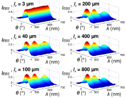
Figure 1 shows representative EBS intensity from the aqueous suspensions of the microspheres with the diameter = 1.5 (standard deviation, S.D. = 0.04 ) ( = 0.93 at ). We varied from 3 to 800 with the fixed (). As increases, the Mie scattering features such as the oscillatory pattern and the slope of the overall decline of intensity with wavelength become obvious and prominent. These spectral features indicate that only a few scattering events give rise to EBS. Increasing reduces the number of particles in the coherence volume that is determined by , hence only lower orders of scattering events contribute to the EBS peak. For example, the spectrum of the sample with resembles that of the diffuse multiple scattering of highly packed media. As increases, the Mie scattering patterns are revealed. Finally, for , the spectral shape remains unchanged, indicating that the parametric condition reaches to the minimal number of scattering events required for EBS.
To explore quantitatively the observations above, we developed a Mie theory-based double scattering model, which provides the backscattering spectrum and the angular profile of EBS from double scattering. The radial intensity probability of double scattering can be expressed as
| (1) |
where is the transverse radial distance between two scatterers, and are the vertical distances from the surface to the scatterers, respectively, , is the phase function of single scattering, is the diameter of the microscophere, and is an attenuation coefficient. was obtained separately by measuring reflectance intensity for various sample thicknesses and calculating the exponential attenuation length. and () were calculated using Mie theory van de Hulst (1995).
We also obtained the angular profile of the EBS peak from of double scattering. can be expressed as an integral transform of the path length distribution of the conjugated time-reversed light paths Akkermans et al. (1986): where is the projection of the wave vector onto the plane orthogonal to the backward direction, is the probability of the radial intensity distribution of EBS photons with the radial vector r pointing from the first to the last points on a conjugated time-reversed light path (r is perpendicular to the incident light), and is the degree of spatial coherence with the first order Bessel function Born and Wolf (1999). If a medium is isotropic, the two-dimensional Fourier integral becomes the Fourier transform of :
| (2) |
with . As a result, the width of the LEBS peak is inversely proportional to .
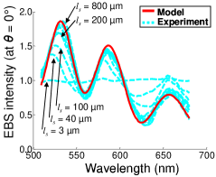
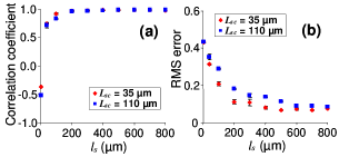
Figure 2 shows that for greater than a few , the spectral shape of approaches the spectrum of the double scattering model and then remains unchanged for . In order to quantify the agreement between the model and the experimental results, we used two complimentary measures: the root mean square (RMS) error and the correlation coefficient. The RMS error measures the overall estimation accuracy while the correlation coefficient measures the capability of the double scattering model to replicate the oscillation characteristics of the experimental spectra. Figure 3 plots the correlation coefficient (Fig. 3(a)) and the RMS error (Fig. 3(b)) as a function of for two different values of ( and ). In both cases, as shown in Fig. 3, the two measures level off for , thus indicating that the double scattering model is in excellent agreement with the experimental data.
Figure 4 compares obtained experimentally with the predictions of the double scattering model for two different values of . We convoluted the profiles of the LEBS peaks with the angular response of the instrument to take into account the finite point-spread function of the detection system and the incident beam divergence. As can be seen from Fig. 4, the double scattering model is in excellent agreement with experimental data and predicts both the angular and spectral profiles of LEBS. As expected from the Fourier transform relationship between and (Eq. (2)), the shorter generates the broader LEBS peak as shown in Fig. 4. These results confirm the hypothesis that in low-coherence regime () the number of scattering events giving rise to EBS reaches its minimum and LEBS is indeed generated by means of double scattering.

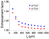
In LEBS, a priori surprisingly, the LEBS peaks from the helicity preserving channel are lower than those from the orthogonal helicity channel. Conventionally, the EBS peaks from the channel are much higher than those from the channel. Figure 5 shows the enhancement factor of LEBS peaks at recorded from a discrete random medium consisting of aqueous suspension of the microspheres (, S.D. = 0.01 ) from the both channels with . From Mie theory, the forward scattering preserves the degree of circular polarization, while the backscattering flips the circular polarization in a manner similar to light reflection from a mirror. In LEBS, the direction of light scattered by one of the scatterers should be close to the forward direction while the direction of light scattered by the other scatterer should be close to the backward direction. Therefore, the enhancement factor from the channel is consistently higher than that from the channel, and the difference in the enhancement factors between the channel and the channel is nearly constant for , supporting the validity of the double scattering model of LEBS.
In conclusion, (i) using low coherence illumination, we were able to create a virtual coherence volume within random media, which rejects longer paths and isolates lower order scatterings in EBS. (ii) Controlling the coherence length of illumination and the optical properties of the discrete random media, we were able to isolate double scattering in EBS. This led for the first time to prove an existing theoretical hypothesis that the minimum number of scattering events needed to generate EBS is double scattering. (iii) We demonstrated that a large number of the independent coherence areas provide statistical information about the optical properties of random media without configuration or ensemble averaging. (iv) We reported experimental results for in weakly scattering disordered media. In this dramatically different regime, can be made to be the shortest length scale except the particle size. (v) In the previous publications Kim et al. (2004, 2005), we showed that LEBS signals from human colon are sensitive to early precancerous alterations in colon cancer. However, the origin of LEBS in colonic mucosa has not been completely understood. Therefore, our finding that EBS originates from the time-reversed paths of double scattering events in weakly scattering media will further facilitate understanding of EBS signals for tissue diagnosis and characterization, providing a potential method about how to analyze the LEBS signals from biological tissue.
Correspondence to:
Young L. Kim at younglae@northwestern.edu
References
- van (1984) M.P. van Albada and A. Lagendijk, Phys. Rev. Lett. 55, 2692 (1985); P.E. Wolf and G. Maret, Phys. Rev. Lett. 55, 2696 (1985); Y. Kuga and A. Ishimaru, J. Opt. Soc. Am. A 1, 831 (1984).
- Wiersma et al. (1995a) D. S. Wiersma, M. P. Vanalbada, B. A. Vantiggelen, and A. Lagendijk, Phys. Rev. Lett. 74, 4193 (1995a).
- Labeyrie et al. (2003) G. Labeyrie, D. Delande, C. A. Muller, C. Miniatura, and R. Kaiser, Phys. Rev. A 67, Art No 033814 (2003).
- Sapienza et al. (2004) R. Sapienza, S. Mujumdar, C. Cheung, A. G. Yodh, and D. Wiersma, Phys. Rev. Lett. 92, Art No 033903 (2004).
- Huang et al. (2001) J. Huang, N. Eradat, M. E. Raikh, Z. V. Vardeny, A. A. Zakhidov, and R. H. Baughman, Phys. Rev. Lett. 86, 4815 (2001).
- Wiersma et al. (1995b) D. S. Wiersma, M. P. van Albada, and A. Lagendijk, Phys. Rev. Lett. 75, 1739 (1995b).
- Mishchenko and Dlugach (1993) M. I. Mishchenko and J. M. Dlugach, Planet. Space Sci. 41, 173 (1993).
- Kim et al. (2004) Y. L. Kim, Y. Liu, V. M. Turzhitsky, H. K. Roy, R. K. Wali, and V. Backman, Opt. Lett. 29, 1906 (2004).
- Kim et al. (2005) Y. L. Kim, Y. Liu, V. M. Turzhitsky, R. K. Wali, H. K. Roy, and V. Backman, Opt. Lett. 30, 741 (2005).
- Yoo et al. (1990) K. M. Yoo, G. C. Tang, and R. R. Alfano, Appl. Opt. 29, 3237 (1990).
- Vreeker et al. (1988) R. Vreeker, M. P. van Albada, R. Sprik, and A. Lagendijk, Phys. Lett. A 132, 51 (1988).
- Tourin et al. (1997) A. Tourin, A. Derode, P. Roux, B. A. van Tiggelen, and M. Fink, Phys. Rev. Lett. 79, 3637 (1997).
- Barabanenkov and Barabanenkov (1999) Y. N. Barabanenkov and M. Y. Barabanenkov, Waves random Media 9, 13 (1999).
- Lenke et al. (2000) R. Lenke, R. Lehner, and G. Maret, Europhys. Lett. 52, 620 (2000).
- Schwartz and Dogariu (2005) C. Schwartz and A. Dogariu, Opt. Lett. 30, 1431 (2005).
- van der Mark et al. (1988) M. B. van der Mark, M. P. van Albada, and A. Lagendijk, Phys. Rev. B 37, 3575 (1988).
- Akkermans et al. (1988) E. Akkermans, P. E. Wolf, R. Maynard, and G. Maret, J. Phys. France 49, 77 (1988).
- Born and Wolf (1999) M. Born and E. Wolf, Principles of optics : electromagnetic theory of propagation, interference and diffraction of light (Cambridge University Press, Cambridge; New York, 1999), 7th ed.
- van de Hulst (1995) H. C. van de Hulst, Light scattering by small particles (Dover Publications, New York, 1995).
- Akkermans et al. (1986) E. Akkermans, P. E. Wolf, and R. Maynard, Phys. Rev. Lett. 56, 1471 (1986).