Intrinsic Low Temperature Paramagnetism in B-DNA
Abstract
We present experimental study of magnetization in -DNA in conjunction with structural measurements. The results show the surprising interplay between the molecular structures and their magnetic property. In the B-DNA state, -DNA exhibits paramagnetic behaviour below 20 K that is non-linear in applied magnetic field whereas in the A-DNA state, remains diamagnetic down to 2 K. We propose orbital paramagnetism as the origin of the observed phenomena and discuss its relation to the existence of long range coherent transport in B-DNA at low temperature.
It is now a common knowledge that the electrical conduction in DNA is intimately linked to experimental factors such as molecules’ base-pair sequence, type of electrodes, surrounding counter ions and number of water molecules Endres ; Adessi ; Alik1 ; Kelley . The experimental accounts to date span a whole spectrum of conduction mechanism from insulators, semi-conductors, metals to proximity induced superconductors Pablo ; Fink ; Porath ; Alik2 . Magnetization is an alternative, non-invasive mean to probe the intrinsic electronic properties of matter, as the measurements do not require any electrode attachments. Unlike the intensive experimental efforts made on electronic transport in DNA molecules, their magnetization has been scarcely explored due to experimental difficulties such as the overwhelming presence of water. Basic questions on the intrinsic magnetic properties of DNA such as the magnitude of its magnetic susceptibility, , have remained unclear. It is widely known that DNA is diamagnetic near room temperature with a sizable anisotropy stemming from the presence of aromatic rings of the base pairs whose magnitude is comparable to that of benzene Maret1 ; Maret2 ; Iizuka . But how does the over-all magnetic state of DNA depend on intrinsic parameters (molecular structure, base-pair sequence) as well as extrinsic parameters such as counter ion types? Does DNA magnetization depend on these parameters in a way reminiscent to the electrical conduction? And if so, what are the consequences and implications for the usage of DNA as molecular wires? To answer these adjuring questions, we have studied the low temperature susceptibility and magnetization of randomly oriented -DNA molecules and its relation to their molecular structure (A- and B-DNA) and counter ion types (Na+ and Mg2+), two parameters known to greatly influence the electronic property of DNA Endres . We find that the magnetization is temperature independent and diamagnetic at high temperatures (100 K and above) regardless of water content in both A- and B-DNA structures. Surprisingly, once the molecules are sufficiently ‘wet’ and thus are found in B-structure, a paramagnetic upturn was observed at lower temperatures that is non-linear in magnetic field in addition to the atomic diamangetic component. Collectively, these observations reveal for the first time, the intrinsic non-diamagnetic state in DNA molecules that is intricately related to their molecular structures.
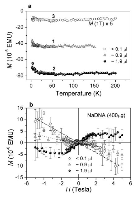
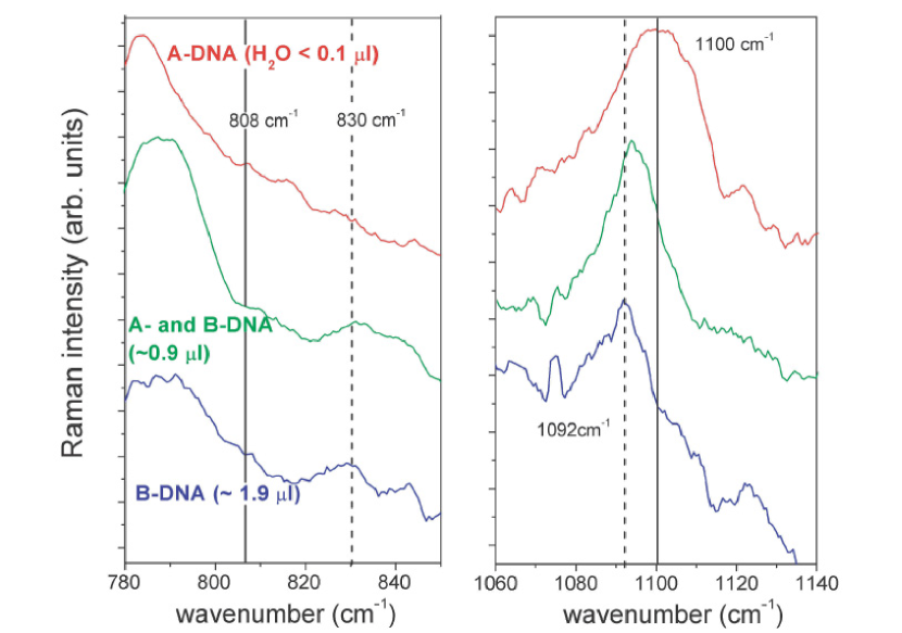
-DNA samples (400g each) in two counter ion types, Na+ (hereafter called NaDNA) and Mg2+ (MgDNA) were prepared in quartz capillary tubes that served as sample holders for both magnetization (QuantumDesign MPMS-R2 SQUID magnetometer) and structural studies (micro-Raman spectrometer) sample . -DNA (16m, 48,502 base-pairs) was chosen specifically because the proximity induced superconductivity and the low temperature negative magnetoresistive behavior were detected previously in these molecules Alik1 . The molecular structure of DNA changes dramatically with surrounding hydration levels. In aqueous environment DNA molecules are in B-DNA structure where base-pairs are stacked parallel to one another with inter-base-pair distance of 3.2 and the helix diameter of 19 . When molecules are dried, the bases become severly tilted off the helix axis and the helical diameter becomes broad (23 ) Chromatin . The molecular structure of the samples was transformed between the dry A-DNA and the wet, natural B-DNA states by adding or removing water from the samples. In their driest states, NaDNA and MgDNA samples contained 0.1 and 0.3 of H2O, respectively. At each stage of re-hydration and/or dehydration the quartz capillaries were sealed to maintain the water content constant and the magnetization and the molecular structure (via micro-Raman spectroscopy) were studied in parallel. Figure 1a shows the magnetization () of NaDNA with 0.1, 0.9 and 1.9 of water as a function of temperature () at 5 Tesla. Originally, the sample contained 0.9 of H2O. Then the water content was increased to 1.9 and finally dried down to 0.1 . By subtracting the water contribution from the total magnetization at 100 K, we have extracted the diamagnetic susceptibility of DNA, = -0.63 0.1 10-6 EMUG-1g-1. Within the experimental accuracy, the diamagnetic susceptibility was found to be independent of water content, that is, = . This value, determined from two NaDNA samples, is in fair agreement with the calculated atomic diamagnetic susceptibility of DNA, -0.52 10-6 EMU G-1g-1. At temperatures below 20 K, the magnetization of NaDNA containing 1.9 and 0.9 of H2O indicate unexpected paramagnetic up-turn that disappeared once the sample was dried to H2O 0.1 . Figure 1b portraying (without H2O contribution) as a function of magnetic field () at = 2 K clearly presents this low temperature paramagnetism. At helium temperature, DNA in aqueous environment exhibits a magnetization crossing-over from diamagnetic to paramagnetic that is non linear in magnetic field. The magnitude of this paramagnetic increase, = , is comparable to that of the diamagnetic contribution of DNA. The corresponding Raman spectra depicting the structural transformation from A- to B structure in NaDNA are presented in Fig. 2 raman . Among the large and highly reliable index of Raman bands corresponding to vibrational modes of DNA geometry and conformations Deng , we concentrate on two bands representing the backbone vibrations to identify the structural state of our samples as described in the figure caption. While with 0.1 of H2O, sample was found almost purely in A-state, with 0.9 , B-DNA as well as a small signature of A-DNA were observed. With further addition of H2O, molecules were found entirely in the B-state. By comparing the molecular structure and the magnetization of NaDNA, it appears that B-DNA is a prerequisite condition for the low temperature paramagnetism in DNA. It needs to be noted, however, that once the relative humidity, (the weight of H2O divided by that of dry DNA) exceeds 0.9, the DNA molecules assume B-DNA state Lindsay . = 0.9 corresponds to 0.36 of H2O in our sample, far less than the nominal amount of 0.9 used here. This observation indicates that H2O is not diffused uniformly due to the sample geometry and the preparation method. The more rigorous investigation on the water content and the structural analysis on NaDNA samples via X-ray diffraction will be reported elsewhere.
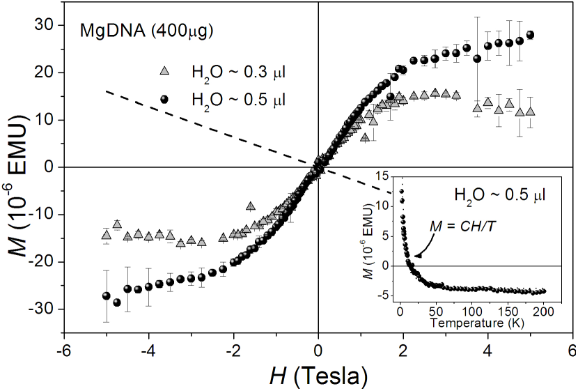
In MgDNA sample, we were unable to remove H2O sufficiently to create predominantly A-DNA state. In fact, the Raman spectra (not shown) of the MgDNA in its driest state (0.3 ) indicated mainly B-DNA bands, and with 0.5 of H2O the molecules were found to be in purely B-DNA state. This is in marked contrast with NaDNA where the presence of A- and B-DNA were both detected at much higher water content. This observation is consistent with a known property of Mg2+, i.e., that prevents the transition from B- to A-DNA more efficiently than Na+ ions Schultz . The magnetization measurements on MgDNA with 0.5 of H2O preceded the measurements with 0.3 . As can be seen from Fig. 3, a purely diamagnetic behaviour at low temperatures was never achieved in MgDNA in line with the observation in NaDNA. In the driest state, only a slight decrease in was detected. Furthermore, the paramagnetic magnetization was found to become independent of water content for H2O values higher than 0.5 (measured up to 2.2 ). Temperature dependence of the magnetization of wet MgDNA follows the Curie law as shown in the inset. Susceptibility at higher temperatures was determined to be -0.8 0.1 10-6 EMU G-1g-1, larger than the value found for NaDNA sample. This difference ( 0.2 10-6 EMU G-1g-1) corresponds to the diamagnetic susceptibility of the residual buffer ions (MgCl2 and NH4-Acetate). It is also noteworthy that the low temperature was found to be 4 times higher in MgDNA than in NaDNA.
The apparition of paramagnetism in B-DNA at low temperature that is non-linear in applied field is robust. There are two possible origins for the observed non-linear paramagnetism: electron spin () and orbital magnetism. Assuming the electron spins (magnetic ions or hydroxyl radicals Debije , for example) to render the observed behaviour, we have fit the paramagnetic component of the magnetization, , to the Brillouin and Curie law. The best fits were obtained for = 1/23/2 with the total number of spins of 1015 for NaDNA and 41015 for MgDNA, respectively. Such high concentrations of spins should be detectable by Electron Paramagnetic Resonance (EPR) provided that the signal line width does not exceed 300G. The examination via EPR EPRexp revealed no such presence in NaDNA at room temperature. The MgDNA sample was examined between room temperature and 4 K. The only EPR absorption signal was detected at = 4.28 which can be attributed to Fe3+ (= 5/2, = 30/7) in an asymmetric crystal field. The number of these spins was determined to be of the order of 1012, far too small to be responsible for the observed EPR . Furthermore, the magnetization of H2O used to humidify the samples was examined separately using SQUID magnetometer. 2 of H2O, was found to contain (5 T, 2 K) less than 10-6 EMU. Therefore these experiments as well as the disappearance of paramagnetic component in A-DNA after the drying process exclude free radicals and magnetic impurities in water and buffer solutions from the possible origins of low temperature paramagnetism.
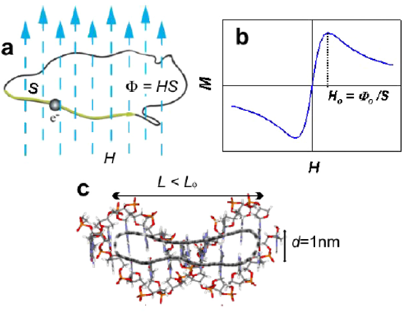
Thus far, the paramagnetism appears to be an intrinsic property unique to B-DNA. An interesting possibility is the existence of persistent current loops along the DNA molecules on a mesoscopic micron scale. The mesoscopic orbital magnetism has been shown theoretically to be paramagnetic and non-linear when repulsive electron-electron interactions dominate over single particle effects Altshuler ; Ambegaokar . The total magnetization of the system then follows , where is the Landau susceptibility, and is the Fermi wave number. The non-linear magnetization reaches its maximum at = (), where = = 4.14 10-7 G cm2 is the magnetic flux quantum and is the maximum surface area enclosed inside the coherent current loop (Fig. 4(a) and (b)). The orbital magnetism associated to the persistent currents has been already observed in mesoscopic rings and 2-D squares and is considered the hallmark of phase coherent transport at low temperatures Levy . In our DNA samples measured at 2K, the magnitude of paramagnetic signal is 2 4 times that of the total diamagnetic susceptibility. At lower temperatures the size of orbital paramagnetic susceptibility is expected to grow rapidly. We estimate the electron coherent length, , using the experimental values from our measurements, = 1 2 Tesla, via = where is the distance between bases of B-DNA molecules. Our calculation yields the electron path on the order of 1 m along the helical axis of the molecules. Such circulation of electrons enclosing a finite flux can be achieved through the combination of intra- and interstrand (across the hydrogen bonds) transfer of electrons on bases (Fig. 4c). Hydrogen bond assisted electronic exchange has already been witnessed in some organic molecules Ferrer . The value of the electron path found here agrees with the coherent length of 1m in -DNA determined by Kasumov et al. Alik1 where proximity induced superconductivity was detected at 1 K. Our observation may also imply a coherent electron transport along the helical length of the molecule at low temperatures, but exclusively in B-DNA, consistent with experimental reports on the DNA electronic conductivity that showed higher conductivity in wet-DNA molecules Tran ; Otsuka . Lastly, from the enhanced size of the low temperature paramagnetic signal as well as the persistence of B-DNA in MgDNA, Mg2+ appears to facilitate the electron transfer inside DNA molecules.
In summary, we have found a low temperature, non-linear paramagnetic behaviour in the B-state of -DNA molecules. This effect is found in both DNA samples prepared with Na+ and Mg2+ counter ions. The paramagnetic susceptibility of molecules prepared with Mg2+ ions is found larger by a factor of 4 compared to the Na+ counterpart. The present results can be interpreted by the existence of a mesoscopic orbital paramagnetism in B-DNA molecules that may be related to the proximity induced superconductivity observed in these molecules. Magnetization of other types and aligned DNA molecules should be examined in order to confirm the orbital origins of this paramagnetism.
We thank F. Livolant, A. Bertin, D. Durand, S. Guéron, J.F. Allemand, D. Bensimon and V. Croquette for stimulating discussions and experimental guidances.
References
- (1) R. G. Endres et al., Rev. Mod. Phys. 76, 195 (2002).
- (2) Ch. Adessi et al., Comp. Nanosci. Nanotech. 2002, 56, (2002).
- (3) A. Yu. Kasumov et al., Appl. Phys. Lett. 84, 1007 (2004).
- (4) S. O. Kelley and J. K. Barton, Science 283, 375 (1999).
- (5) P. J. de Pablo et al., Phys. Rev. Lett 85, 4992 (2000).
- (6) H. W. Fink, C. Schnenberger, Nature 398, 407 (1999).
- (7) T. Porath et al., Nature, 403, 635 (2000).
- (8) A. Kasumov et al., Science 291, 280 (2001).
- (9) G. Maret et al., Biopolymers 22, 2727, (1983).
- (10) G. Maret et al., Phys. Rev. Lett. 35, 397 (1975).
- (11) E. Iizuka and Y. Kondo, Mol. Cryst. Liq. Cyrst. 51, 285 (1979).
- (12) Samples were obtained from New England Biolabs and Amersham Bioscience (500 g/ml with 10mM Tris-HCl and 1mM EDTA). NaDNA: The original solution was diluted in 10 ml of 60/40 H2O/Isopropanol solution containing 0.3 M of NaCl for co-precipitation. The solution was then centrifuged at 15 kG and at 4 oC for 35 minutes. The precipitate was rinced in 70 Ethanol and was centrifuged again at 15 kG and at 4 oC for 15 minutes. This procedure was repeated twice to remove the excess Na+ ions. MgDNA: The original solution was replaced by 9 mM MgCl2/20 mM NH4-acetate buffer solution by dialysis. This solution was concentrated via centrifuging through Microcon (Millipore) filter, down to 5 mg/ml, then lyophilized for 2 hours at room temperature. The H2O content of the samples were controlled by injection (Milli-Q distilled and deionized H2O, 18 M) or by evaporation at 45 - 55 oC ( 48 hours for NaDNA and over one week for MgDNA samples). Quartz capillary tube sample holders were obtained from Heraeus (2 mm O.D. and 1 mm I.D.). The H2O amount was determined using a precision scale ( 0.1 g). Samples were suspended by capillary force at the mid-height of the capillary tube.
- (13) A- and B-DNA structures can be found in, for example, K. E. van Holde, Chromatin, Springer Series in Molecular Biology, Springer-Verlag, Paris (1988).
- (14) H. Deng et al., Biopolymers, 50, 656 (1999).
- (15) The Raman spectra were obtained with 514.5 nm excitation line of an Ar+-Kr+ laser in confocal micro-Raman configuration with 10 magnification. The scattered light was analysed using a Jobin-Yvon triple grating spectrometer (T64000) consisting of a holographic notch filter and liquid nitrogen cooled CCD detector. The radiation power at source was between 3 and 10 mW. Raman spectra were taken at several regions within the sample. Spectra shown here are the accumulated averages of 10 exposures of 30-60 seconds each. The effective spectral resolution was less than 1 cm-1.
- (16) S. M. Lindsay et al., Biopolymers 27, 1015 (1988).
- (17) J. Schultz et al., Biophys. J. 66, 810 (2001).
- (18) M. G. Debije et al., Radiat. Res., 154, 163 (2000).
- (19) Bruker spectrometer equipped with an Oxford cryostat was operated at 10GHz operation frequency, 100kHz modulation frequency and 20 G modulation amplitude.
- (20) The number of spins was determined by comparing the intensity of the spectra to that of a calibrated CuSO5H2O single crystal.
- (21) B. L. Alsthuler, et al., Phys. Rev. Lett., 66, 88 (1991).
- (22) V. Ambegaokar and U. Eckern, Phys. Rev. Lett., 65, 381 (1990).
- (23) L. P. Lévy, et al., Physica B, 189, 204 (1993).
- (24) J. R. Ferrer et al., Chem. Mater. 13, 2447 (2001).
- (25) P. Tran et al., Phys. Rev. Lett., 85, 1564 (2000).
- (26) Y. Otsuka et al., Jap. Journ. Appl. Phys., 41, 891 (2002).