Electrophoresis of DNA on a disordered two-dimensional substrate
Abstract
We propose a new method for electrophoretic separation of DNA in which adsorbed polymers are driven over a disordered two-dimensional substrate which contains attractive sites for the polymers. Using simulations of a model for long polymer chains, we show that the mobility increases with polymer length, in contrast to gel electrophoresis techniques, and that separation can be achieved for a range of length scales. We demonstrate that the separation mechanism relies on steric interactions between polymer segments, which prevent substrate disorder sites from trapping more than one DNA segment each. Since thermal activation does not play a significant role in determining the polymer mobility, band broadening due to diffusion can be avoided in our separation method.
pacs:
87.15.Tt, 87.14.Gg, 87.15.AaI Introduction
The development of new methods for efficiently separating charged biopolymers by length has been an area of significant recent activity due to the fact that strategies for genome sequencing are based on sorting DNA fragments by size Viovy . Simple charge-based sorting is not possible because the increase in electrostatic force on longer molecules with greater total charge is exactly offset by a corresponding increase in hydrodynamic drag Slater86 . Instead, separation is achieved using techniques such as gel electrophoresis, in which longer molecules are slowed relative to shorter ones due to interactions with cross links in the gel. Gel and capillary electrophoretic techniques are limited to DNA strands less than base pairs (bp) in length Schwinefus ; the mobility saturates for longer strands, and sufficiently large strands fail to pass through the gel at all. There is a need for separation of strands up to bp, and thus new techniques which can sort longer molecules are of particular interest Huang96Ashton . The motion of elastic strings through random media is also of general interest for a wide range of systems including magnetic domain wall motion, vortex lattice motion in superconductors, and charge density waves.
Several recent proposals for electrophoretic techniques move away from the traditional media of gels and polymers and instead take advantage of advances in nanolithography to create microstructured devices for separation Volkmuth ; Dukeetc ; Han2etc ; Turner ; Chou99 ; Han99 ; Rousseau97 ; Bakajin01 ; Huang02 ; Huang022 ; Cabodi02 . The sorting effectiveness of these techniques is limited by the relative size of the nanofabricated structure and the polymers to be sorted, making it necessary to fabricate a separate device for each size range of interest. In contrast, Seo et al. Seo proposed an adsorption-based separation technique that could permit the sorting of polymers which vary in size by three orders of magnitude. When the ionic strength of the buffer solution is altered Menes , the DNA is partially adsorbed onto a clean surface, forming a series of loops which extend into the solution and trains which are adsorbed on the substrate. It has been proposed that separation occurs because the longer polymers have a larger number of train segments, and thus experience a greater retardation of their motion Pernodet00 ; Luo .
The quasi-two dimensional geometry considered in Ref. Seo is very appealing for separation purposes, in part because adsorbed polymers spread out significantly into flat ‘pancakes’ deGennes compared to their coiled three-dimensional configurations, permitting better coupling to length differences. The sorting mechanism in Ref. Seo precludes complete adsorption, however, since there is no separation for fully desorbed or fully adsorbed polymers. This limits the length range that can be processed, since if the surface is strongly attractive to DNA, long DNA chains fully adsorb and separation by length is lost. If instead the surface weakly attracts DNA, short chains desorb from the surface and cannot be separated Seo04 .
Here we propose an alternative sorting technique for long DNA strands in which the polymers are fully adsorbed on the surface. To permit separation, we spatially modify the surface, but instead of of using posts or other impenetrable barriers, we consider randomly spaced pinning sites which temporarily retard the motion of the polymer, yet still allow it to pass through. Such pinning could be created via the manipulation of lipid bilayer membranes Hovis2etc or surface patterning Workman . We show that in this geometry, longer polymers are more mobile than shorter ones, in contrast to typical separation methods where longer polymers move more slowly. This avoids the jamming or clogging associated with long polymers in other techniques. The steric interaction between polymer segments causes the longer polymers to be less well pinned by the random disorder than the short polymers, and allows separation by length to occur.
To demonstrate our separation mechanism, we use a simulation model that we have developed for long DNA fragments. Many of the existing simulation models for electrophoretic processes are best suited for shorter polymers Olveraetc . Since we are concerned with polymers up to 300 m in length, we do not attempt to simulate each atom in the polymer. Instead, we adopt a bead-spring model in which the polymer is represented by multiple beads Rouse53 which are each spaced many persistence lengths apart. There is an entropic resistance to the stretching of the polymer segment between two beads, which is represented by a finitely extensible nonlinear spring (FENE) potential Warner that replaces the internal degrees of freedom of the polymer molecule Doyle97 . An essential assumption of this model is that the polymer segment between beads is significantly longer than the polymer persistence length. This is in contrast to bead-stick models Kramer , where the distance between beads is ten or less actual chemical segments.
II Simulation
We employ Brownian dynamics Ermak , permitting us to use time steps of order 0.1 ns, orders of magnitude greater than the sub-fs time steps required in all-atom molecular dynamics. In this technique, the solvent is treated statistically rather than explicitly Rudisill92 . The dimensionless force on bead in a chain base pairs long represented by beads is given by
| (1) |
where is the spring force along the chain, represents the excluded volume between beads, is the force from a disordered substrate, is the electrophoretic force, and is a thermal noise term. Distances are measured in terms of , the root mean square length of the spring. Forces are expressed in terms of . We can neglect hydrodynamic interactions since they are screened due to the proximity to the solid substrate Maier ; Bakajin98 . Electroosmotic effects can be controlled in the usual way by means of a high concentration buffer Han99 ; Seo ; Huang022 ; Han02 . We assume that the Debye length is considerably smaller than the distance between the beads in our model.
The force between bead and neighboring beads is given by
| (2) |
where is the elongation of the spring, is the distance vector between two neighboring beads, is the equilibrium spring length, is the maximum allowable elongation, and the Hookean spring constant . This phenomenological spring potential Warner has the properties that it is equivalent to a Hookean spring for small , but becomes infinite at finite spring elongation. The persistence length of double-stranded DNA is ÅReese . The Kuhn length , giving m, where we assume that the ionic strength of the buffer solution is sufficient to screen electrostatic repulsion between sections of the chain Patel . Each base pair is 0.34 nm long so one Kuhn length contains 300 bp Long . Since we will be using a Gaussian chain model for the excluded volume interactions, which requires the chain segments between beads to be represented statistically and not deterministically, the number of Kuhn lengths between beads must be sufficiently large (well above the bead-rod limit of ), so we take m. This gives and m.
The excluded volume term accounts for the repulsive interaction between polymer segments when they approach each other Akkermans01 ; Kumar03 , an effect which is more pronounced in two dimensions than in three dimensions Slater95 . The excluded volume interaction for beads and a unitless distance apart is taken after that used in Ref. Jendrejack , which is based on the energy penalty due to overlap of two Gaussian coils, and has the form
| (3) |
where
| (4) |
Here the size parameter Jendrejack , while the excluded volume parameter is taken to be . This gives
| (5) |
The substrate roughness is represented by finite range parabolic pinning traps of radius , strength , and density . The pin size was chosen to be close to the Kuhn length. The force on bead from pin a distance away is given by
| (6) |
where is the Heaviside step function.
The electrophoretic force on each bead from an applied electric field is
| (7) |
Here, is the charge per bead, where the charge per unit length of DNA in solution is C/m, or ÅVolkmuth . is the Langevin thermal noise term representing the Brownian forces. It is a delta-correlated white noise process which obeys and the fluctuation-dissipation theorem Kubo66 . Here is a Kronecker delta tensor and is the Dirac delta function. Time is measured in units of and we take . is the friction coefficient characterizing the viscous interaction between the bead and the solvent. We use the experimentally measured value of for polymers diffusing in a bilayer, Ns/m Olson . Theoretically, for a polymer moving in three dimensions, , where is the effective bead radius and is the solvent viscosity. We note that a theoretical expression for the friction coefficient in the case of a particle confined to a membrane suspended in a solvent has been developed by Saffman Saffman75 , where only a weak dependence of on effective bead radius is obtained.
III Results
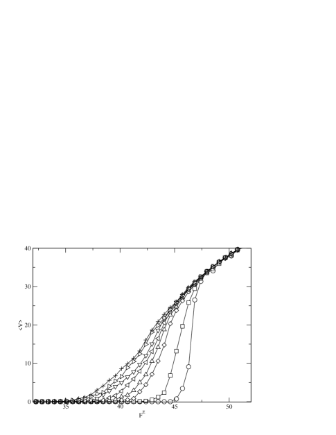
We first consider the velocity of the polymers over the rough substrate as a function of polymer length . We sweep the electric field strength and find the average velocity at each field value during 200 repetitions of the sweep. In Fig. 1 we plot the velocity-force curves for polymers of length ranging from to 100, where is the number of beads used to model the polymer, in a sample with pinning density and strength at room temperature. In physical units, this length range is m to 160m, and it includes -phage DNA, which has a contour length of m Hsieh . A velocity of 10 corresponds to 0.3 m/s, and an applied field of 30 corresponds to 4.16 V/cm. After the polymers depin, there is a range of driving force over which we find a nonlinear velocity-force characteristic. Within this range the shorter polymers move more slowly than the longer polymers for a given electric field strength.
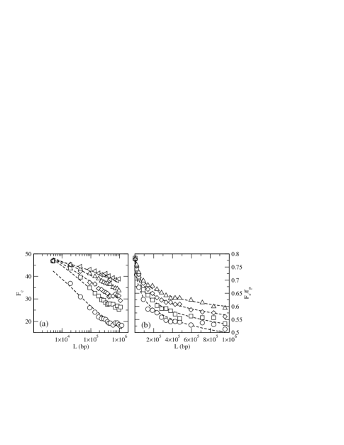
Short polymers are better pinned by the underlying disorder than long polymers, and thus a higher driving force must be applied before the short polymers begin to move over the substrate. In Fig. 2 we illustrate the dependence of the critical depinning force on polymer length for a range of pinning strengths and densities. Here we define as the driving force at which . We have chosen this definition since in our technique, physical separation of the polymers will occur when the polymers are moving with different velocities. In an experiment, if the electric field were held at a value between for polymers of two lengths, the two polymers will be separated since one is strongly mobile while the other is nearly immobile. As shown in Fig. 2, in each case we find that drops logarithmically with , as indicated by the dashed lines. As the pinning density is reduced from to , shown in Fig. 2(a), the depinning force drops and the variation of with becomes steeper, meaning that the separation resolution is enhanced. At the same time, the length range over which effective separation can be achieved drops, and thus there is a trade-off which must be considered depending on the range of sizes that are to be separated. We show the scaled length dependence of in Fig. 2(b) for pinning strengths to 80. The pinning effectiveness drops slightly faster than the pinning strength, as indicated by the fact that the curves do not fall on top of each other. For the weakest pins, the separation effectiveness washes out above a length of .
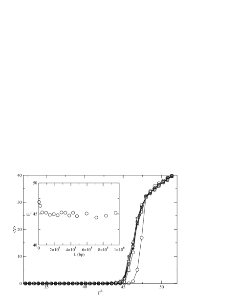
The excluded volume interactions play the key role in the separation mechanism. What is happening physically can be understood as follows. Consider a polymer composed of only a single bead. This polymer can be completely trapped by a single pinning site. Next, consider a polymer composed of two beads. Although it is possible for the polymer to find two adjacent pinning sites such that both beads are pinned, it is more likely that one bead will be pinned while the other is still free. In this case the force from only one pin will have to hold two beads still against the electrophoretic force. If the excluded volume interactions are removed, both beads can fit inside the pin, doubling the effective pinning force. As the polymer becomes longer and is composed of more beads, the relative fraction of the polymer that is pinned decreases, provided that each pin can capture only one bead. This results in the decreased threshold for depinning and the increased mobility of the longer polymers relative to the short ones. The importance of the excluded volume interaction is that it enforces a pin occupancy of at most one bead per pin. Thus, the excluded volume interaction is what produces the decrease in with polymer length. We test this by running a series of simulations without the excluded volume interaction. The depinning force for this case is shown in the inset to Fig. 3, where it is clear that has no significant dependence on . The corresponding velocity-force curves are shown in Fig. 3, where it can clearly be seen that the curves lie on top of each other except for the very shortest polymers.

We stress that the separation mechanism at work here is significantly different than that which occurs in the case of impenetrable obstacles such as cross links in gels or nanofabricated posts. This can be seen by observing images of the moving polymers. A representative set of images for polymers of different length is shown in Fig. 4. The chains are moving toward the top of the figure in the direction. Rather than forming hairpin structures, the polymers frequently form a bundle on their advancing end, and sometimes drag one or two tail segments.
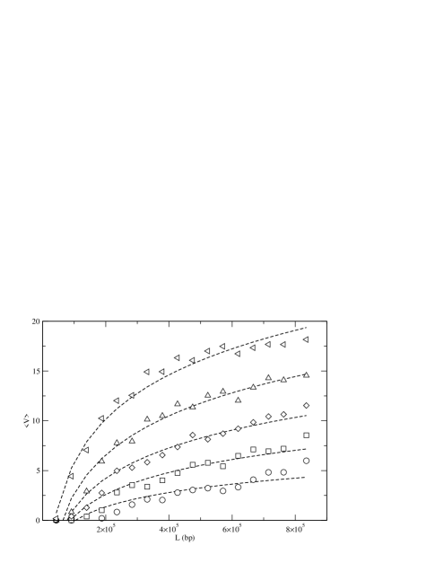
For separation purposes, the polymer velocity must depend on length. This can be achieved if the depinning force of the polymers is length dependent. To demonstrate this explicitly, we run a series of simulations in which the driving force is held at a fixed value, and measure the average velocity . The results are plotted in Fig. 5 for five different values of in a sample with and . The velocity increases logarithmically with polymer length, as indicated by the dashed lines, and velocity variations of an order of magnitude can be achieved.

We note that in traditional gel electrophoresis techniques, diffusion is an important limiting effect, since the polymers are moving through the gel relatively slowly and depend on thermal fluctuations to help them translocate through the gel. This effect is particularly pronounced for long polymers, which have extreme difficulty passing through the gel at all. In contrast, in the technique proposed here, the polymers are much more mobile than they would be in a gel. The configurations and depinning of the polymers are dominated by the strong electric fields and pinning imposed, and thermal effects play essentially no role in the separation. We observe no significant thermal diffusion in our system at all. As a result, diffusive broadening of the bands can be prevented. The bands do still broaden due to the intrinsic randomness of the pinning, which causes the progress of the polymers over the substrate to be somewhat variable. To illustrate the magnitude of this broadening, in Fig. 6(a) we plot the total distance traveled by the polymers under different drives applied for a fixed period of time for 100 realizations of disorder. Error bars indicate the average maximum and minimum distances traveled by polymers of a particular length. Higher resolution can be obtained by allowing the polymers to move a larger distance through the gel, as illustrated in Fig. 6(b).

We quantify the separation power of our technique by measuring the resolution as a function of polymer length. The resolution is affected by both the selectivity and efficiency of the separation for a given length difference Petersen . The selectivity is proportional to the difference in mobility for polymers of different length,
| (8) |
where is the average velocity for a polymer of length . Fig. 7(a) shows that the selectivity for the same system in Fig. 6(a) does not vary with and is highest for the shortest polymers, in the same region where Fig. 5 indicates that the versus curve has the steepest slope. The efficiency is proportional to the width of the band observed after the polymers have traveled a distance ,
| (9) |
As can be seen in Fig. 7(b), increases with both and , consistent with the decrease in the size of the error bars at higher and shown in in Fig. 6(a). The resolution is defined as
| (10) |
where is the mean efficiency for the polymer lengths being compared. We plot versus in Fig. 7(c). The resolution depends more strongly on the selectivity than on the efficiency, and as a result we find that is highest for the shortest polymers and is not a strong function of . The resolution can be improved by allowing the polymers to travel a longer distance, as in Fig. 6(b). We compare the resolution for shorter and longer distances traveled in Fig. 7(d), where we find not only an enhancement of for the longer travel distance, but also a shift in the peak value of towards longer polymers. This suggests that the technique could be optimized for separation of the desired range of by adjusting the distance traveled by the polymers.
IV Conclusion
In summary, we have used a model developed for the simulation of long DNA segments to demonstrate a new length separation mechanism for polymers adsorbed to a disordered two-dimensional substrate. Longer polymers are more mobile than short polymers, and the depinning force decreases logarithmically with polymer length. Correspondingly, the polymer velocity increases logarithmically with length. The separation mechanism arises due to the excluded volume interaction between chain segments, which serves to reduce the effectiveness of the random pinning for longer polymers. One possible experimental system in which our proposed separation mechanism could be realized is solid-supported cationic lipid membranes, where DNA is confined to two dimensions but free to diffuse in plane Maier . The pinning could be produced in the form of disorder on the supporting substrate, which would perturb the bilayer and interfere with the free diffusion of the DNA. Such disorder could potentially be produced by an experimental technique as simple as not fully cleaning the substrate before depositing the bilayer. Our proposed separation mechanism offers several advantages over existing techniques. (1) It may not be necessary to use elaborate nanofabrication methods to produce the pinning. (2) The technique can be used to separate extremely long strands of DNA which will not pass through conventional gels. (3) It may be possible to achieve high throughput since the polymers do not need to work their way around fixed impassible obstacles, but are instead only temporarily hindered by the pinning sites, and can thus achieve much higher overall mobilities than are possible in a gel, particularly for long polymers. (4) Since thermal effects do not play a significant role in the separation technique, thermal broadening of the bands by diffusion should be strongly suppressed. The resolution limitation caused by band broadening from the intrinsic disorder of the substrate can be reduced by allowing the polymers to travel a longer distance during separation.
V Acknowledgments
This work was supported by the U.S. Department of Energy under Contract No. W-7405-ENG-36.
References
- (1) J.-L. Viovy, Rev. Mod. Phys. 72, 813 (2000), and references therein.
- (2) G.W. Slater and J. Noolandi, Biopolymers 25, 431 (1986).
- (3) J.J. Schwinefus and M.D. Morris, Macromolecules 32, 3678 (1999).
- (4) Z. Huang, J.T. Petty, B. O’Quinn, J.L. Longmire, N.C. Brown, J.H. Jett, and R.A. Keller, Nucl. Acid Res. 24, 4202 (1996); R. Ashton, C. Padala, and R.S. Kane, Curr. Opin. Biotech. 14, 497 (2003).
- (5) W. Volkmuth and R.H. Austin, Nature (London) 358, 600 (1992);
- (6) T.A.J. Duke and R.H. Austin, Phys. Rev. Lett. 80, 1552 (1998); D. Ertaş, ibid. 80, 1548 (1998).
- (7) S.W. Turner, A.M. Perez, A. Lopez, and H.G. Craighead, J. Vac. Sci. Technol. B 16, 3835 (1998); J. Han, S.W. Turner, and H.G. Craighead, Phys. Rev. Lett. 83, 1688 (1999); J. Han and H.G. Craighead, Science 288, 1026 (2000); D. Nykypanchuk, H.H. Strey, and D.A. Hoagland, ibid. 297, 987 (2002).
- (8) C.-F. Chou, O. Bakajin, S.W.P. Turner, T.A.J. Duke, S.S. Chan, E.C. Cox, H.G. Craighead, and R.H. Austin, Proc. Natl. Acad. Sci. 96, 13762 (1999).
- (9) J. Han and H.G. Craighead, J. Vac. Sci. Technol. A 17, 2142 (1999).
- (10) J. Rousseau, G. Drouin, and G.W. Slater, Phys. Rev. Lett. 79, 1945 (1997).
- (11) O. Bakajin, T.A.J. Duke, J. Tegenfeldt, C.-F. Chou, S.S. Chan, R.H. Austin, and E.C. Cox, Anal. Chem. 73, 6053 (2001).
- (12) L.R. Huang, J.O. Tegenfeldt, J.J. Kraeft, J.C. Sturm, R.H. Austin, and E.C. Cox, Nature 20, 1048 (2002).
- (13) L.R. Huang, P. Silberzan, J.O. Tegenfeldt, E.C. Cox, J.C. Sturm, R.H. Austin, and H. Craighead, Phys. Rev. Lett. 89, 178301 (2002).
- (14) M. Cabodi, Y.-F. Chen, S.W. Turner, H.G. Craighead, and R.H. Austin, Electrophoresis 23, 3496 (2002).
- (15) S.W.P. Turner, M. Cabodi, and H.G. Craighead, Phys. Rev. Lett. 88, 128103 (2002).
- (16) Y.-S. Seo, V.A. Samuilov, J. Sokolov, M. Rafailovich, B. Tinland, J. Kim, and B. Chu, Electrophoresis 23, 2618 (2002).
- (17) R. Menes, P. Pincus, R. Pittman, and N. Dan, Europhys. Lett. 44, 393 (1998).
- (18) N. Pernodet, V. Samuilov, K. Shin, J. Sokolov, M.H. Rafailovich, D. Gersappe, and B. Chu, Phys. Rev. Lett. 85, 5651 (2000).
- (19) H. Luo and D. Gersappe, Electrophoresis 23, 2690 (2002).
- (20) P.-G. de Gennes, Scaling Concepts in Polymer Physics (Cornell Univ. Press, Ithaca, NY, 1979).
- (21) Y.-S. Seo, H. Luo, V.A. Samuilov, M.H. Rafailovich, J. Sokolov, D. Gersappe, and B. Chu, Nano Lett. 4, 659 (2004).
- (22) L.A. Kung, L. Kam, J.S. Hovis, and S.G. Boxer, Langmuir 16, 6773 (2000); R.N. Orth , J. Kameoka, W.R. Zipfel, B. Ilic, W.W. Webb, T.G. Clark, and H.G. Craighead, Biophys. J. 85, 3066 (2003); K. Morigaki, K. Kiyosue, and T. Taguchi, Langmuir 20, 7729 (2004); C.K. Yee, M.L. Amweg, and A.N. Parikh, J. Am. Chem. Soc. 126, 13962 (2004).
- (23) R.K. Workman and S. Manne, Langmuir 18, 661 (2002).
- (24) M. Olvera de la Cruz, J.M. Deutsch, and S.F. Edwards, Phys. Rev. A 33, 2047 (1986); J.M. Deutsch, Phys. Rev. Lett. 59, 1255 (1987); J.M. Deutsch, Science 240, 922 (1988); J.M. Deutsch and T.L. Madden, J. Chem. Phys. 90, 2476 (1989); E.O. Schaffer and M. Olvera de la Cruz, Macromolecules 22, 1351 (1989).
- (25) P.E. Rouse, J. Chem. Phys. 21, 1272 (1953).
- (26) H.R. Warner, Ind. Eng. Chem. Fundamentals 11, 379 (1972).
- (27) P.S. Doyle, E.S.G. Shaqfeh, and A.P. Gast, J. Fluid Mech. 334, 251 (1997).
- (28) G.S. Grest and K. Kremer, Phys. Rev. A 33, R3628 (1986).
- (29) D.L. Ermak and J.A. McCammon, J. Chem. Phys. 69, 1352 (1978).
- (30) J.W. Rudisill and P.T. Cummings, J. Non-Newt. Fluid Mech. 41, 275 (1992).
- (31) B. Maier and J.O. Rädler, Phys. Rev. Lett. 82, 1911 (1999).
- (32) O.B. Bakajin, T.A.J. Duke, C.F. Chou, S.S. Chan, R.H. Austin, and E.C. Cox, Phys. Rev. Lett. 80, 2737 (1998).
- (33) J. Han and H.G. Craighead, Anal. Chem. 74, 394 (2002).
- (34) H.R. Reese and B.H. Zimm, J. Chem. Phys. 92, 2650 (1990).
- (35) P.D. Patel and E.S.G. Shaqfeh, J. Chem. Phys. 118, 2941 (2003).
- (36) D. Long and J.L. Viovy, Phys. Rev. E 53, 803 (1996).
- (37) R.L.C. Akkermans and W.J. Briels, J. Chem. Phys. 114, 1020 (2001).
- (38) K.S. Kumar and J.R. Prakash, Macromolecules 36, 7842 (2003).
- (39) G.W. Slater and S.Y. Wu, Phys. Rev. Lett. 75, 164 (1995).
- (40) R.M. Jendrejack, J.J. de Pablo, and M.D. Graham, J. Chem. Phys. 116, 7752 (2002).
- (41) R. Kubo, Rep. Prog. Phys. 29, 255 (1966).
- (42) D.J. Olson , J.M. Johnson, P.D. Patel, E.S.G. Shaqfeh, S.G. Boxer, and G.G. Fuller, Langmuir 17, 7396 (2001).
- (43) P.G. Saffman and M. Delbruck, Proc. Nat. Acad. Sci. USA 72, 3111 (1975).
- (44) C.-C. Hsieh, L. Li, and R.G. Larson, J. Non-Newt. Fluid Mech. 113, 147 (2003).
- (45) S.L. Petersen and N.E. Ballou, Anal. Chem. 64, 1676 (1992).